吉林大学学报(医学版) ›› 2025, Vol. 51 ›› Issue (4): 976-983.doi: 10.13481/j.1671-587X.20250414

• 基础研究 • 上一篇    下一篇

叶黄素对兔颞下颌关节软骨组织中PI3K/AKT信号通路和软骨细胞自噬的调控作用及其机制

安玮1,2,买买提吐逊·吐尔地null1,2,姚志涛1,2()   

  1. 1.新疆医科大学第一附属医院 附属口腔医院口腔颌面创伤正颌外科, 新疆 乌鲁木齐 830000
    2.新疆维吾尔自治区口腔医学研究所,新疆 乌鲁木齐 830000
  • 收稿日期:2024-11-21 接受日期:2025-04-10 出版日期:2025-07-28 发布日期:2025-08-25
  • 通讯作者: 姚志涛 E-mail:443433061@qq.com
  • 作者简介:安 玮(1987-),女,山西省运城市人,主治医师,在读博士研究生,主要从事口腔颌面部创伤与正颌及颞下颌关节疾病方面的研究。
  • 基金资助:
    新疆维吾尔自治区科技厅自然科学基金项目(2022D01C251)

Regulatory effect of lutein on PI3K/AKT signaling pathway and chondrocyte autophagy in mandibular joint cartilage tissue of rabbits and its mechanism

Wei AN1,2, MAIMAITITUXUN·Tuerdi1,2,Zhitao YAO1,2()   

  1. 1.Department of Orthognathic Surgery for Oral and Maxillofacial Trauma,Affiliated Stomatology Hospital,First Affiliated Hospital,Xinjiang Medical University,Urumqi 830000,China
    2.Institute of Stomatology,Xinjiang Uyghur Autonomous Region,Urumqi 830000,China
  • Received:2024-11-21 Accepted:2025-04-10 Online:2025-07-28 Published:2025-08-25
  • Contact: Zhitao YAO E-mail:443433061@qq.com

摘要:

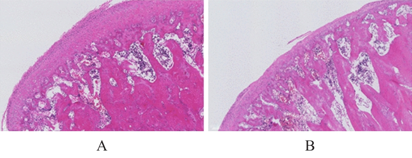
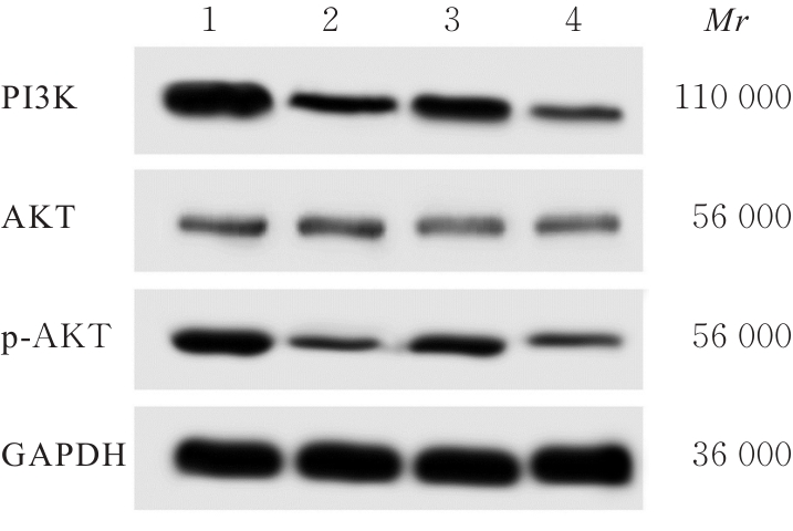
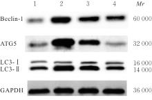
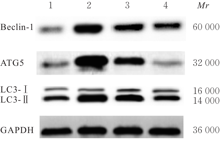
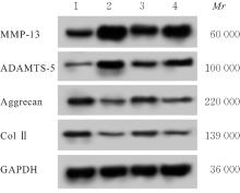
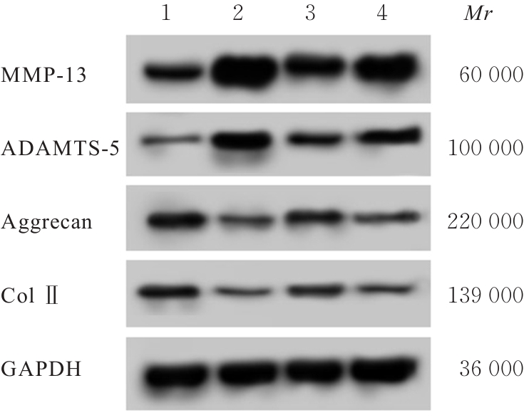
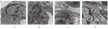
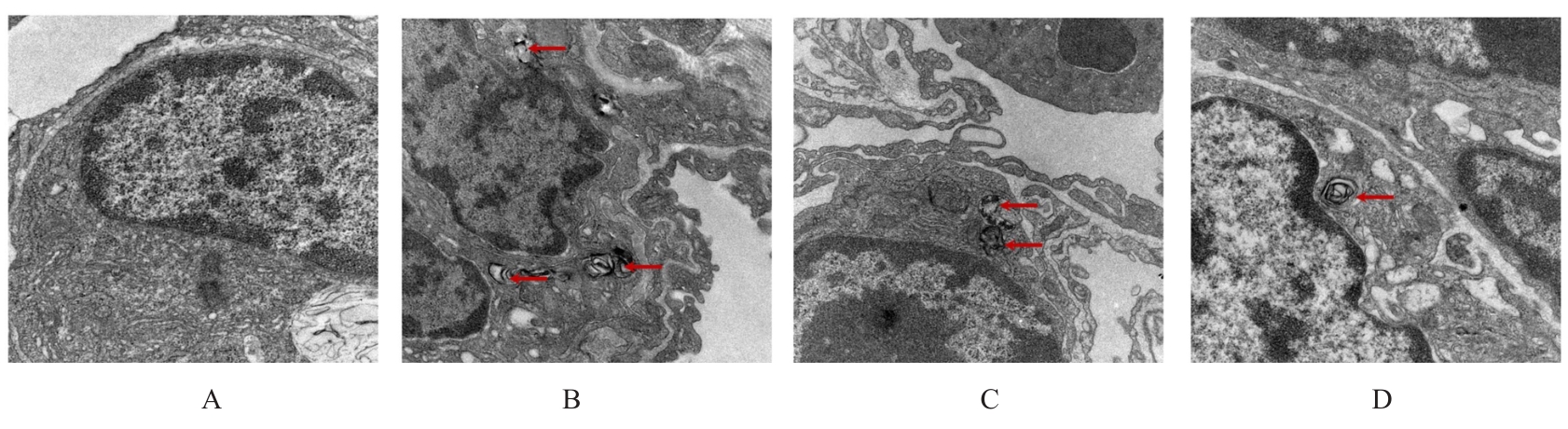
目的 探讨叶黄素对创伤性颞下颌关节强直(TMJA)兔颞下颌关节软骨组织中磷脂酰肌醇3-激酶(PI3K)/蛋白激酶B(AKT)信号通路和软骨细胞自噬的调控作用,并阐明其作用机制。 方法 32只雄性新西兰大白兔分为假手术组、模型组、叶黄素组和3-甲基腺嘌呤(3-MA)(PI3K/AKT信号通路抑制剂)+叶黄素组,每组8只。模型组、叶黄素组和3-MA+叶黄素组建立兔TMJA模型,假手术组兔仅暴露组织不进行手术。叶黄素组兔给予10 mg·kg-1叶黄素,3-MA+叶黄素组兔给予15 mg·kg-1 3-MA和10 mg·kg-1叶黄素。所有药物均于手术后24 h开始通过兔耳缘静脉进行注射,每周1次,连续注射3个月。完成后取手术侧兔颞下颌关节软骨组织,HE染色评估各组兔颞下颌关节软骨组织病理形态表现,Western blotting法检测各组兔颞下颌关节软骨组织中PI3K、AKT、磷酸化AKT(p-AKT)、Beclin-1、自噬相关蛋白5(ATG5)、微管相关蛋白1轻链3-Ⅰ(LC3-Ⅰ)、微管相关蛋白1轻链3-Ⅱ(LC3-Ⅱ)、自噬受体蛋白(P62)、基质金属蛋白酶13(MMP-13)、解聚蛋白酶5(ADAMTS-5)、蛋白聚糖(aggrecan)和Ⅱ型胶原(ColⅡ)蛋白表达水平,透射电子显微镜法检测各组兔颞下颌关节软骨组织中自噬小体数。 结果 与假手术组比较,模型组兔颞下颌关节软骨组织病理评分升高(P<0.05)。与假手术组比较,模型组兔颞下颌关节软骨组织中PI3K、p-AKT、aggrecan和ColⅡ蛋白表达水平降低(P<0.05),Beclin-1、ATG5、P62、MMP-13和ADAMTS-5蛋白表达水平及LC3-Ⅱ/LC3-Ⅰ比值升高(P<0.05);与模型组比较,叶黄素组兔颞下颌关节软骨组织中PI3K、p-AKT、aggrecan和Col Ⅱ蛋白表达水平升高(P<0.05),Beclin-1、ATG5和p62蛋白表达水平及LC3-Ⅱ/LC3-Ⅰ比值降低(P<0.05);与叶黄素组比较,3-MA+叶黄素组兔颞下颌关节软骨组织中PI3K、p-AKT、Beclin-1、ATG5和P62蛋白表达水平及LC3-Ⅱ/LC3-Ⅰ比值降低(P<0.05),MMP-13和ADAMTS-5蛋白表达水平升高(P<0.05),aggrecan和Col Ⅱ蛋白表达水平降低(P<0.05)。与假手术组比较,模型组兔颞下颌关节软骨组织中自噬小体数增多(P<0.05);与模型组比较,叶黄素组兔颞下颌关节软骨组织中自噬小体数减少(P<0.05);与叶黄素组比较,3-MA+叶黄素组兔颞下颌关节软骨组织中自噬小体数减少(P<0.05)。 结论 叶黄素通过调控PI3K/AKT信号通路和软骨细胞自噬改善TMJA兔的颞下颌关节软骨损伤。

关键词: 叶黄素, 磷脂酰肌醇3-激酶, 蛋白激酶B, 颞下颌关节强直, 软骨细胞自噬

Abstract:

Objective To discuss the regulatory effect of lutein on the phosphatidylinositol-3-kinase (PI3K)/protein kinase B (AKT) signaling pathway and chondrocyte autophagy in mandibular joint cartilage tissue of rabbits with traumatic temporomandibular joint ankylosis (TMJA), and to clarify its mechanism. Methods Thirty-two male New Zealand white rabbits were divided into sham operation group, model group, lutein group, and 3-MA (PI3K/AKT signaling pathway inhibitor) + lutein group, and there were 8 rabbits in each group. The rabbit model of TMJA was established in model group, lutein group, and 3-MA+lutein group, while the rabbits in sham operation group only underwent tissue exposure without surgery. The rabbits in lutein group were administered 10 mg·kg-1 lutein, and those in 3-MA+lutein group were administered 15 mg·kg-1 3-MA and 10 mg·kg?1 lutein. All the drugs were injected via the marginal ear vein starting 24 h after surgery, once a week for 3 consecutive months. After completion, the cartilage tissue on the surgical side was collected. HE staining was used to evaluate the patho morphology of the mandibular joint cartilage tissue of the rabbits in various groups; Western blotting method was used to detect the expression levels of PI3K, AKT, phosphorylated AKT (p-AKT), Beclin-1, autophagy-related protein 5 (ATG5), microtubule-associated protein 1 light chain 3-Ⅰ (LC3-Ⅰ), microtubule-associated protein 1 light chain 3-Ⅱ(LC3-Ⅱ), autophagy receptor protein (P62), matrix metalloproteinase 13 (MMP-13), a disintegrin and metalloproteinase with thrombospondin motifs-5 (ADAMTS-5), aggrecan, and type Ⅱ collagen (Col Ⅱ) proteins in temporomandibular joint cartilage tissue of the rabbits in various groups; transmission electron microscopy was used to detect the numbers of autophagosomes in temporomandibular joint cartilage tissue of the rabbits in various groups. Results Compared with sham operation group, the pathological score of mandibular joint cartilage tissue of the rabbits in model group was increased (P<0.05). The Western blotting results showed that compared with sham operation group, the expression levels of PI3K, p-AKT, aggrecan, and Col Ⅱ proteins in mandibular joint cartilage tissue of the rabbits in model group were decreased (P<0.05), while the expression levels of Beclin-1, ATG5, P62, MMP-13, ADAMTS-5 proteins and ratio of LC3-Ⅱ/LC3-Ⅰ were increased (P<0.05). Compared with model group, the expression levels of PI3K, p-AKT, aggrecan, and Col Ⅱ proteins in mandibular joint cartilage tissue of the rabbits in lutein group were increased (P<0.05), while the expression levels of Beclin-1, ATG5, P62 protein and ratio of LC3-Ⅱ/LC3-Ⅰ were decreased (P<0.05). Compared with lutein group, the expression levels of PI3K, p-AKT, Beclin-1, ATG5, and ratio of LC3-Ⅱ/LC3-Ⅰ, and P62 proteins in mandibular joint cartilage tissue of the rabbits in 3-MA+ lutein group were decreased (P<0.05), while the expression levels of MMP-13 and ADAMTS-5 proteins were increased (P<0.05), and the expression levels of aggrecan and Col Ⅱ proteins were decreased (P<0.05). The transmission electron microscope observation results showed that compared with sham operation group, the number of autophagosomes in temporomandibular joint cartilage tissue of the rabbits in model group was increased (P<0.05); compared with model group, the number of autophagosomes in temporomandibular joint cartilage tissue of the rabbits in lutein group was decreased (P<0.05); compared with lutein group, the number of autophagosomes in temporomandibular joint cartilage tissue of the rabbits in 3-MA+lutein group was decreased (P<0.05). Conclusion Lutein improves the mandibular joint cartilage tissue damage in the TMJA rabbits by regulating the PI3K/AKT signaling pathway and chondrocyte autophagy.

Key words: Lutein, Phosphatidylinositol 3-kinase, Protein kinase B, Temporomandibular joint ankylosis, Chondrocyte autophagy

中图分类号: 

  • R782.6