吉林大学学报(医学版) ›› 2025, Vol. 51 ›› Issue (5): 1194-1203.doi: 10.13481/j.1671-587X.20250505

• 基础研究 • 上一篇    

KLK5过表达对裸鼠皮下移植瘤生长及顺铂敏感性的影响

闫荣免1,孙新婷1,关欣1,程谕2,韩丽英1()   

  1. 1.吉林大学第二医院妇产科,吉林 长春 130041
    2.佳木斯大学附属第一医院妇产科,黑龙江 佳木斯 154002
  • 收稿日期:2025-03-24 接受日期:2025-06-09 出版日期:2025-09-28 发布日期:2025-11-05
  • 通讯作者: 韩丽英 E-mail:hanly@jlu.edu.cn
  • 作者简介:闫荣免(2000-),女,河南省南阳市人,在读硕士研究生,主要从事妇科肿瘤和盆底脏器脱垂诊治方面的研究。
  • 基金资助:
    吉林省科技厅自然科学基金项目(YDZJ202301ZYTS054)

Effects of KLK5 overexpression on growth of subcutaneous xenograft tumor and cisplatin sensitivity in nude mice

Rongmian YAN1,Xinting SUN1,Xin GUAN1,Yu CHENG2,Liying HAN1()   

  1. 1.Department of Obstetrics and Gynecology,Second Hospital,Jilin University,Changchun 130041,China
    2.Department of Obstetrics and Gynecology,First Hospital,Jiamusi University,Jiamusi 154002,China
  • Received:2025-03-24 Accepted:2025-06-09 Online:2025-09-28 Published:2025-11-05
  • Contact: Liying HAN E-mail:hanly@jlu.edu.cn

摘要:

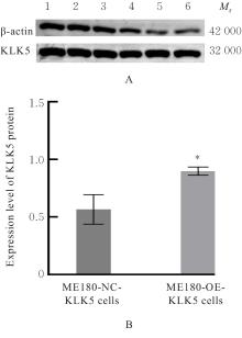
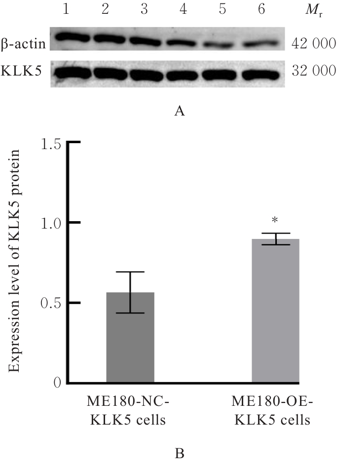
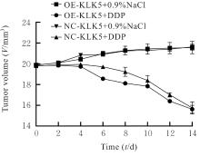
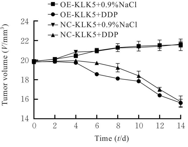
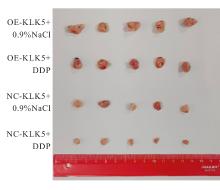
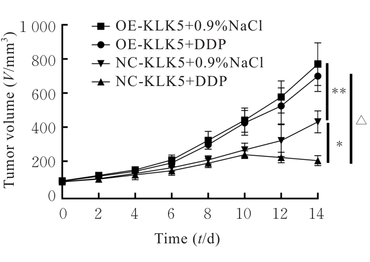
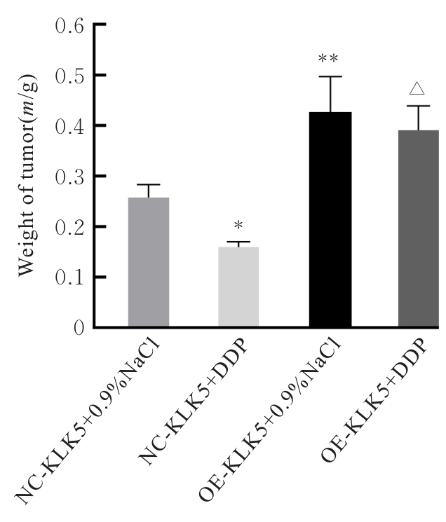
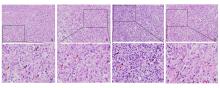
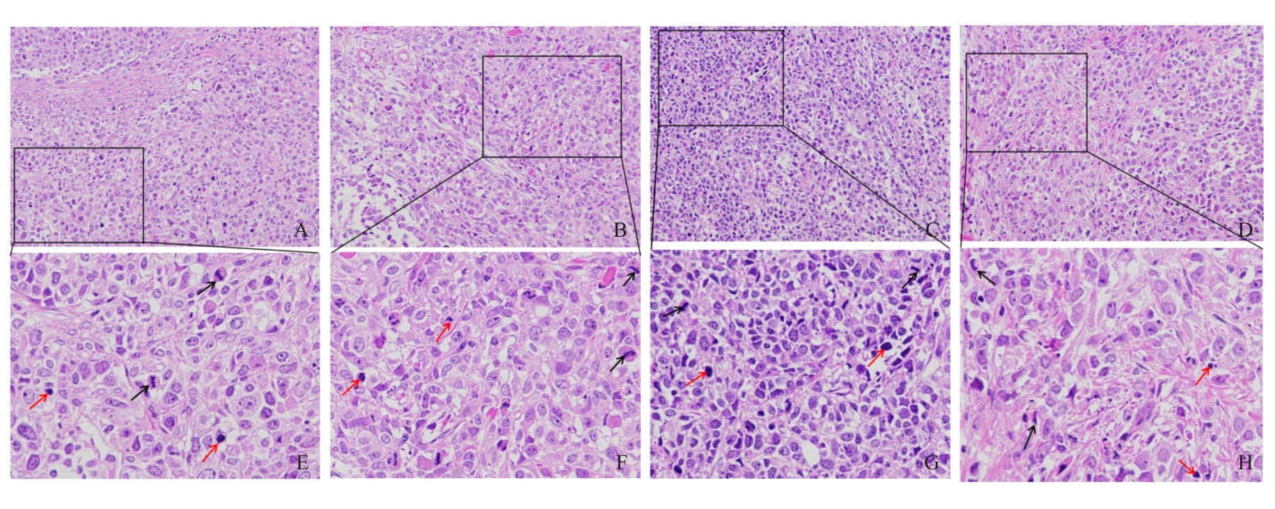
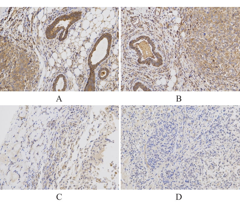
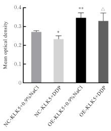
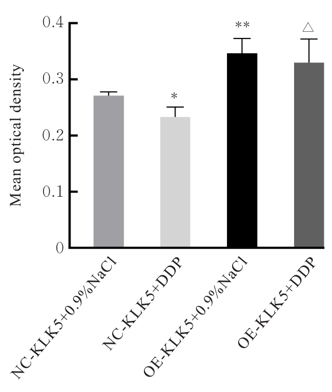
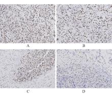
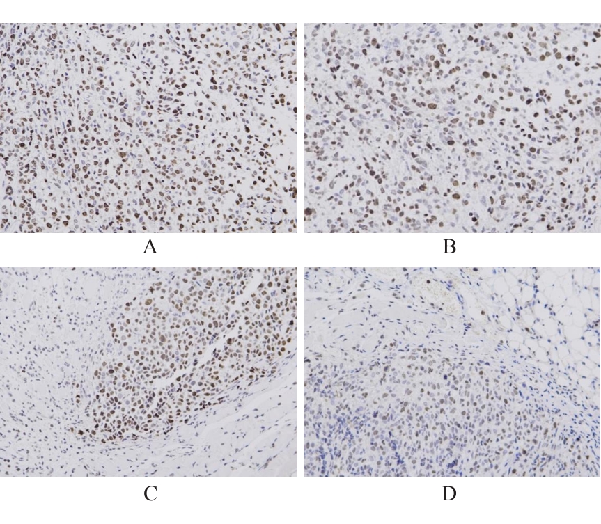
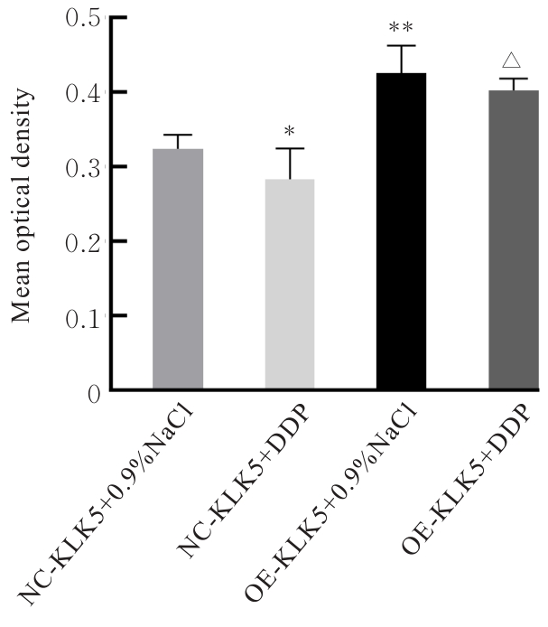
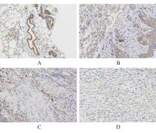
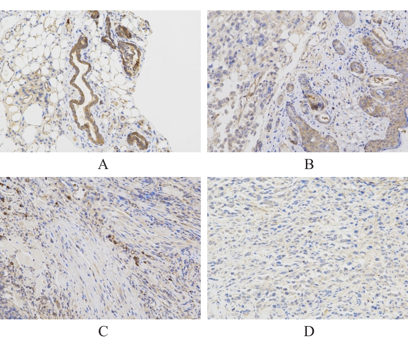
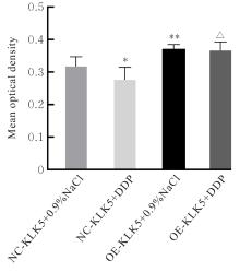
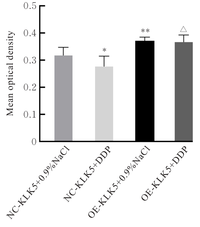
目的 探讨激肽释放酶5(KLK5)过表达对宫颈癌细胞的增殖、侵袭及顺铂(DDP)敏感性的影响,并阐明其作用机制。 方法 采用Western blotting法验证KLK5稳定转染过表达的宫颈癌细胞(ME180-OE-KLK5)。取对数生长期的宫颈癌ME180-NC-KLK5和OE-KLK5细胞,分别将其接种于裸鼠皮下建立皮下移植瘤模型,造模成功后裸鼠随机分为生理盐水对照组(NC-KLK5+0.9%NaCl组)、DDP治疗组(NC-KLK5+DDP组)、KLK5过表达组(OE-KLK5+0.9%NaCl组)和KLK5过表达联合DDP组(OE-KLK5+DDP组),每组5只。NC-KLK5+DDP组和OE-KLK5+DDP组裸鼠按照5 mg·kg-1的比例腹腔注射DDP;NC-KLK5+0.9%NaCl组和OE-KLK5+0.9%NaCl组裸鼠按照0.01 mL·g-1的比例腹腔注射生理盐水。每2 d称裸鼠质量,并记录瘤体的长径和短径,计算肿瘤体积,绘制瘤体生长曲线,于第14天给药结束后24 h,处死裸鼠,剥离瘤体并称质量。采用HE染色法观察各组裸鼠肿瘤组织病理形态表现,免疫组织化学染色法观察各组裸鼠肿瘤组织中KLK5、Ki67和基质金属蛋白酶9(MMP-9)蛋白表达水平。 结果 与ME180-NC-KLK5细胞比较,ME180-OE-KLK5细胞中KLK5蛋白表达水平升高(P<0.05)。皮下移植瘤种植后第1周,各组裸鼠进食和活动状态良好,体质量逐渐增长。第2周开始进入给药阶段,NC-KLK5+0.9%NaCl组裸鼠进食和活动状态及体质量较第1周无明显变化;与NC-KLK5+0.9%NaCl组比较,NC-KLK5+DDP组裸鼠开始出现食欲减退,体质量不增长,活动状态减弱;第3周药物治疗期间,NC-KLK5+0.9%NaCl组裸鼠进食及活动状态较第2周无明显变化,开始出现体质量不增长;与NC-KLK5+0.9%NaCl组比较,NC-KLK5+DDP组裸鼠进食和活动状态明显减弱,体质量降低。与NC-KLK5+0.9%NaCl组比较,NC-KLK5+DDP组裸鼠移植瘤体积减小(P<0.01);与NC-KLK5+DDP组比较,OE-KLK5+DDP组裸鼠移植瘤体积明显增大(P<0.001);与NC-KLK5+0.9%NaCl组比较,OE-KLK5+0.9%NaCl组裸鼠移植瘤体积增大(P<0.001);与OE-KLK5+0.9%NaCl组比较,OE-KLK5+DDP组裸鼠移植瘤体积差异无统计学意义(P>0.05)。与NC-KLK5+0.9%NaCl组比较,NC-KLK5+DDP组裸鼠移植瘤质量降低(P<0.05);与NC-KLK5+DDP组比较,OE-KLK5+DDP组裸鼠瘤质量明显升高(P<0.001);与NC-KLK5+0.9%NaCl组比较,OE-KLK5+0.9%NaCl组裸鼠瘤质量升高(P<0.001);与OE-KLK5+0.9%NaCl组比较,OE-KLK5+DDP组裸鼠瘤体质量差异无统计学意义(P>0.05)。与NC-KLK5+0.9%NaCl组比较,OE-KLK5+0.9%NaCl组裸鼠移植瘤细胞核异质性更强;OE-KLK5+DDP组和NC-KLK5+DDP组裸鼠移植瘤细胞出现形态学改变,表现为细胞核固缩、碎裂,肿瘤细胞体积缩小以及出现坏死和凋亡等。与NC-KLK5+DDP组比较,OE-KLK5+DDP组裸鼠移植瘤坏死程度更明显。与NC-KLK5+0.9%NaCl组比较,NC-KLK5+DDP组裸鼠移植瘤组织中KLK5、Ki67和MMP-9蛋白表达水平降低(P<0.05);与NC-KLK5+DDP组比较,OE-KLK5+DDP组裸鼠移植瘤组织中KLK5、Ki67和MMP-9蛋白表达水平升高(P<0.001);与NC-KLK5+0.9%NaCl组比较,OE-KLK5+0.9%NaCl组裸鼠移植瘤组织中KLK5、Ki67和MMP-9蛋白表达水平升高(P<0.001);与OE-KLK5+0.9%NaCl组比较,OE-KLK5+DDP组裸鼠移植瘤组织中KLK5、Ki67和MMP-9蛋白表达水平差异无统计学意义(P>0.05)。 结论 KLK5过表达可促进DDP处理的宫颈癌ME180细胞裸鼠皮下移植瘤的生长,上调移植瘤组织中Ki-67和MMP-9蛋白表达,降低移植瘤对DDP的敏感性。

关键词: 激肽释放酶5, 宫颈肿瘤, 顺铂, 基质金属蛋白酶9, ME180细胞, 移植瘤

Abstract:

Objective To discuss the effects of kallikrein 5 (KLK5) overexpression on the proliferation, invasion and cisplatin (DDP) sensitivity of cervical cancer cells, and to clarify its mechanism. Methods Western blotting method was used to verify the stable transfection and overexpression of KLK5 in the cervical cancer cell (ME180-OE-KLK5). The cervical cancer ME180-NC-KLK5 and ME180-OE-KLK5 cells in logarithmic growth phase were subcutaneously inoculated into the nude mice to establish the subcutaneous xenograft models. After successful modeling, the mice were randomly divided into normal saline control group (NC-KLK5+0.9% NaCl group), DDP treatment group (NC-KLK5+DDP group), KLK5 overexpression group(OE-KLK5+0.9% NaCl group) and KLK5 overexpression combined with DDP group(OE-KLK5+DDP group), with 5 mice in each group. The nude mice in NC-KLK5+DDP group and OE-KLK5+DDP group were given intraperitoneal injection of DDP at a dose of 5 mg·kg?1; the nude mice in NC-KLK5+0.9% NaCl group and OE-KLK5+0.9% NaCl group were given intraperitoneal injection of normal saline at a dose of 0.01 mL·g?1. The body weights of nude mice were measured every 2 d, and the long diameter and short diameter of the tumors were recorded to calculate the tumor volume and plot the tumor growth curve. At 24 h after the last administration on day 14, the nude mice were sacrificed, and the tumors were dissected and weighed. HE staining method was used to observe the pathomorphology of tumor tissue in the nude mice in various groups; immunohistochemistry staining method was used to observe the expression levels of KLK5, Ki67 and matrix metalloproteinase-9 (MMP-9) proteins in the tumor tissues of the nude mice in various groups. Results Compared with ME180-NC-KLK5 cells, the expression level of KLK5 protein in ME180-OE-KLK5 cells was increased (P<0.05). In the first week after subcutaneous xenograft inoculation, the nude mice in various groups showed good feeding and activity status, and their body weights gradually increased. The drug administration phase started from the second week. During the drug treatment period, the feeding and activity status as well as body weight of the nude mice in NC-KLK5+0.9%NaCl group showed no significant changes compared with the first week; compared with NC-KLK5+0.9%NaCl group, the nude mice in NC-KLK5+DDP group began to show loss of appetite, no increase in body weight, and decreased activity. During the drug treatment period in the third week, the feeding and activity status of the nude mice in NC-KLK5+0.9%NaCl group showed no significant changes compared with the second week, while they began to show no increase in body weight; compared with NC-KLK5+0.9%NaCl group, the feeding and activity status of the nude mice in NC-KLK5+DDP group were significantly weakened, and their body weights decreased. Compared with NC-KLK5+0.9%NaCl group, the volume of xenograft tumor in NC-KLK5+DDP group was decreased (P<0.01); compared with NC-KLK5+DDP group, the volume of xenograft tumor OE-KLK5+DDP group was significantly increased (P<0.001); compared with NC-KLK5+0.9%NaCl group, the volume of xenograft tumor of the nude mice in OE-KLK5+0.9%NaCl group was increased (P<0.001); compared with OE-KLK5+0.9%NaCl group, the volume of xenograft tumors in the nude mice in OE-KLK5+DDP group showed no statistically significant difference (P>0.05). Compared with NC-KLK5+0.9%NaCl group, the weight of xenograft tumor of the nude mice in NC-KLK5+DDP group was decreased (P<0.05); compared with NC-KLK5+DDP group, the weight of xenograft tumors of the nude mice in OE-KLK5+DDP group was significantly increased (P<0.001); compared with NC-KLK5+0.9%NaCl group, the weight of xenograft tumor of the nude mice in OE-KLK5+0.9%NaCl group was increased (P<0.001); compared with OE-KLK5+0.9%NaCl group, the weight of xenograft tumors of the nude mice in OE-KLK5+DDP group showed no statistically significant difference (P>0.05). Compared with NC-KLK5+0.9%NaCl group, the xenograft tumor cells of the nude mice in OE-KLK5+0.9%NaCl group showed greater nuclear heterogeneity; the xenograft tumor cells of the nude mice in OE-KLK5+DDP group and NC-KLK5+DDP group showed cytomorphological changes, manifested as nuclear pyknosis and fragmentation, reduced cell volume, and the appearance of necrosis and apoptosis. Compared with NC-KLK5+DDP group, the degree of necrosis in xenograft tumor of the nude mice in OE-KLK5+DDP group was more pronounced. Compared with NC-KLK5+0.9%NaCl group, the expression levels of KLK5, Ki67 and MMP-9 proteins in xenograft tumor tissue of the nude mice in NC-KLK5+DDP group were decreased (P<0.05); compared with NC-KLK5+DDP group, the expression levels of KLK5, Ki67, and MMP-9 proteins in xenograft tumor tissue of the nude mice in OE-KLK5+DDP group were increased (P<0.001); compared with NC-KLK5+0.9%NaCl group, the expression levels of KLK5, Ki67 and MMP-9 proteins in xenograft tumor tissue of the nude mice in OE-KLK5+0.9%NaCl group were increased (P<0.001); compared with OE-KLK5+0.9%NaCl group, the expression levels of KLK5, Ki67 and MMP-9 in xenograft tumor tissue of the nude mice in OE-KLK5+DDP group showed no statistically significant differences (P>0.05). Conclusion KLK5 overexpression can promote the growth of subcutaneous xenograft tumors of cervical cancer ME180 cells treated with DDP, up-regulate the expressions of Ki67 and MMP-9 in the xenograft tumor tissue, and reduce the sensitivity of the xenograft tumor to DDP.

Key words: Kallikrein 5, Uterine cervical neoplasm, Cisplatin, Matrix metalloproteinase-9, ME180 cells, Xenograft tumor

中图分类号: 

  • R737.33