吉林大学学报(医学版) ›› 2023, Vol. 49 ›› Issue (1): 8-14.doi: 10.13481/j.1671-587X.20230102

• 基础研究 •    下一篇

SO2对2型糖尿病大鼠心肌纤维化的改善作用及其机制

赵俊雄1,吴倩2,聂连桂1,刘盛权1,江振涛3,陈坚4,肖婷5,杨军1()   

  1. 1.南华大学衡阳医学院附属第一医院心内科,湖南 衡阳 421001
    2.南华大学衡阳医学院附属 第二医院全科,湖南 衡阳 421001
    3.南华大学衡阳医学院附属南华医院心内科,湖南 衡阳 421001
    4.南华大学衡阳医学院附属南华医院重症医学科,湖南 衡阳 421001
    5.广东医科大学 附属龙华区中心医院心内科,广东 深圳 518110
  • 收稿日期:2022-04-05 出版日期:2023-01-28 发布日期:2023-02-03
  • 通讯作者: 杨军 E-mail:yangjunketizu@163.com
  • 作者简介:赵俊雄(1995-),男,云南省昆明市人,在读硕士研究生,主要从事心血管系统疾病临床和基础方面的研究。
  • 基金资助:
    国家自然科学基金项目(81870230);湖南省卫健委临床重大专项项目(20201913);湖南省自然科学基金委员会科卫联合项目(2021JJ70035);湖南省自然科学基金委员会青年基金项目(2021JJ40499);广东省深圳市龙华区医疗卫生机构区级科研项目(2020035)

Ameliorative effect of SO2 on myocardial fibrosis in type 2 diabetes mellitus rats and its mechanism

Junxiong ZHAO1,Qian WU2,Liangui NIE1,Shengquan LIU1,Zhentao JIANG3,Jian CHEN4,Ting XIAO5,Jun YANG1()   

  1. 1.Department of Cardiology,First Affiliated Hospital,Hengyang Medical School,University of South China,Hengyang 421001,China
    2.Department of General Medicine,Second Affiliated Hospital,Hengyang Medical School,University of South China,Hengyang 421001,China
    3.Department of Cardiology,Affiliated Nanhua Hospital,Hengyang Medical School,University of South China,Hengyang 421001,China
    4.Department of Intensive Care Unit,Affiliated Nanhua Hospital,Hengyang Medical School,University of South China,Hengyang 421001,China
    5.Department of Cardiology,Affiliated Longhua Central Hospital,Guangdong Medical University,Shenzhen 518110,China
  • Received:2022-04-05 Online:2023-01-28 Published:2023-02-03
  • Contact: Jun YANG E-mail:yangjunketizu@163.com

摘要:

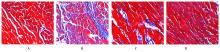
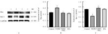
目的 探讨二氧化硫(SO2)对链脲佐菌素(STZ)和高糖高脂饮食诱导的2型糖尿病(T2DM)大鼠心肌纤维化的改善作用及对细胞凋亡和转化生长因子β1(TGF-β1)/Smad同源物2/3(Smad2/3)通路的影响,阐明SO2改善糖尿病心肌纤维化可能的分子机制。 方法 40只雄性SD大鼠随机分为对照组、T2DM组、T2DM+SO2组和SO2组。除对照组和SO2组外,T2DM组和T2DM+SO2组大鼠采用腹腔注射STZ(35 mg·kg-1)和高糖高脂饮食建立T2DM大鼠模型,T2DM+SO2组和SO2组大鼠腹腔注射外源性SO2供体[亚硫酸钠(Na2SO3)/亚硫酸氢钠(NaHSO3),85 mg·kg-1·d-1],共8周,采用Masson染色观察各组大鼠心肌组织纤维化病理形态表现,并计算各组大鼠心肌胶原容积分数,采用Western blotting法检测各组大鼠心肌组织中相关蛋白表达水平。 结果 与对照组比较,T2DM组大鼠心肌组织形态结构紊乱,心肌胶原纤维形成明显增加,心肌胶原容积分数明显升高(P<0.01),心肌组织中α平滑肌肌动蛋白(α-SMA)、Ⅲ型胶原(ColⅢ)、B细胞淋巴瘤2(Bcl-2)相关X蛋白(Bax)、TGF-β1、Smad2和Smad3蛋白表达水平明显升高(P<0.01),Bcl-2蛋白表达水平明显降低(P<0.01);与T2DM组比较,T2DM+SO2组大鼠心肌组织形态结构有所改善,心肌胶原纤维形成明显减少,心肌胶原容积分数明显降低(P<0.01),心肌组织中α-SMA、Col Ⅲ、Bax、TGF-β1、Smad2和Smad3蛋白表达水平明显降低(P<0.01),Bcl-2蛋白表达水平明显升高(P<0.01)。与对照组比较,SO2组大鼠上述各项指标差异均无统计学意义(P>0.05)。 结论 SO2可通过抑制促凋亡相关蛋白和TGF-β1/Smad2/3通路蛋白表达,缓解T2DM大鼠心肌纤维化。

关键词: 二氧化硫, 转化生长因子β1, Smad同源物2, Smad同源物3, 细胞凋亡, 心肌纤维化, 2型糖尿病

Abstract:

Objective To investigate the ameliorative effects of sulfur dioxide (SO2) on myocardial fibrosis in the type 2 diabetes mellitus (T2DM) rats induced by streptozocin (STZ) and high glucose and high fat diet and the effects on apoptosis and transforming growth factor-β1(TGF-β1)/Smad family member 2/3(Smad2/3) pathway, and to clarify the possible molecular mechanisms of SO2 in ameliorating diabetic myocardial fibrosis. Methods Forty male SD rats were randomly divided into control group, T2DM group, T2DM+SO2 group, and SO2 group. Except for control group and SO2 group, the rats in T2DM and T2DM+SO2 groups were used to establish the T2DM rat models by intraperitoneal injection of STZ (35 mg·kg-1) and high glucose and high fat diet, and the rats in T2DM+SO2 and SO2 groups were injected intraperitoneally with exogenous SO2 donors [sodium sulfite(Na2SO3)/sodium bisulfite(NaHSO3),85 mg·kg-1·d-1] for 8 weeks; the pathomorphology of myocardial fibrosis of the rats in various groups was observed by Masson staining, and the myocardial collagen volume fractions of the rats in various groups were calculated, and the expression levels of relevant proteins in myocardial tissue of the rats in various groups were detected by Western blotting method. Results Compared with control group, the myocardial tissue of the rats in T2DM group showed morphology and structural disorders, the myocardial collagen fiber formation was significantly increased, the myocardial collagen volume fraction was significantly increased (P<0.01), the expression levels of α-smooth muscle actin(α-SMA) and type Ⅲ collagen(Col Ⅲ),B-cell lymphoma-2(Bcl-2) associated X protein(Bax), TGF-β1, Smad2, and Smad3 proteins were significantly increased (P<0.01), and the expression level of Bcl-2 protein was significantly decreased (P<0.01). Compared with T2DM group, the morphological structure of myocardial tissue of the rats in T2DM+SO2 group was improved, the myocardial collagen fiber formation and the myocardial collagen volume fraction were significantly decreased(P<0.01), the expression levels of α-SMA, Col Ⅲ, Bax, TGF-β1, Smad2, and Smad3 proteins were significantly decreased(P<0.01),and the expression level of Bcl-2 protein was significantly increased (P<0.01). Compared with control group, there were no statistically significant differences in the above indexes of the rats in SO2 group rats(P>0.05). Conclusion SO2 can alleviate myocardial fibrosis in the T2DM rats by inhibiting the expressions of pro-apoptosis-related proteins and TGF-β1/Smad2/3 pathway proteins.

Key words: Sulfur dioxide, Transforming growth factor-β1, Smad family member 2, Smad family mumber 3, Apoptosis, Myocardial fibrosis, Type 2 diabetes mellitus

中图分类号: 

  • R587.1