Journal of Jilin University(Medicine Edition) ›› 2022, Vol. 48 ›› Issue (6): 1448-1454.doi: 10.13481/j.1671-587X.20220610
• Research in basic medicine • Previous Articles Next Articles
Preparation method of platelet-rich fibrin and hydroxyapatite complex and property evaluation
Qingyu ZHANG1,Tingrui XU1,Junjun JIAO2,Degeng XIA2,Tianyi ZHANG2,Li ZHANG1( ),Ning MA2()
- 1.Department of Emergency, Stomatology Hospital, Jilin University, Changchun 130021, China
2.Department of Periodontics, Stomatology Hospital, Jilin University, Changchun 130021, China
-
Received:2022-06-29Online:2022-11-28Published:2022-12-07 -
Contact:Li ZHANG,Ning MA E-mail:zhang_li99@jlu.edu.cn;man@jlu.edu.cn
CLC Number:
- R782.1
Cite this article
Qingyu ZHANG,Tingrui XU,Junjun JIAO,Degeng XIA,Tianyi ZHANG,Li ZHANG,Ning MA. Preparation method of platelet-rich fibrin and hydroxyapatite complex and property evaluation[J].Journal of Jilin University(Medicine Edition), 2022, 48(6): 1448-1454.
share this article

Fig. 1
Preparation of PRF/HA complex"
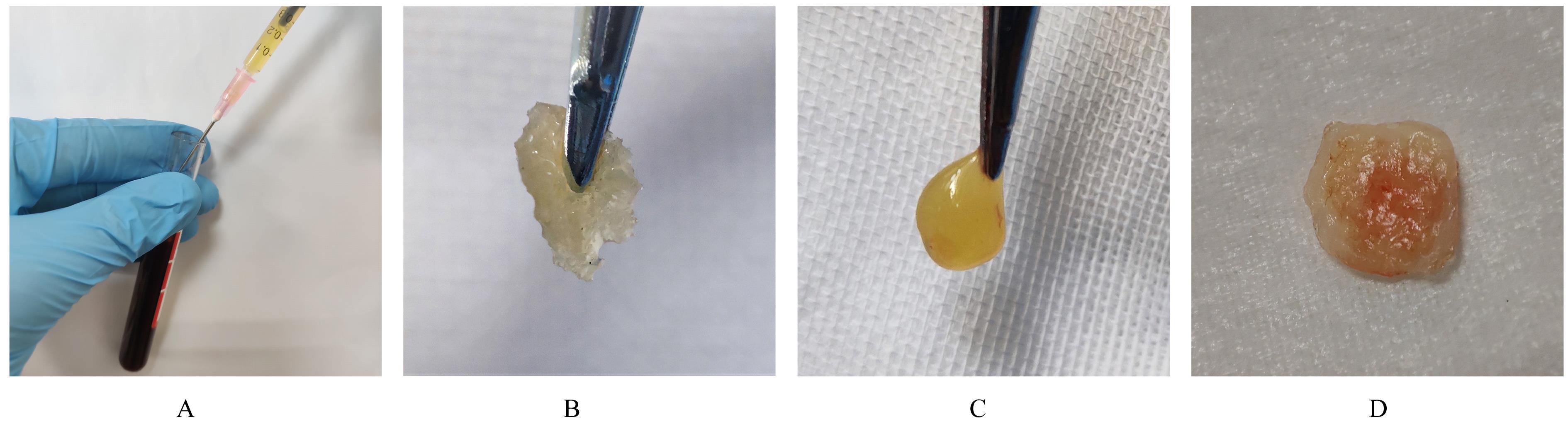

Fig.2
Microstructures of cross-section of PRF/HA complex observed by SEM"
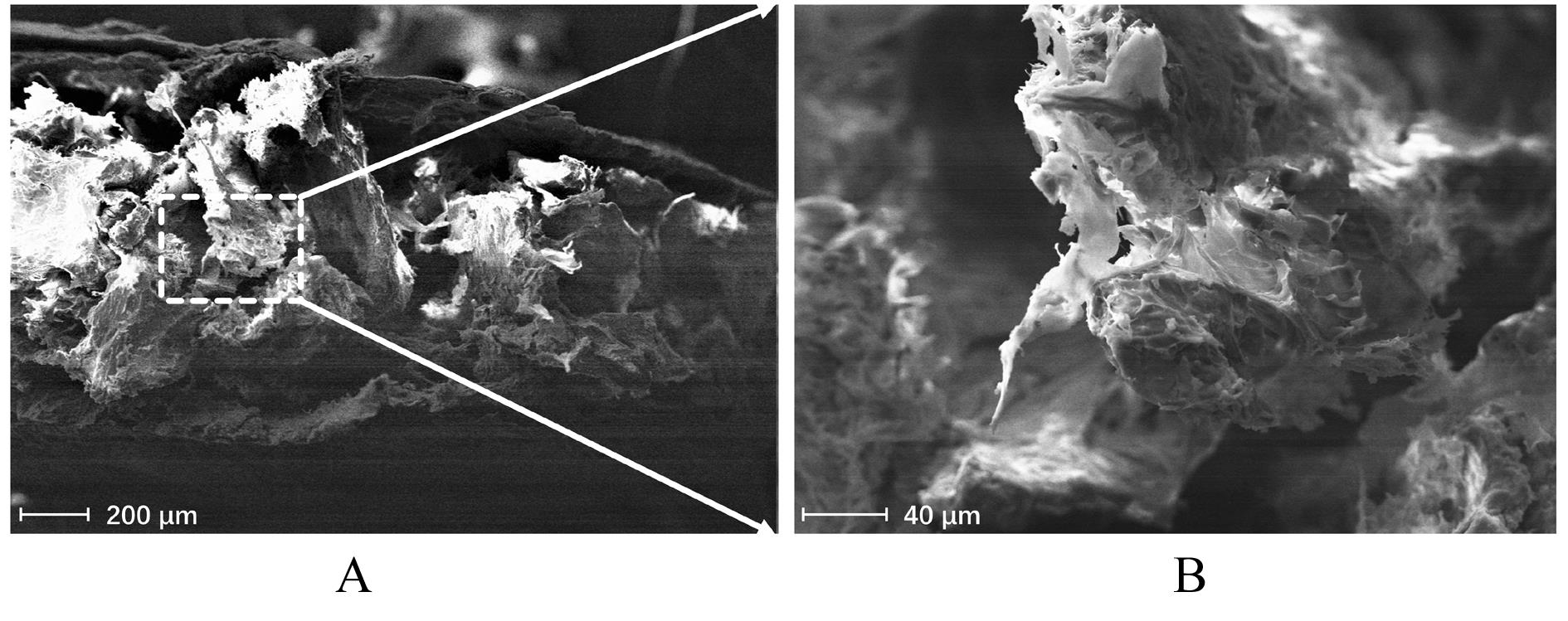
Fig. 3
Microstructures of PRF/HA complex after immersed in SBF for different time points observed by SEM (Bar=5 μm)"
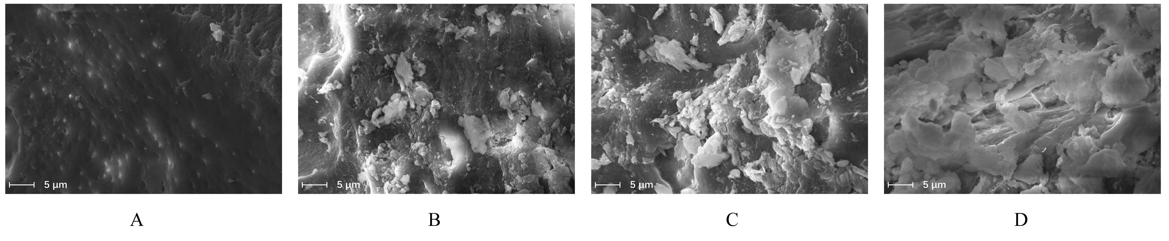
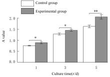
Fig. 4
Proliferation activities of MC3T3-E1 in various groups"
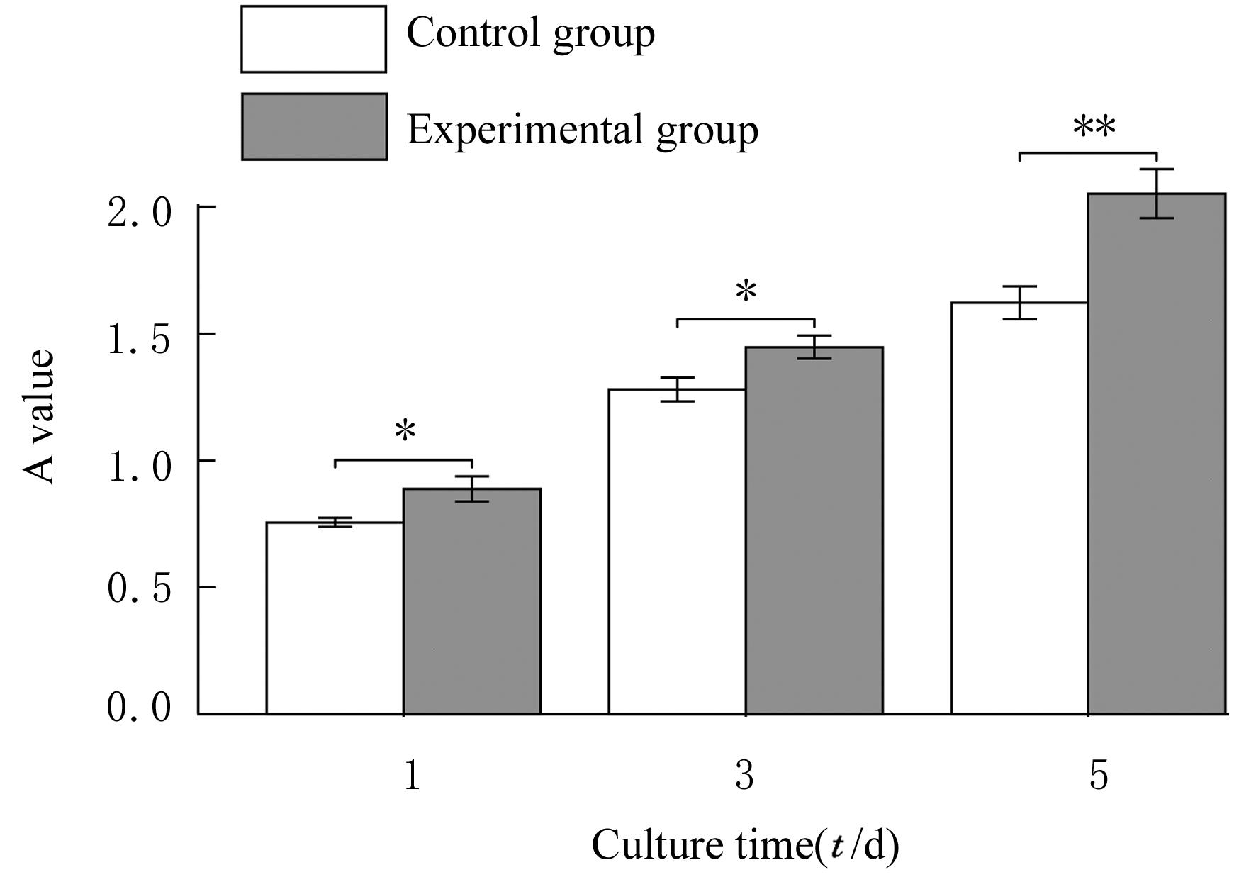
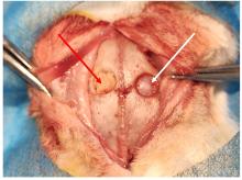
Fig. 5
Establishment of cranial defect model and implantation of material of white rabbits"


Fig. 6
X-ray images of cranial defect of white rabbits 4 and 8 weeks after operation in two groups"
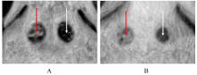
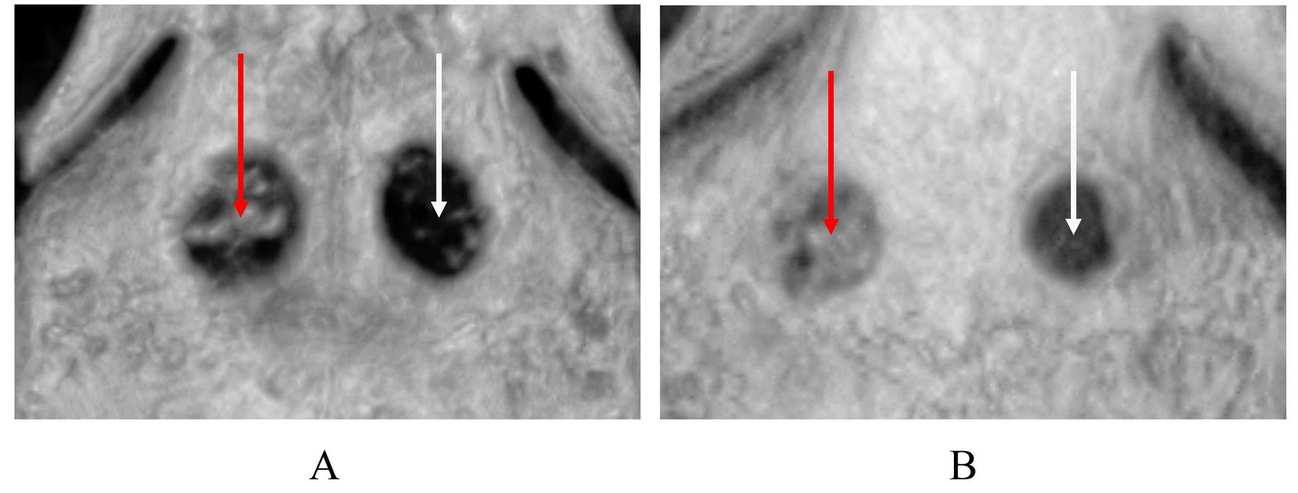
Fig. 7
CBCT images of cranial defect of white rabbits 4 and 8 weeks after operation"
| 1 | SCHORN L, FIENITZ T, DE DONNO F, et al. Critical-size defect augmentation using sintered and non-sintered bovine bone matrix - an experimental controlled study in minipigs[J]. J Oral Maxillofac Surg, 2021, 79(9): 1866-1873. |
| 2 | DUMITRESCU C R, NEACSU I A, SURDU V A, et al. Nano-hydroxyapatite vs. xenografts: synthesis, characterization, and in vitro behavior[J]. Nanomaterials (Basel), 2021, 11(9): 2289. |
| 3 | Moradi A, Pakizeh M, Ghassemi T. A review on bovine hydroxyapatite; extraction and characterization[J]. Biomed Phys Eng Express,2021,8(1):012001. |
| 4 | BATAS L, TSALIKIS L, STAVROPOULOS A. PRGF as adjunct to DBB in maxillary sinus floor augmentation: histological results of a pilot split-mouth study[J]. Int J Implant Dent, 2019, 5(1): 14. |
| 5 | LU D Z, ZHANG Y B, DONG W, et al. Effectiveness of strontium-doped brushite, bovine-derived hydroxyapatite and synthetic hydroxyapatite in rabbit sinus augmentation with simultaneous implant installation[J]. J Biomed Mater Res B Appl Biomater, 2020, 108(8): 3402-3412. |
| 6 | CECERSKA-HERYĆ E, GOSZKA M, SERWIN N, et al. Applications of the regenerative capacity of platelets in modern medicine[J]. Cytokine Growth Factor Rev, 2022, 64: 84-94. |
| 7 | AGRAWAL A A. Evolution, current status and advances in application of platelet concentrate in periodontics and implantology[J]. World J Clin Cases, 2017, 5(5): 159-171. |
| 8 | GHANAATI S, BOOMS P, ORLOWSKA A, et al. Advanced platelet-rich fibrin: a new concept for cell-based tissue engineering by means of inflammatory cells[J]. J Oral Implantol, 2014, 40(6): 679-689. |
| 9 | FARSHIDFAR N, AMIRI M A, JAFARPOUR D,et al.The feasibility of injectable PRF (I-PRF) for bone tissue engineering and its application in oral and maxillofacial reconstruction: from bench to chairside[J]. Biomater Adv, 2022, 134: 112557. |
| 10 | GARCÍA-GARETA E, COATHUP M J, BLUNN G W.Osteoinduction of bone grafting materials for bone repair and regeneration[J]. Bone, 2015, 81: 112-121. |
| 11 | KIM S Y, LEE Y J, CHO W T, et al. Preliminary animal study on bone formation ability of commercialized particle-type bone graft with increased operability by hydrogel[J]. Materials (Basel), 2021, 14(16): 4464. |
| 12 | LI Q, YU T, WANG F, et al. Endothelial progenitor cells with stem cells enhance osteogenic efficacy[J]. Am J Transl Res, 2020, 12(6): 2409-2424. |
| 13 | FUJIOKA-KOBAYASHI M, MIRON R J, HERNANDEZ M, et al. Optimized platelet-rich fibrin with the low-speed concept: growth factor release, biocompatibility, and cellular response[J].J Periodontol, 2017, 88(1): 112-121. |
| 14 | MIRON R J, FUJIOKA-KOBAYASHI M, HERNANDEZ M, et al. Injectable platelet rich fibrin (i-PRF): opportunities in regenerative dentistry? [J]. Clin Oral Investig, 2017, 21(8): 2619-2627. |
| 15 | SHAH R, GOWDA T M, THOMAS R, et al. Biological activation of bone grafts using injectable platelet-rich fibrin[J]. J Prosthet Dent, 2019, 121(3): 391-393. |
| 16 | KOKUBO T, YAMAGUCHI S. Simulated body fluid and the novel bioactive materials derived from it[J]. J Biomed Mater Res A, 2019, 107(5): 968-977. |
| 17 | ZADPOOR A A. Relationship between in vitro apatite-forming ability measured using simulated body fluid and in vivo bioactivity of biomaterials[J]. Mater Sci Eng C Mater Biol Appl, 2014, 35: 134-143. |
| 18 | KOKUBO T, TAKADAMA H. How useful is SBF in predicting in vivo bone bioactivity? [J]. Biomaterials, 2006, 27(15): 2907-2915. |
| 19 | OLIVEIRA É R, NIE L, PODSTAWCZYK D, et al. Advances in growth factor delivery for bone tissue engineering[J]. Int J Mol Sci, 2021, 22(2): 903. |
| 20 | MIRON R J, ZUCCHELLI G, PIKOS M A, et al. Use of platelet-rich fibrin in regenerative dentistry: a systematic review[J]. Clin Oral Investig, 2017, 21(6): 1913-1927. |
| 21 | DELGADO-RUIZ R A, CALVO-GUIRADO J L, ROMANOS G E. Critical size defects for bone regeneration experiments in rabbit calvariae: systematic review and quality evaluation using ARRIVE guidelines[J].Clin Oral Implants Res,2015,26(8): 915-930. |
| 22 | FEKRAZAD R, SADEGHI GHUCHANI M, ESLAMINEJAD M B, et al. The effects of combined low level laser therapy and mesenchymal stem cells on bone regeneration in rabbit calvarial defects[J]. J Photochem Photobiol B, 2015, 151: 180-185. |
| [1] | Xingzhu CHEN,Mingyue YU,Shuang LIU,Jianing LI,Zunxuan XIE,Jinyao LIU,Yuyan LIU. Remineralization property of fluoride loaded poly (propylene carbonate) dental patch and its cytotoxicity on fibroblast L929 cells of mice [J]. Journal of Jilin University(Medicine Edition), 2022, 48(6): 1429-1436. |
| [2] | Xianshun XIE,Wei WANG,Haibing JIANG. Effect of miR-431-3p on proliferation and apoptosis of gastric cancer cells and its mechanism of targeted regulation of CTDP1 gene expression [J]. Journal of Jilin University(Medicine Edition), 2022, 48(6): 1555-1565. |
| [3] | Hongxia SUN,Chunxu LIU,Xuejun AN,Guanghua CUI,Jingyu WANG,Shuangxi TONG,Xiaoqiu YANG. Effect of Schisandra chinensis polysaccharide on proliferation and apoptosis of human bladder cancer T24 cells and its mechanisms [J]. Journal of Jilin University(Medicine Edition), 2022, 48(5): 1216-1222. |
| [4] | Qian ZHANG,Jing LI. Inhibitory effect of TLR4 gene overexpression on autophagy of gastric cancer cells and its mechanism [J]. Journal of Jilin University(Medicine Edition), 2022, 48(5): 1238-1246. |
| [5] | Xiaomei HUANG,Hongwei WANG,Zhenyan HAN,Qiuyuan WEI,Sujing HUANG. Effects of lncRNA GHET1 on biological behaviors of trophoblast cells in preeclampsia through Wnt/β-catenin signaling pathway [J]. Journal of Jilin University(Medicine Edition), 2022, 48(5): 1324-1332. |
| [6] | Degeng XIA,Qingyu ZHANG,Junjun JIAO,Tingrui XU,Tianyi ZHANG,Yang ZHONG,Zhulan ZHAO,Ning MA,Li ZHANG. Multidisciplinary aesthetic restoration of anterior dental area of patient with failed treatment experience: A case report and literature review [J]. Journal of Jilin University(Medicine Edition), 2022, 48(4): 1058-1064. |
| [7] | Dongxiao BIAN,Xingfu BAO,Min HU. Promotion effect of rabbit acellular cartilage matrix particles combined with SD rat adipose tissue-derived stem cells on endochondral osteogenesis [J]. Journal of Jilin University(Medicine Edition), 2022, 48(4): 883-891. |
| [8] | Zhijuan WANG,Mingshu ZHANG,Liping YE. Effects of platelet-derived growth factor D on proliferation, migration and invasion of lung cancer H1299 cells through ERK signaling pathway and their mechanisms [J]. Journal of Jilin University(Medicine Edition), 2022, 48(4): 898-904. |
| [9] | Yandi MA,Xiangyun LU,Shangfeng HE,Xueyan YU,Yunhua HU,Haixia GAO,Yunzhao CHEN,Jie YU,Wenjie WANG,Feng LI,Xiaobin CUI. Expression of m6A methylation binding protein YTHDF2 in esophageal carcinoma tissue and its effect on proliferation and migration of esophageal carcinoma cells [J]. Journal of Jilin University(Medicine Edition), 2022, 48(4): 962-970. |
| [10] | Guowu WANG,Yuan YAO,Yu ZHANG,Na XU,Fang LIU. Inhibitory effect of miR-152 on proliferation and invasion of endometrial carcinoma cells by reducing low-density lipoprotein receptor expression [J]. Journal of Jilin University(Medicine Edition), 2022, 48(3): 591-599. |
| [11] | Jie PAN, Dezhou WANG, Wenzhi SONG. Repair effect of new nHA/PLGA nanocomposite microspheres on skull defects of rats [J]. Journal of Jilin University(Medicine Edition), 2022, 48(3): 615-621. |
| [12] | Guanhu LI,Qingxu LANG,Chunyan LIU,Qin LIU,Mengrou GENG,Xiaoqian LI,Zhenqi WANG. Inhibitory effect of valproic acid combined with X-ray irradiation on proliferation of breast cancer MDA-MB-231 cells and its mechanism [J]. Journal of Jilin University(Medicine Edition), 2022, 48(3): 622-629. |
| [13] | Peiyu YAN,Aichen ZHANG,Hong ZHANG,Yang LI,Mengmeng ZHANG,Mengze LUO,Ying PAN. Therapeutic effect of adipose-derived mesenchymal stem cells on premature ovarian failure model rats and its mechanism [J]. Journal of Jilin University(Medicine Edition), 2022, 48(3): 648-656. |
| [14] | Cuilan LIU,Fengai HU,Jing LIU,Dan WANG,Changyun QIU,Dunjiang LIU,Di ZHAO. Effect of adiponectin receptor agonist AdiopRon on biological behaviors of glioma cells and its mechanism [J]. Journal of Jilin University(Medicine Edition), 2022, 48(3): 702-710. |
| [15] | Zhuangzhi WU,Xiaoning HE,Siqi CHEN. Inhibitory effect of miR-124-3p on proliferation and invasion of oral squamous cell carcinoma cells and its mechanism [J]. Journal of Jilin University(Medicine Edition), 2022, 48(3): 718-727. |
|