Journal of Jilin University(Medicine Edition) ›› 2024, Vol. 50 ›› Issue (3): 749-758.doi: 10.13481/j.1671-587X.20240319
• Research in clinical medicine • Previous Articles
Bioinformatics analysis based on immune-related genes and immune cell infiltration of in-stent restenosis after percutaneous coronary intervention
Yufei FENG1,Shan JIN2,3,Yubing WANG2,3,Yinfei LU1,Lijuan PANG2,3,4( ),Kejian LIU1()
- 1.Department of Cardiology,First Affiliated Hospital,Shihezi University,Shihezi 832002,China
2.Department of Pathology,School of Medical Sciences,Shihezi University,Shihezi 832000,China
3.Department of Pathology,First Affiliated Hospital,Shihezi University,Shihezi 832000,China
4.Department of Pathology,Zhanjiang Central People’s Hospital,Zhanjiang City,Guangdong Province,Zhanjiang 524000,China
-
Received:2023-06-09Online:2024-05-28Published:2024-07-01 -
Contact:Lijuan PANG,Kejian LIU E-mail:ocean123456@163.com;25931884@qq.com
CLC Number:
- R392.12
Cite this article
Yufei FENG,Shan JIN,Yubing WANG,Yinfei LU,Lijuan PANG,Kejian LIU. Bioinformatics analysis based on immune-related genes and immune cell infiltration of in-stent restenosis after percutaneous coronary intervention[J].Journal of Jilin University(Medicine Edition), 2024, 50(3): 749-758.
share this article
Tab.1
Sample informations of GSE46560 dataset"
| Group | Sample | Source name | Sample type | Organism | Genotype | Age(year) |
|---|---|---|---|---|---|---|
Non-ISR | GSM1132406 | Blood | mRNA | Homo sapien | Male | 55 |
| GSM1132407 | Blood | mRNA | Homo sapien | Female | 64 | |
| GSM1132408 | Blood | mRNA | Homo sapien | Male | 75 | |
| GSM1132409 | Blood | mRNA | Homo sapien | Male | 34 | |
| GSM1132410 | Blood | mRNA | Homo sapien | Male | 78 | |
| GSM1132411 | Blood | mRNA | Homo sapien | Male | 56 | |
ISR | GSM1132412 | Blood | mRNA | Homo sapien | Female | 78 |
| GSM1132413 | Blood | mRNA | Homo sapien | Male | 58 | |
| GSM1132414 | Blood | mRNA | Homo sapien | Male | 50 | |
| GSM1132415 | Blood | mRNA | Homo sapien | Male | 56 | |
| GSM1132416 | Blood | mRNA | Homo sapien | Male | 60 |
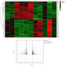
Fig. 1
DEGs of ISR in GSE46560 dataset"

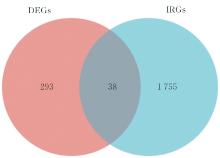
Fig. 2
DEIRGs of ISR in GSE46560 dataset"
Tab. 2
Top 10 DEIRGs of ISR in GSE46560 dataset"
| Rank | Gene | Log2FC | P |
|---|---|---|---|
| 1 | IL-1R1 | -0.729 984 494 | 0.001 589 458 |
| 2 | CCR3 | -0.848 170 488 | 0.009 625 487 |
| 3 | CD19 | 0.821 019 788 | 0.015 127 227 |
| 4 | SLPI | -1.580 539 076 | 0.016 843 607 |
| 5 | IFNA6 | 0.609 176 376 | 0.023 118 319 |
| 6 | TNFRSF9 | -0.586 127 630 | 0.023 564 212 |
| 7 | SYK | -0.692 433 588 | 0.029 734 058 |
| 8 | IL-17RA | -0.549 992 248 | 0.032 917 596 |
| 9 | IL-10RB | -0.572 143 770 | 0.042 174 389 |
| 10 | IFNAR1 | -0.782 222 264 | 0.048 843 759 |
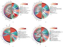
Fig. 3
GO functional and KEGG signaling pathway enrichment analysis on DEIRGs"
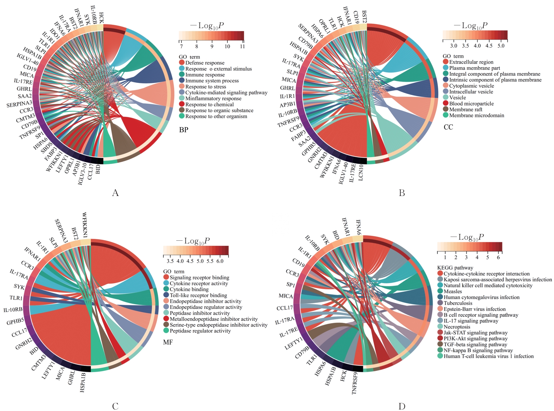

Fig. 4
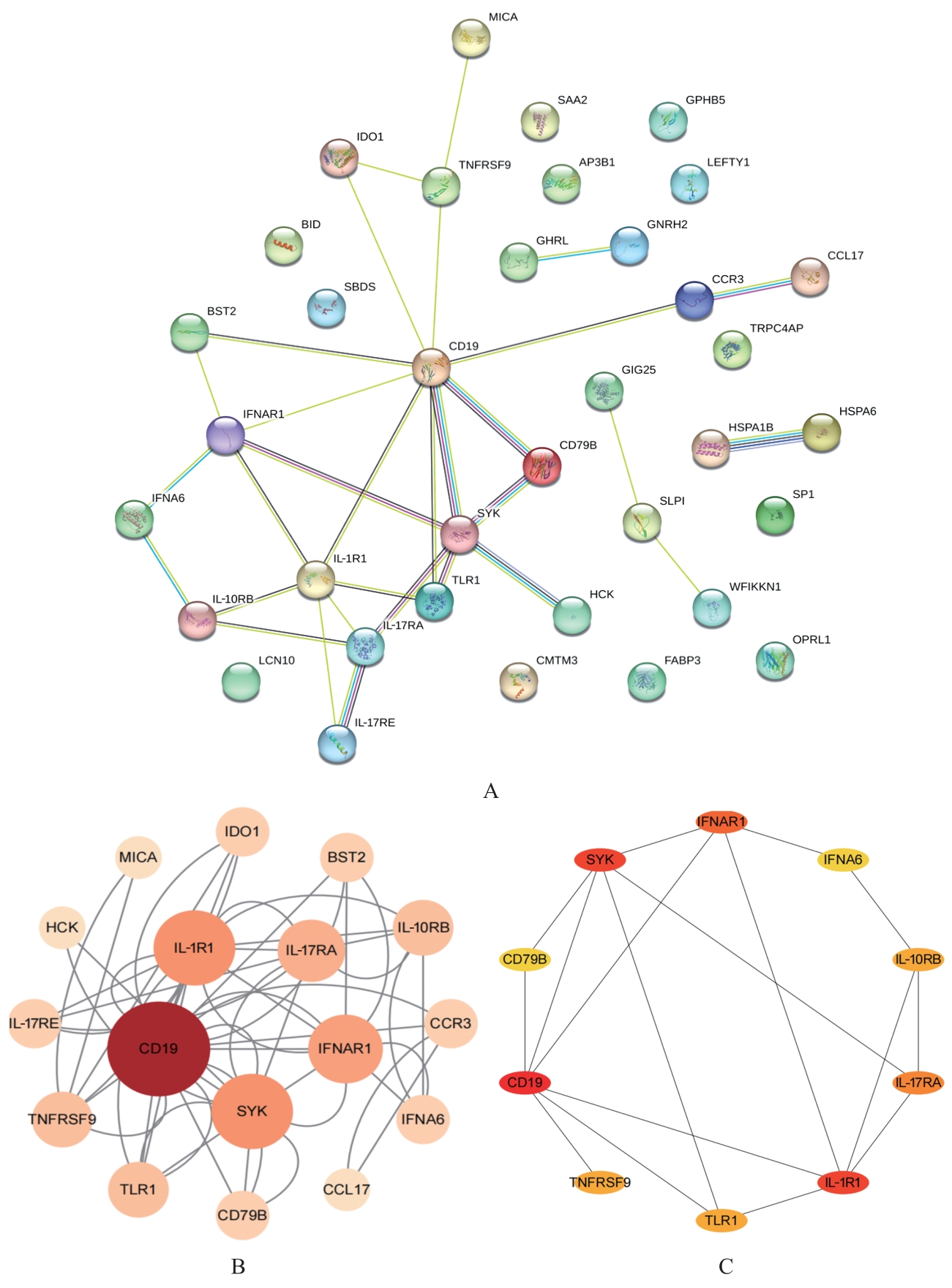
PPI network map and DEIRGs visualization and top 10 Hub genes"
Tab.3
Top 10 Hub genes screened by CytoHubba plugin"
| Rank | Gene | Betweenness |
|---|---|---|
| 1 | CD19 | 140.833 330 |
| 2 | IL-1R1 | 49.333 332 |
| 3 | SYK | 47.166 668 |
| 4 | IFNAR1 | 30.500 000 |
| 5 | TNFRSF9 | 30.000 000 |
| 6 | CCR3 | 30.000 000 |
| 7 | IL-17RA | 12.666 667 |
| 8 | IL-10RB | 4.833 334 |
| 9 | SLPI | 2.000 000 |
| 10 | IFNA6 | 1.666 667 |

Fig. 5
Validation of CD19 mRNA expression amount and evaluation on diagnostic value"
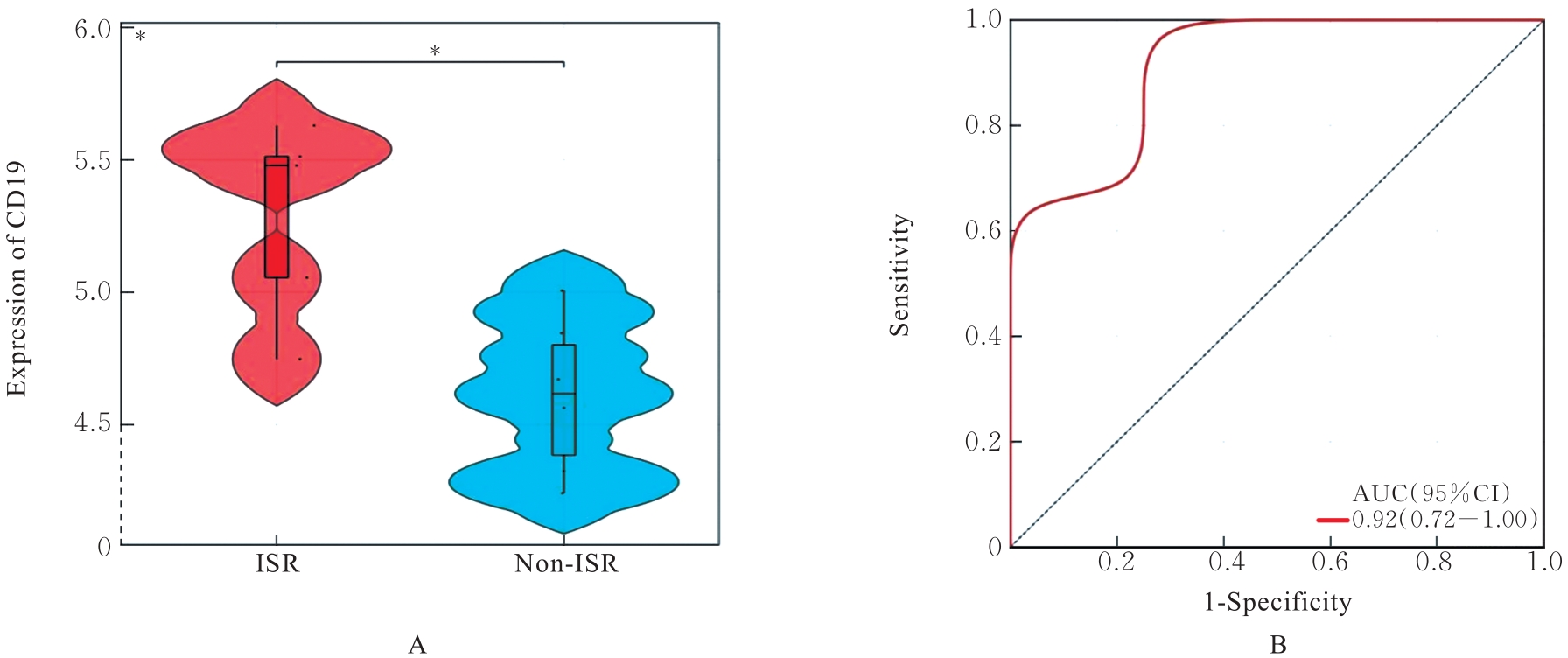
Fig. 6
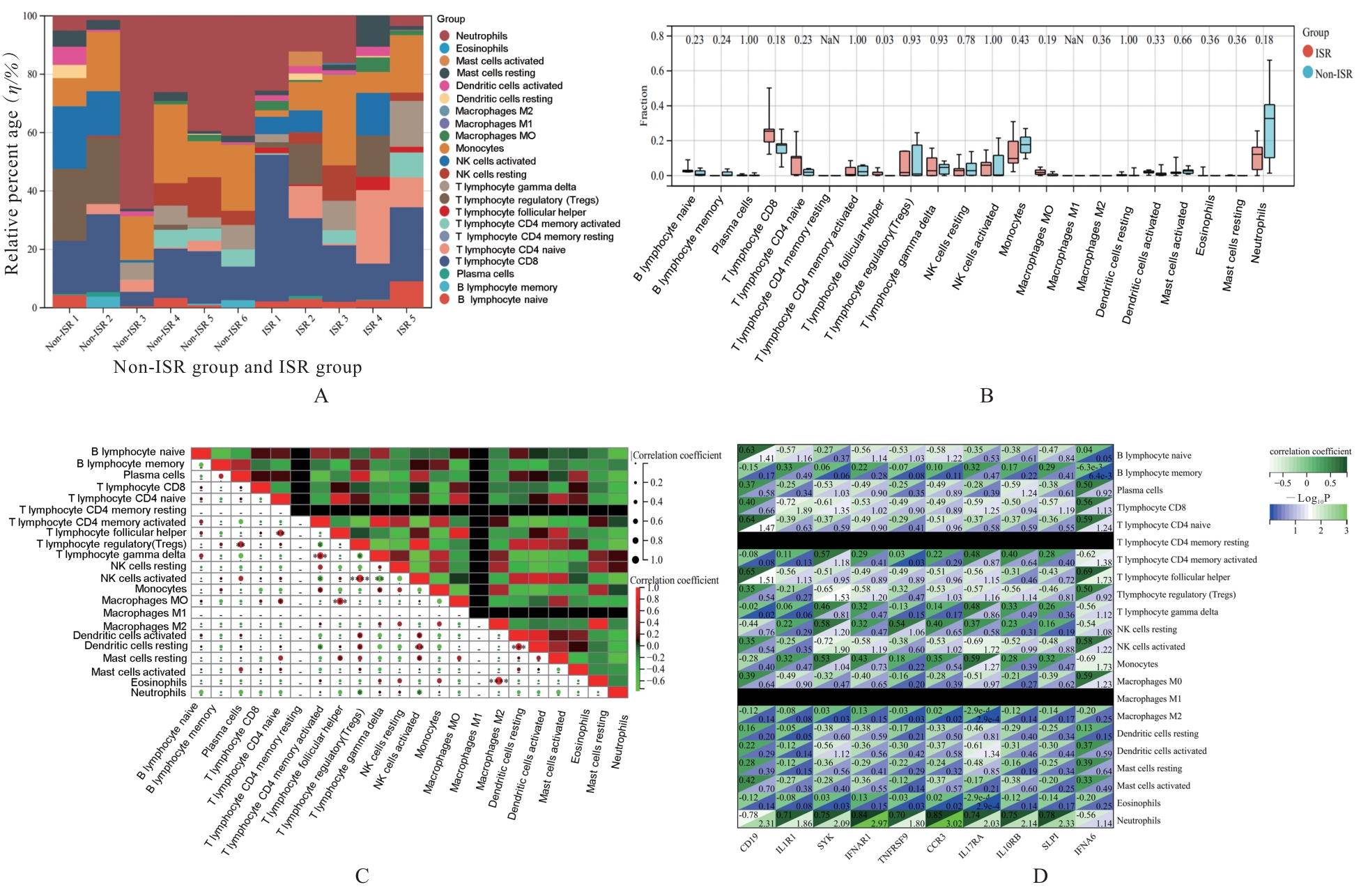
Immune cell infiltration analysis"
| 1 | HOARE D, BUSSOOA A, NEALE S, et al. The future of cardiovascular stents: bioresorbable and integrated biosensor technology[J]. Adv Sci, 2019, 6(20): 1900856. |
| 2 | ALFONSO F, GONZALO N, RIVERO F, et al. The year in cardiovascular medicine 2020: interventional cardiology[J]. Eur Heart J, 2021, 42(10): 985-1003. |
| 3 | ULLRICH H, OLSCHEWSKI M, MÜNZEL T, et al. Coronary In-stent restenosis: predictors and treatment[J]. Dtsch Arztebl Int, 2021, 118(38): 637-644. |
| 4 | YERASI C, CASE B C, FORRESTAL B J, et al. Drug-coated balloon for De Novo Coronary artery disease: JACC state-of-the-art review[J]. J Am Coll Cardiol, 2020, 75(9): 1061-1073. |
| 5 | NICCOLI G, MONTONE R A, SABATO V, et al. Role of allergic inflammatory cells in coronary artery disease[J]. Circulation, 2018, 138(16): 1736-1748. |
| 6 | BANAI S, FINKELSTEIN A, ALMAGOR Y, et al. Targeted anti-inflammatory systemic therapy for restenosis: The Biorest Liposomal Alendronate with Stenting Study (BLAST)-a double blind, randomized clinical trial[J]. Am Heart J, 2013, 165(2): 234-240. |
| 7 | YAO Y, YAN Z Y, LIAN S L, et al. Prognostic value of novel immune-related genomic biomarkers identified in head and neck squamous cell carcinoma[J]. J Immunother Cancer, 2020, 8(2): e000444. |
| 8 | 范吉林, 朱婷婷, 田晓玲, 等. 基于房颤中circRNA-miRNA-mRNA网络构建和免疫细胞浸润的生物信息学分析[J]. 吉林大学学报(医学版), 2022, 48(6): 1535-1545. |
| 9 | FAN J X, CAO S, CHEN M Y, et al. Investigating the AC079305/DUSP1axis as oxidative stress-related signatures and immune infiltration characteristics in ischemic stroke[J]. Oxid Med Cell Longev, 2022, 2022: 8432352. |
| 10 | 张德洪, 郑明珠, 李家秋, 等. 基于MSR1 mRNA和蛋白在泛癌组织中表达的生物信息学分析及其意义[J]. 吉林大学学报(医学版), 2023, 49(2): 425-439. |
| 11 | 赵志娟, 孟 莲, 刘春霞. 基于融合基因作用于横纹肌肉瘤的miRNA-mRNA调控网络的生物信息学分析[J]. 吉林大学学报(医学版), 2022, 48(1): 154-162. |
| 12 | GIUSTINO G, COLOMBO A, CAMAJ A, et al. Coronary In-stent restenosis: JACC state-of-the-art review[J]. J Am Coll Cardiol, 2022, 80(4): 348-372. |
| 13 | COLLET C, GRUNDEKEN M J, ASANO T, et al. State of the art: coronary angiography[J]. EuroIntervention, 2017, 13(6): 634-643. |
| 14 | DEMYANETS S, TENTZERIS I, JARAI R, et al. An increase of interleukin-33 serum levels after coronary stent implantation is associated with coronary in-stent restenosis[J]. Cytokine, 2014, 67(2): 65-70. |
| 15 | SCHILLINGER M, MINAR E. Restenosis after percutaneous angioplasty: the role of vascular inflammation[J]. Vasc Health Risk Manag, 2005, 1(1): 73-78. |
| 16 | DEL PORTO F, CIFANI N, PROIETTA M, et al. Lag3+ regulatory T lymphocytes in critical carotid artery stenosis[J]. Clin Exp Med, 2019, 19(4): 463-468. |
| 17 | DEL PORTO F, CIFANI N, PROIETTA M, et al. Regulatory T CD4+ CD25+ lymphocytes increase in symptomatic carotid artery stenosis[J]. Ann Med, 2017, 49(4): 283-290. |
| 18 | LI Q Q, LI Y T, HUANG W, et al. Loss of lipocalin 10 exacerbates diabetes-induced cardiomyopathy via disruption of Nr4a1-mediated anti-inflammatory response in macrophages[J]. Front Immunol, 2022, 13: 930397. |
| 19 | HAOHAN S, PUSSADHAMMA B, JUMNAINSONG A, et al. Association of major histocompatibility complex class I related chain A/B positive microparticles with acute myocardial infarction and disease severity[J]. Diagnostics, 2020, 10(10): 766. |
| 20 | GORCZYNSKI R M. IL-17 signaling in the tumor microenvironment[J]. Adv Exp Med Biol, 2020, 1240: 47-58. |
| 21 | DI SALVO T G, YANG K C, BRITTAIN E, et al. Right ventricular myocardial biomarkers in human heart failure[J]. J Card Fail, 2015, 21(5): 398-411. |
| 22 | ALIMADADI A, MUNROE P B, JOE B, et al. Meta-analysis of dilated cardiomyopathy using cardiac RNA-seq transcriptomic datasets[J]. Genes, 2020, 11(1): 60. |
| 23 | ZHANG B, YAO R J, HU C, et al. Epigallocatechin gallate mediated sandwich-like coating for mimicking endothelium with sustained therapeutic nitric oxide generation and heparin release[J]. Biomaterials, 2021, 269: 120418. |
| 24 | LU H K, HUANG Y, LIANG X Y, et al. Pinellia ternata attenuates carotid artery intimal hyperplasia and increases endothelial progenitor cell activity via the PI3K/Akt signalling pathway in wire-injured rats[J]. Pharm Biol, 2020, 58(1): 1184-1191. |
| 25 | TANG Z H, WANG Y, FAN Y B, et al. Suppression of c-Cbl tyrosine phosphorylation inhibits neointimal formation in balloon-injured rat arteries[J]. Circulation, 2008, 118(7): 764-772. |
| 26 | KHAN R, AGROTIS A, BOBIK A. Understanding the role of transforming growth factor-beta1 in intimal thickening after vascular injury[J]. Cardiovasc Res, 2007, 74(2): 223-234. |
| 27 | WANG S X, LINCOLN T M, MURPHY-ULLRICH J E. Glucose downregulation of PKG-I protein mediates increased thrombospondin1-dependent TGF-{beta}activity in vascular smooth muscle cells[J]. Am J Physiol Cell Physiol, 2010, 298(5): C1188-C1197. |
| 28 | LI X C, DING Y, ZI M T, et al. CD19, from bench to bedside[J]. Immunol Lett, 2017, 183: 86-95. |
| 29 | WENG R Q, LIU S D, GU X D, et al. Clonal diversity of the B cell receptor repertoire in patients with coronary in-stent restenosis and type 2 diabetes[J]. Open Life Sci, 2021, 16(1): 884-898. |
| 30 | FUJIMOTO M, POE J C, HASEGAWA M, et al. CD19 regulates intrinsic B lymphocyte signal transduction and activation through a novel mechanism of processive amplification[J]. Immunol Res, 2000, 22(2/3): 281-298. |
| 31 | FUJIMOTO M, FUJIMOTO Y, POE J C, et al. CD19 regulates Src family protein tyrosine kinase activation in B lymphocytes through processive amplification[J]. Immunity, 2000, 13(1): 47-57. |
| 32 | DEVEZA R C, ELKINS M, SARAGIOTTO B T. PEDro systematic review update: exercise for coronary heart disease[J]. Br J Sports Med, 2017, 51(9): 755-756. |
| 33 | CHOE N, KWON J S, KIM Y S, et al. The microRNA miR-34c inhibits vascular smooth muscle cell proliferation and neointimal hyperplasia by targeting stem cell factor[J]. Cell Signal, 2015, 27(6): 1056-1065. |
| 34 | CROTTY S. T follicular helper cell biology: a decade of discovery and diseases[J]. Immunity, 2019, 50(5): 1132-1148. |
| 35 | PREITE S, HUANG B, CANNONS J L, et al. PI3K orchestrates T follicular helper cell differentiation in a context dependent manner: implications for autoimmunity[J]. Front Immunol, 2018, 9: 3079. |
| [1] | Zixu YANG,Chang SU,Boyuan WANG,Chong LIU,Minghe LI. Bioinformatics analysis on molecular subtypes and clinical characteristics of head and neck squamous cell carcinoma based on genes associated with lactate metabolism [J]. Journal of Jilin University(Medicine Edition), 2024, 50(1): 198-207. |
| [2] | Hui YE,Zhe SUN,Liting ZHOU,Wen QI,Lin YE. Bioinformatics analysis on differentially expressed genes in lung adenocarcinoma based on GEO and TCGA Databases [J]. Journal of Jilin University(Medicine Edition), 2023, 49(6): 1491-1503. |
| [3] | Haikang CUI,Xudong ZHANG,Xiaoning LI,Xi YANG,Lan YANG,Wenjie ZHANG. Bioinformatics analysis on predition effect of subtypes of cell pyroptosis and APOD on prognosis of gastric cancer patients [J]. Journal of Jilin University(Medicine Edition), 2023, 49(5): 1268-1279. |
| [4] | Yaqi XU,Yanyu WANG,Wenjing ZHANG,Mei HAN,Huaxia MU,Xi YANG,Weixiao BU,Zikun TAO,Yujia KONG,Fuyan SHI,Suzhen WANG. Bioinformatics analysis on screening of key genes of hepatitis B virus-related hepatocellular carcinoma and its relationship with prognosis [J]. Journal of Jilin University(Medicine Edition), 2023, 49(5): 1243-1252. |
| [5] | Zhiyuan XIAO,Bing SONG,Xinyu MA,Lianhui JIN,Tong ZHENG,Fang CHAI. Bioinformatics analysis on expression of apoptosis related gene CD44 in thyroid carcinoma tissue and its relationship with tumor invasion and immune cell infiltration [J]. Journal of Jilin University(Medicine Edition), 2023, 49(2): 473-481. |
| [6] | Haiwei QUAN,Yali ZHANG,Han WANG,Yijiu AI,Yutong ZOU,Wei XIA. Detection of expression level of miR-4286 in exosome in peripheral blood of patients with acute myeloid leukemia and its diagnostic value [J]. Journal of Jilin University(Medicine Edition), 2023, 49(2): 460-466. |
| [7] | Xiaoyan WANG,Yihong HU,Yucheng HAN,Xianqiong ZOU. Bioinformatics analysis on mechanism of COMMD7 in occurrence and development of brain low-grade glioma [J]. Journal of Jilin University(Medicine Edition), 2023, 49(2): 414-424. |
| [8] | Jilin FAN,Tingting ZHU,Xiaoling TIAN,Sijia LIU,Jing SU,Shiliang ZHANG. Bioinformatics analysis based on circRNA-miRNA-mRNA network construction and immune cell infiltration in atrial fibrillation [J]. Journal of Jilin University(Medicine Edition), 2022, 48(6): 1535-1545. |
| [9] | Ying ZHAO,Danyu ZHAO,Chao LIU. Bioinformatics analysis on prognostic evaluation value of TXNDC11 gene in pan-cancer and its immunity regulation [J]. Journal of Jilin University(Medicine Edition), 2022, 48(1): 142-153. |
| [10] | Weihang LI,Ziyi DING,Dong WANG,Yikai PAN,Yuhui LIU,Shilei ZHANG,Jing LI,Ming YAN. Bioinformatics analysis based on screening of core driving genes in osteosarcoma and construction of gene model for prediction of survival time of patients [J]. Journal of Jilin University(Medicine Edition), 2021, 47(6): 1570-1580. |
| [11] | Lan ZHENG,Songzhe PIAO,Ran XU,Xinyue WANG,Yixuan WANG,Zhenhua LIN,Yang YANG. Bioinformatics analysis based on effect of expression levels of m6A regulators on immune infiltration and prognosis of uterine corpus endometrial carcinoma [J]. Journal of Jilin University(Medicine Edition), 2021, 47(2): 438-452. |
| [12] | CUI Yan, SHI Yongfeng, GUO Ziyuan, LIU Bin, WANG Jinpeng, ZHAO Lei, WANG Junnan, PIAO Jinhua. Application of intravascular ultrasound in analysis on influencing factors of prognosis in patients with different coronary artery in-stent restenosis [J]. Journal of Jilin University Medicine Edition, 2016, 42(04): 746-752. |
| [13] | HE Meng-zi,ZHAO Yin-long,LIU Xiao-dong,MA Shu-mei,LIU Xin. Screening and prediction of differential expression miRNA and their target genes in papillary thyroid carcinoma tissue [J]. Journal of Jilin University Medicine Edition, 2013, 39(1): 99-103. |
| [14] | YU Jing-Hong, LI Huan, SUN Wei-Qi, XU Xiao-Lei, GAO Jiu-Chun. Analysis of levels of |microelements as biomarker ininjury induced by formaldehyde [J]. J4, 2009, 35(6): 1065-1067. |
|
||