Journal of Jilin University(Medicine Edition) ›› 2024, Vol. 50 ›› Issue (6): 1481-1490.doi: 10.13481/j.1671-587X.20240601
• Research in basic medicine •
Inhibitory effect of diosmetin on ferroptosis of GC-2 spermatocytes induced by RSL3 in mice and its mechanism
Baolian MA,Xiaoxue HU,Xiaowen AI,Yonglan ZHANG( )
)
- Department of Pharmacology,School of pharmacy and Bioengineering,Chongqing University of Technology,Chongqing 400054,China
-
Received:2023-11-28Online:2024-11-28Published:2024-12-10 -
Contact:Yonglan ZHANG E-mail:lanzy2015@cqut.edu.cn
CLC Number:
- R966
Cite this article
Baolian MA,Xiaoxue HU,Xiaowen AI,Yonglan ZHANG. Inhibitory effect of diosmetin on ferroptosis of GC-2 spermatocytes induced by RSL3 in mice and its mechanism[J].Journal of Jilin University(Medicine Edition), 2024, 50(6): 1481-1490.
share this article
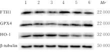
Tab.4
Expression levels of FTH1, GPX4, and HO-1 proteins in GC-2 cells after treated with RSL3 for different time"
| Group | FTH1 | GPX4 | HO-1 |
|---|---|---|---|
| Time after treated with RSL3 | |||
| 0 h | 1.00±0.00 | 1.00±0.00 | 1.00±0.00 |
| 6 h | 0.84±0.12 | 0.63±0.16** | 1.09±0.06 |
| 12 h | 0.72±0.01* | 0.68±0.05** | 1.23±0.03* |
| 24 h | 0.84±0.15 | 0.65±0.11** | 0.56±0.32* |
| 36 h | 0.78±0.02 | 0.83±0.05 | 0.47±0.18** |
| 48 h | 0.85±0.11 | 1.10±0.03 | 0.42±0.23** |
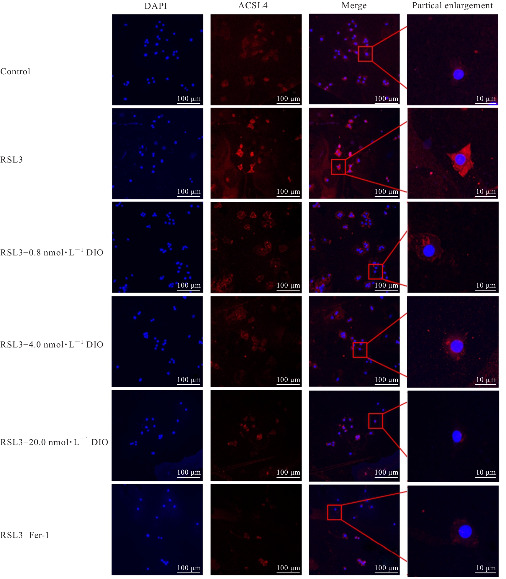
Tab.5
Activities of SOD, levels of MDA, and ratios of GSH/GSSG in GC-2 cells in various groups"
| Group | SOD [λB/(U·mg-1)] | MDA[mB/(mol·g-1)] | Ratio of GSH/GSSG |
|---|---|---|---|
| Control | 355.69±24.05 | 1.00±0.00 | 1.00±0.00 |
| RSL3 | 269.64±39.82* | 1.85±0.35** | 0.36±0.04* |
| RSL3+0.8 nmol·L-1 DIO | 373.40±28.41△ | 1.42±0.48 | 0.79±0.31 |
| RSL3+4.0 nmol·L-1 DIO | 402.08±39.52△△ | 1.26±0.22 | 0.96±0.18△ |
| RSL3+20.0 nmol·L-1 DIO | 425.60±25.37△△ | 1.08±0.21△ | 1.20±0.23△△ |
| RSL3+Fer-1 | 377.03±22.71△ | 1.04±0.20△△ | 1.28±0.32△△ |
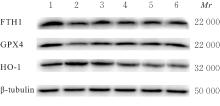
Tab.6
Expression levels of ferroptosis-related proteins in GC-2 cells in various groups"
| Group | FTH1 | GPX4 | HO-1 |
|---|---|---|---|
| Control | 1.00±0.00 | 1.00±0.00 | 1.00±0.00 |
| RSL3 | 0.54±0.05* | 0.74±0.05** | 1.38±0.16* |
| RSL3+0.8 nmol·L-1 DIO | 1.06±0.18△ | 0.93±0.01△ | 1.05±0.03△ |
| RSL3+4.0 nmol·L-1 DIO | 1.21±0.34△△ | 0.98±0.06△ | 1.00±0.09△ |
| RSL3+20.0 nmol·L-1 DIO | 1.12±0.20△△ | 0.93±0.01△ | 0.95±0.21△△ |
| RSL3+Fer-1 | 1.10±0.12△ | 0.95±0.06△ | 0.83±0.16△△ |
| 1 | 石 敏, 桂耀庭, 龙 霞, 等. 小鼠精母细胞在体外分化为精子细胞[J]. 生殖与避孕, 2007, 27(3): 173-177. |
| 2 | ZHAO Y, ZHANG H, CUI J G, et al. Ferroptosis is critical for phthalates driving the blood-testis barrier dysfunction via targeting transferrin receptor[J]. Redox Biol, 2023, 59: 102584. |
| 3 | TANG D L, KROEMER G. Ferroptosis[J]. Curr Biol, 2020, 30(21): R1292-R1297. |
| 4 | RYTER S W. Heme oxgenase-1, a cardinal modulator of regulated cell death and inflammation[J]. Cells, 2021, 10(3): 515. |
| 5 | DAI C S, CHEN X, LI J B, et al. Transcription factors in ferroptotic cell death[J]. Cancer Gene Ther, 2020, 27(9): 645-656. |
| 6 | LIU Y, WAN Y C, JIANG Y, et al. GPX4: the hub of lipid oxidation, ferroptosis, disease and treatment[J]. Biochim Biophys Acta Rev Cancer, 2023, 1878(3): 188890. |
| 7 | WANG J K, ZHANG Z H, SHI F Q, et al. PM2.5 caused ferroptosis in spermatocyte via overloading iron and disrupting redox homeostasis[J]. Sci Total Environ, 2023, 872: 162089. |
| 8 | RADAELLI E, ASSENMACHER C A, VERRELLE J, et al. Mitochondrial defects caused by PARL deficiency lead to arrested spermatogenesis and ferroptosis[J]. eLife, 2023, 12: e84710. |
| 9 | HASHEMI M M, BEHNAMPOUR N, NEJABAT M, et al. Impact of seminal plasma trace elements on human sperm motility parameters[J]. Rom J Intern Med, 2018, 56(1): 15-20. |
| 10 | 蔡双莲, 吴 峥, 吴 进, 等. 天然黄酮香叶木素类及其衍生物的合成与生物活性研究[J]. 有机化学, 2012, 32(3): 560-566. |
| 11 | CHOI J, LEE D H, PARK S Y, et al. Diosmetin inhibits tumor development and block tumor angiogenesis in skin cancer[J]. Biomed Pharmacother, 2019, 117: 109091. |
| 12 | LEE H N, SUNG J, KIM Y, et al. Inhibitory effect of diosmetin on inflammation and lipolysis in coculture of adipocytes and macrophages[J]. J Food Biochem, 2020, 44(7): e13261. |
| 13 | YANG K, LI W F, YU J F, et al. Diosmetin protects against ischemia/reperfusion-induced acute kidney injury in mice[J]. J Surg Res, 2017, 214: 69-78. |
| 14 | LIU J F, FU L, YIN F, et al. Diosmetin maintains barrier integrity by reducing the expression of ABCG2 in colonic epithelial cells[J]. J Agric Food Chem, 2023, 71(23): 8931-8940. |
| 15 | JANISZEWSKA E, KOKOT I, KMIECIAK A, et al. Are there associations between seminal plasma advanced oxidation protein products and selected redox-associated biochemical parameters in infertile male patients?A preliminary report[J]. Cells, 2022, 11(22): 3667. |
| 16 | CHEN J Y, YANG X, FANG X X, et al. The role of ferroptosis in chronic diseases[J]. Zhejiang Da Xue Xue Bao Yi Xue Ban, 2020, 49(1): 44-57. |
| 17 | YANG W S, STOCKWELL B R. Ferroptosis: death by lipid peroxidation[J]. Trends Cell Biol, 2016, 26(3): 165-176. |
| 18 | ZHENG J S, CONRAD M. The metabolic underpinnings of ferroptosis[J]. Cell Metab, 2020, 32(6): 920-937. |
| 19 | BERSUKER K, HENDRICKS J M, LI Z P, et al. The CoQ oxidoreductase FSP1 acts parallel to GPX4 to inhibit ferroptosis[J]. Nature, 2019, 575(7784): 688-692. |
| 20 | MAIORINO M, CONRAD M, URSINI F. GPx4, lipid peroxidation, and cell death: discoveries, rediscoveries, and open issues[J]. Antioxid Redox Signal, 2018, 29(1): 61-74. |
| 21 | CARR J, LA P, DENNERY P. Heme oxygenase-1 is required for iron homeostasis and mitochondrial respiration[J]. Free Radic Biol Med, 2018, 128: S81. |
| 22 | YANATORI I, RICHARDSON D R, TOYOKUNI S, et al. The iron chaperone poly(rC)-binding protein 2 forms a metabolon with the heme oxygenase 1/cytochrome P450 reductase complex for heme catabolism and iron transfer[J]. J Biol Chem, 2017, 292(32): 13205-13229. |
| 23 | NYBAKKEN G, GRATZINGER D. Myelodysplastic syndrome macrophages have aberrant iron storage and heme oxygenase-1 expression[J]. Leuk Lymphoma, 2016, 57(8): 1893-1902. |
| 24 | ROCHA E R, BERGONIA H A, GERDES S, et al. Bacteroides fragilis requires the ferrous-iron transporter FeoAB and the CobN-like proteins BtuS1 and BtuS2 for assimilation of iron released from heme[J]. Microbiol Open, 2019, 8(4): e00669. |
| 25 | LUO P, LIU D D, ZHANG Q, et al. Celastrol induces ferroptosis in activated HSCs to ameliorate hepatic fibrosis via targeting peroxiredoxins and HO-1[J]. Acta Pharm Sin B, 2022, 12(5): 2300-2314. |
| 26 | AJOOLABADY A, ASLKHODAPASANDHOKMABAD H, LIBBY P, et al. Ferritinophagy and ferroptosis in the management of metabolic diseases[J]. Trends Endocrinol Metab, 2021, 32(7): 444-462. |
| 27 | HU W Y, ZHOU C T, JING Q G, et al. FTH promotes the proliferation and renders the HCC cells specifically resist to ferroptosis by maintaining iron homeostasis[J]. Cancer Cell Int, 2021, 21(1): 709. |
| 28 | OMIYA S, ITO J, OTSU K. Labile iron derived from autophagy-mediated ferritin degradation in cardiomyocytes under pressure overload increases myocardial oxidative stress and develops heart failure in mice[J]. Eur Heart J, 2021, 42(): ehab724.0747. |
| 29 | SHAO Y, ZUO X. PTPRC inhibits ferroptosis of osteosarcoma cells via blocking TFEB/FTH1 signaling[J]. Mol Biotechnol, 2024, 66(10): 2985-2994. |
| 30 | LIU F, DU Z Y, HE J L, et al. FTH1 binds to Daxx and inhibits Daxx-mediated cell apoptosis[J]. Mol Biol Rep, 2012, 39(2): 873-879. |
| 31 | ZHANG R N, PAN T, XIANG Y, et al. Curcumenol triggered ferroptosis in lung cancer cells via lncRNA H19/miR-19b-3p/FTH1 axis[J]. Bioact Mater, 2021, 13: 23-36. |
| 32 | ZHAO L H, JIN L, YANG B. Diosmetin alleviates S. aureus-induced mastitis by inhibiting SIRT1/GPX4 mediated ferroptosis[J]. Life Sci, 2023, 331: 122060. |
| [1] | Junping WEI,Dajia FU,Qingwen MENG,Daofei LIN,Yanzai LIN. Effect of bone marrow mesenchymal stem cell-derived exosomes on myocardial fibrosis in rats induced by isoproterenol and its mechanism [J]. Journal of Jilin University(Medicine Edition), 2024, 50(5): 1348-1357. |
| [2] | Xiaoyong PENG,Yu ZHU,Shuangbo ZHANG,Yingguo ZHU,Tao LI,Liangming LIU,Jianmin WANG,Guangming YANG. Alleviative effect of fluid resuscitation on damage of structure injury of vascular cells after blast injury complicated with hemorrhagic shock in rats by inhibiting ferroptosis of vascular tissue [J]. Journal of Jilin University(Medicine Edition), 2024, 50(5): 1227-1234. |
| [3] | Yanjue YE,Ziyi TANG,Yupei TAN,Shiying YANG,Yong LIU,Li YIN. Effect of azathioprine on ferroptosis in spermatocytes of mice induced by RSL3 [J]. Journal of Jilin University(Medicine Edition), 2024, 50(5): 1217-1226. |
| [4] | Fangyang JIANG,Jing XIAO,He CHANG,Mingyang SUN,Wenjing ZHANG,Guangfu LYU,He LIN,Zhe LIN,Xiaowei HUANG,Yuchen WANG. Effect of polygonatum odoratum polysaccharide on acute kidney injury in mice induced by cisplatin and its ferroptosis mechanism [J]. Journal of Jilin University(Medicine Edition), 2024, 50(5): 1235-1242. |
| [5] | Yingqun NI,Mao YANG,Di YANG,Chenglin GUO,Wenjun ZHU,Yaqin YU,Qin LU,Jinzhi LUO,Chunqin WU,Zhaohui FANG. Screening of key differentially expressed genes involved in osteogenic differentiation of lower limb vascular smooth muscle cells and validation [J]. Journal of Jilin University(Medicine Edition), 2024, 50(3): 620-627. |
| [6] | Ruipeng ZHANG,Jie LI. Resistance and regeneration effects of lncRNA GPRC5D-AS1 on muscle atrophy of myocytes in mice induced by dexamethasone and its mechanism [J]. Journal of Jilin University(Medicine Edition), 2023, 49(6): 1457-1465. |
| [7] | Renyi YANG,Shuwang PENG,Yongheng WANG,Yuxuan DONG,Shanshan DUAN. Construction of ferroptosis prognostic risk model of thyroid cancer and bioinformatics analysis on its potential mechanism [J]. Journal of Jilin University(Medicine Edition), 2023, 49(2): 402-413. |
| [8] | Qiaoling YANG,Lu FU,Yu SHI,Yanjue YE,Rifeng LU,Yong LIU,Li YIN. Inhibitory effect of rosiglitazone on ferroptosis of renal tubular epithelial cells in mice with acute renal injury induced by lipopolysaccharide and its mechanism [J]. Journal of Jilin University(Medicine Edition), 2023, 49(2): 351-359. |
| [9] | Xuechun DU,Baosheng LI,Shuwei QIAO,Yanzhen OU,Zhen LI,Weiyan MENG. Effect of Porphyromonas gingivalis-LPS on expression levels of ferroptosis-related factors in macrophages [J]. Journal of Jilin University(Medicine Edition), 2022, 48(5): 1148-1155. |
| [10] | Shuwei QIAO,Baosheng LI,Xiaoyu LI,Zixuan WANG,Birong LI,Bo DONG,Weiyan MENG. Expression levels of SLC7A11 and GPX4 in human periodontal ligament fibroblasts under inflammation and their significance [J]. Journal of Jilin University(Medicine Edition), 2022, 48(4): 922-928. |
| [11] | Pingli WANG,Shenwei ZHANG,Jiang LI. Inhibitory effect of diosmetin on myocardial hypertrophy induced by isoproterenol in mice and its mechanism [J]. Journal of Jilin University(Medicine Edition), 2021, 47(4): 958-964. |
|
||


