Journal of Jilin University(Medicine Edition) ›› 2025, Vol. 51 ›› Issue (6): 1464-1474.doi: 10.13481/j.1671-587X.20250603
• Research in basic medicine • Previous Articles
Optimization of preparation method for D-galactose-induced mouse model of aging-related cognitive dysfunction
Han SUN1,Weilun SUN1,Huifeng WANG1,Wenli MA1,Huali XU1,Wenwen FU1,2( )
)
- 1.Department of Pharmacology,School of Pharmacy,Jilin University,Changchun 130021,China
2.Pharmacology Laboratory Center,School of Pharmacy,Jilin University,Changchun 130021,China
-
Received:2025-06-24Accepted:2025-07-18Online:2025-11-28Published:2025-12-15 -
Contact:Wenwen FU E-mail:fww@jlu.edu.cn
CLC Number:
- R965.1
Cite this article
Han SUN,Weilun SUN,Huifeng WANG,Wenli MA,Huali XU,Wenwen FU. Optimization of preparation method for D-galactose-induced mouse model of aging-related cognitive dysfunction[J].Journal of Jilin University(Medicine Edition), 2025, 51(6): 1464-1474.
share this article
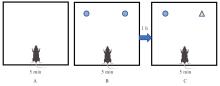
Fig. 1
Illustration of novel objection recognition experiment"
Tab. 1
Primer sequences of PCR"
| Primer | Sequence(5'-3') |
|---|---|
| IL-1β | F: TGCCACCTTTTGACAGTGAT R: TGTGCTGCTGCGAGATTTGA |
| IL-18 | F: CAACGATGATGCACTTGCAGA R: TGACTCCAGCTTATCTCTTGGT |
| TNF-α | F: AGGCACTCCCCCAAAAGATG R: CCACTTGGTGGTTTGTGAGTG |
| IL-4 | F: GGGTTGCCAAGCCTTATCGG R: AGACACCTTGGTCTTGGAGCTTATT |
| GAPDH | F: GGAGAGTGTTTCCTCGTCCC R: ATGAAGGGGTCGTTGATGGC |

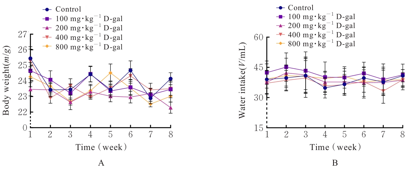
Fig. 2
Body weights and water intakes of mice in various groups"
Tab.2
Forelimb grip strengthes and pole-climbing time of mice in various groups"
| Group | Grip strength (F/N) | Pole-climbing time (t/s) | ||||
|---|---|---|---|---|---|---|
| (week) 4 | 6 | 8 | (week) 4 | 6 | 8 | |
| Control | 87.53±6.75 | 87.42±4.31 | 86.76±5.14 | 5.93±1.60 | 8.36±3.86 | 7.11±2.73 |
| D-gal | ||||||
| 100 mg·kg-1 | 83.54±4.10 | 85.65±4.62 | 80.04±5.45 | 6.12±2.33 | 8.42±3.74 | 11.37±2.42 |
| 200 mg·kg-1 | 85.57±4.53 | 82.24±3.96 | 77.84±5.36** | 6.96±2.28 | 10.82±7.76 | 14.05±4.41* |
| 400 mg·kg-1 | 83.62±5.61 | 89.50±4.97 | 78.05±6.71* | 5.32±1.27 | 7.30±4.73 | 10.56±6.15 |
| 800 mg·kg-1 | 86.33±3.43 | 85.56±2.98 | 81.57±5.26 | 5.41±2.42 | 8.08±3.22 | 9.78±8.34 |
Tab.3
Total distances and recognition indexes of mice in various groups in novel objection recognition experiment (n=10, x±s)"
| Group | Total distance (l/cm) | Recognition index (η/%) | ||||
|---|---|---|---|---|---|---|
| (week) 4 | 6 | 8 | (week) 4 | 6 | 8 | |
| Control | 77.42±14.93 | 71.25±18.91 | 76.68±35.37 | 59.65±23.02 | 48.19±13.11 | 65.54±9.30 |
| D-gal | ||||||
| 100 | 83.57±19.89 | 73.03±20.90 | 75.96±27.37 | 54.39±14.74 | 46.01±13.77 | 54.06±9.49 |
| 200 mg·kg-1 | 85.95±23.83 | 67.40±15.37 | 75.81±31.01 | 59.21±16.96 | 46.64±15.27 | 43.46±11.37* |
| 400 mg·kg-1 | 84.51±16.15 | 76.07±17.35 | 75.35±40.42 | 58.67±16.45 | 46.76±18.49 | 44.19±10.36* |
| 800 mg·kg-1 | 86.86±12.73 | 71.79±17.11 | 77.22±30.89 | 60.46±7.93 | 48.90±16.71 | 57.58±15.47 |
Tab.4
Spontaneous alternation rates of mice in various groups in Y maze test (n=10, x±s, η/%)"
| Group | Spontaneous alternation rate | |||
|---|---|---|---|---|
| (week) 4 | 6 | 8 | ||
| Control | 63.84±9.81 | 64.76±4.90 | 68.78±3.60 | |
| D-gal | ||||
| 100 mg·kg-1 | 61.51±7.80 | 60.35±4.11 | 60.23±7.25* | |
| 200 mg·kg-1 | 62.94±4.89 | 60.63±8.37 | 55.78±5.14** | |
| 400 mg·kg-1 | 64.98±9.70 | 64.74±6.74 | 60.89±5.25* | |
| 800 mg·kg-1 | 69.19±7.21 | 60.12±10.12 | 58.73±6.06** | |
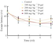
Fig. 3
Escape latencies of mice in various groups during training"
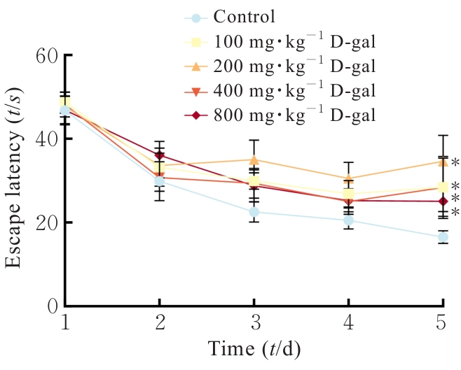
Fig. 4
Representative moving paths of mice in various groups during probe trial"
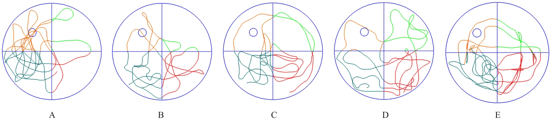
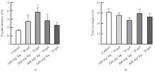
Fig. 5
Escape latency and time spent in target quadrant of mice in various groups during probe trial"
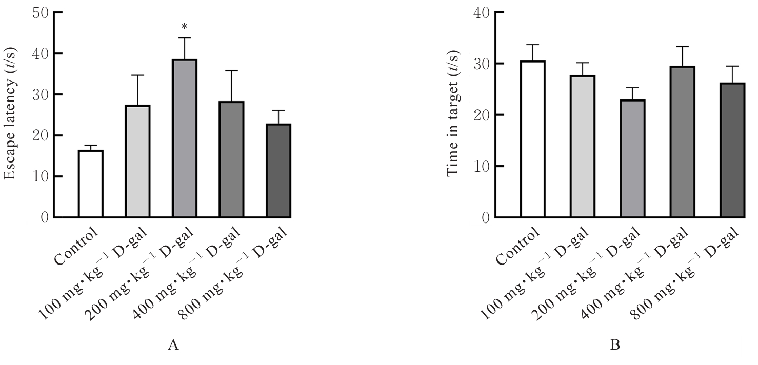
Fig. 6
Pathomorphology of brain tissue of mice in two groups detected by HE staining (×200)"
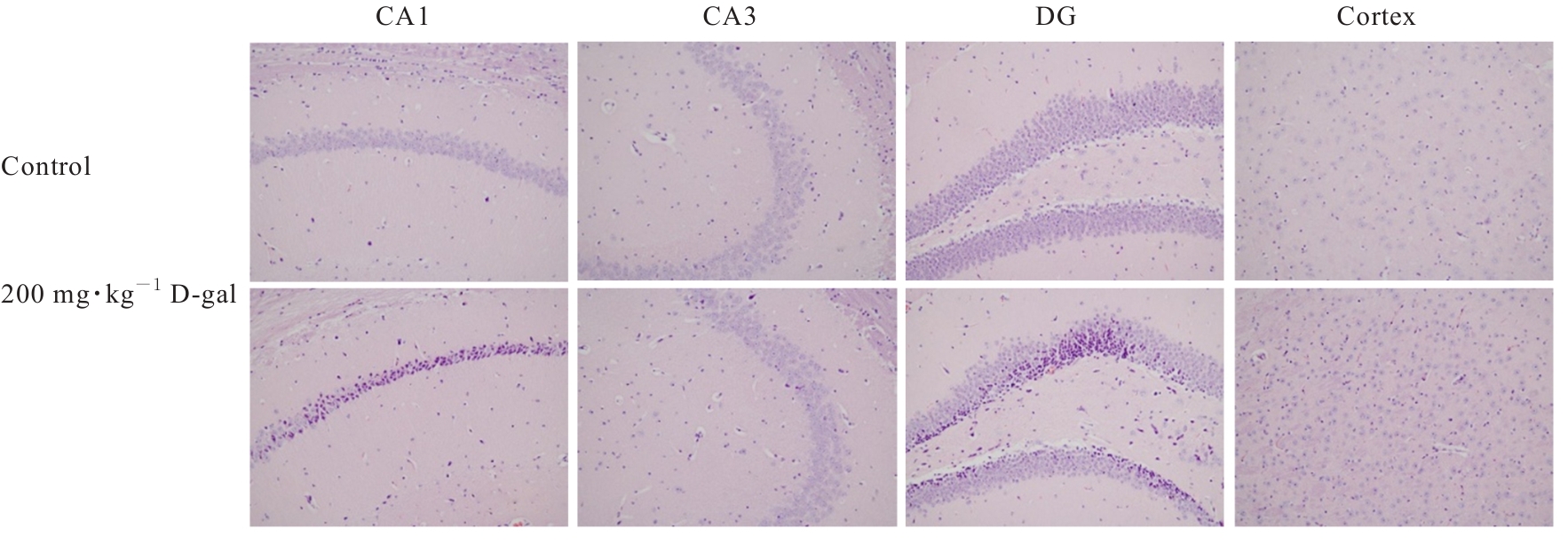
Fig. 7
Pathomorphology of brain tissue of mice in two groups detected by Nissl staining (×200)"
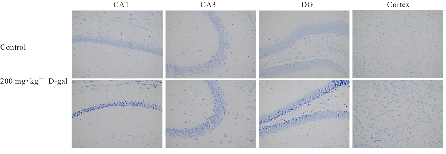
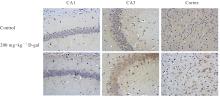
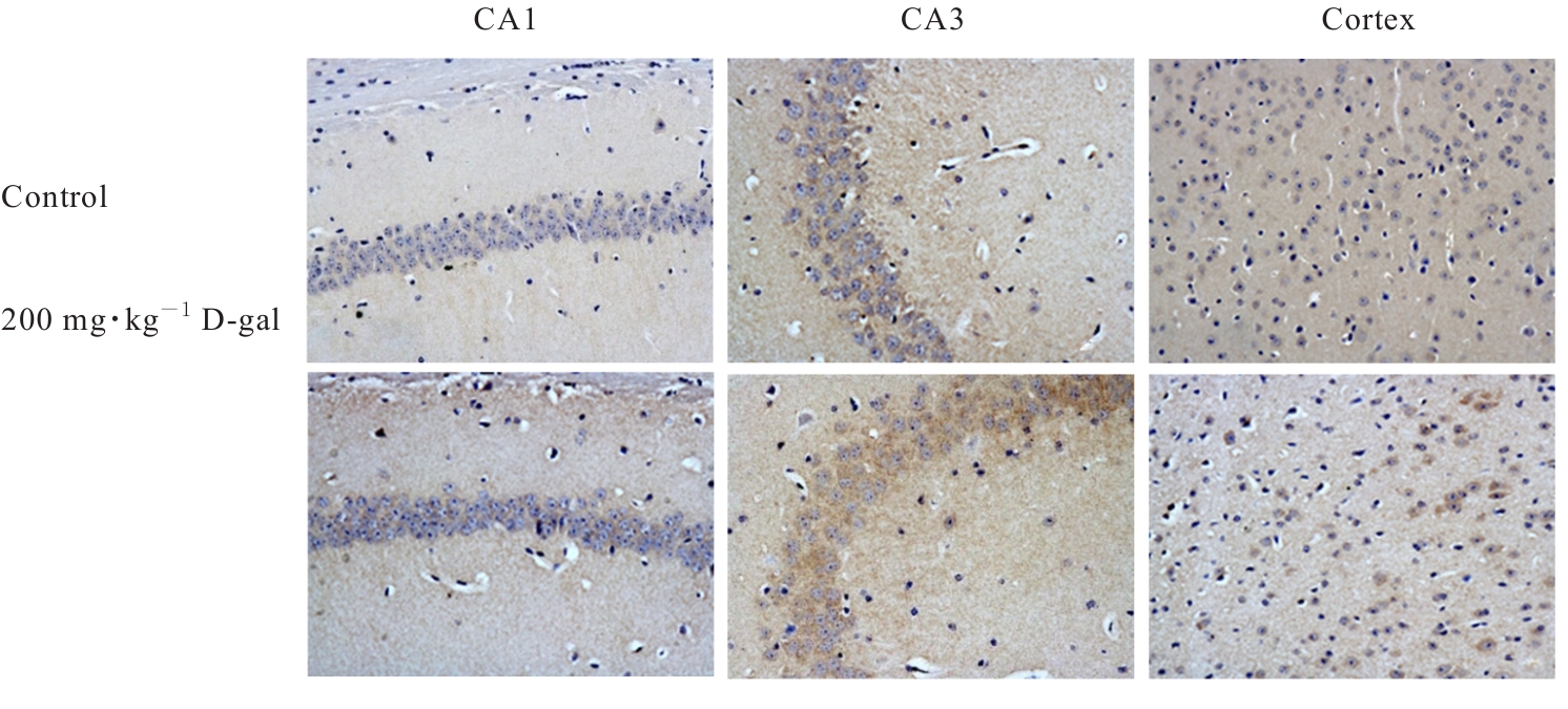
Fig. 8
Expression of aging marker of β-gal protein in brain tissue of mice in two groups (Immunohistochemistry,×400)"
Tab.5
Expression levels of IL-1β,IL-18,TNF-α and IL- 4 mRNA in brain tissue of mice in two groups"
| Group | IL-1β | IL-18 | TNF-α | IL-4 | |
|---|---|---|---|---|---|
| Control | 1.00±0.00 | 1.00±0.00 | 1.00±0.00 | 1.00±0.00 | |
| 200 mg·kg-1 D-gal | 3.20±0.44** | 2.53±0.46* | 2.16±0.57** | 0.60±0.05** |

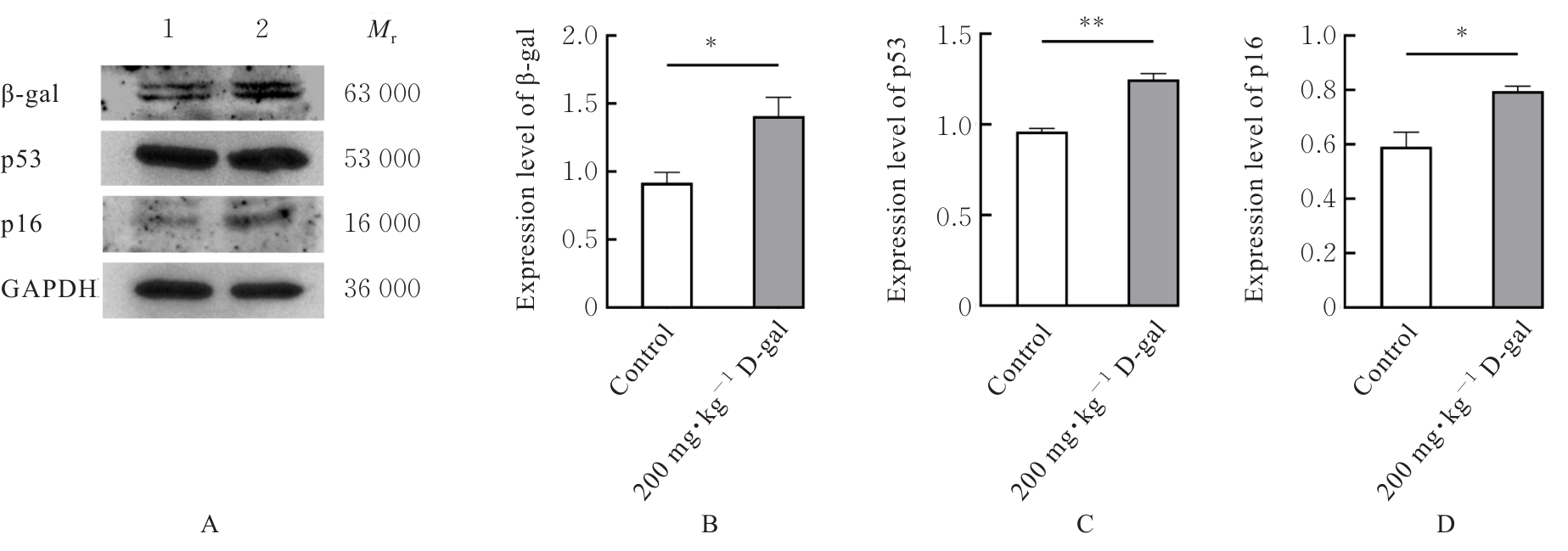
Fig. 9
Electrophoregram(A) and histograms(B-D) of expressions of β-gal, p53, and p16 proteins in hippocampus tissue of mice in two groups"
| [1] | BO-HTAY C, SHWE T, HIGGINS L, et al. Aging induced by D-galactose aggravates cardiac dysfunction via exacerbating mitochondrial dysfunction in obese insulin-resistant rats[J]. Geroscience, 2020, 42(1): 233-249. |
| [2] | 魏孟伶, 贺绘宇, 王 娟, 等. 中老年人生物学衰老与认知功能的关系研究[J]. 现代预防医学, 2025, 52(2): 342-348. |
| [3] | LÓPEZ-OTÍN C, BLASCO M A, PARTRIDGE L, et al. Hallmarks of aging: an expanding universe[J]. Cell, 2023, 186(2): 243-278. |
| [4] | SUN W W, ZHU J H, QIN G Y, et al. Lonicera Japonica polysaccharides alleviate D-galactose-induced oxidative stress and restore gut microbiota in ICR mice[J]. Int J Biol Macromol, 2023, 245: 125517. |
| [5] | JALBERT J J, DAIELLO L A, LAPANE K L. Dementia of the Alzheimer type[J]. Epidemiol Rev, 2008, 30: 15-34. |
| [6] | 吴 薇. D-半乳糖致小鼠脑衰老早期阶段星形胶质细胞的活化及其作用机制[D]. 南京: 南京医科大学, 2010. |
| [7] | 赵凡凡, 周玉枝, 高 丽, 等. D-半乳糖致衰老大鼠模型的研究进展[J]. 药学学报, 2017, 52(3): 347-354. |
| [8] | 马佳丽, 宋莉艳, 李观纬, 等. D-gal诱导小鼠衰老模型建立的适宜浓度探索与模型评价[J]. 健康之路, 2016, 15(11): 5. |
| [9] | AZMAN K F, SAFDAR A, ZAKARIA R. D-galactose-induced liver aging model: Its underlying mechanisms and potential therapeutic interventions[J]. Exp Gerontol, 2021, 150: 111372. |
| [10] | SUN K Y, YANG P Y, ZHAO R, et al. Matrine attenuates D-galactose-induced aging-related behavior in mice via inhibition of cellular senescence and oxidative stress[J]. Oxid Med Cell Longev, 2018, 2018: 7108604. |
| [11] | KUMAR H, BHARDWAJ K, VALKO M, et al. Antioxidative potential of Lactobacillus sp. in ameliorating D-galactose-induced aging[J]. Appl Microbiol Biotechnol, 2022, 106(13): 4831-4843. |
| [12] | GONZALES M M, GARBARINO V R, POLLET E, et al. Biological aging processes underlying cognitive decline and neurodegenerative disease[J]. J Clin Invest, 2022, 132(10): e158453. |
| [13] | WYSS-CORAY T. Ageing, neurodegeneration and brain rejuvenation[J]. Nature, 2016, 539(7628): 180-186. |
| [14] | LI M Q, ZHU M Z, QUAN W, et al. Acteoside palliates d-galactose induced cognitive impairment by regulating intestinal homeostasis[J]. Food Chem, 2023, 421: 135978. |
| [15] | ZHANG J J, HU R Y, CHEN K C, et al. 20(S)- protopanaxatriol inhibited D-galactose-induced brain aging in mice via promoting mitochondrial autophagy flow[J]. Phytother Res, 2023, 37(7): 2827-2840. |
| [16] | HE W, SONG H, YANG Z B, et al. Beneficial effect of GABA-rich fermented milk whey on nervous system and intestinal microenvironment of aging mice induced by D-galactose[J]. Microbiol Res, 2024, 278: 127547. |
| [17] | 仝佳祥, 陈 杨, 李 璇, 等. 枸杞叶改善D-半乳糖致亚急性衰老小鼠学习与记忆能力的效应部位与作用机制研究[J]. 世界科学技术-中医药现代化, 2024, 26(1): 48-60. |
| [18] | AN J R, LIU J T, GAO X M, et al. Effects of liraglutide on astrocyte polarization and neuroinflammation in db/db mice: focus on iron overload and oxidative stress[J]. Front Cell Neurosci, 2023, 17: 1136070. |
| [19] | CHEN Y X, YANG H, WANG D S, et al. Gastrodin relieves cognitive impairment by regulating autophagy via PI3K/AKT signaling pathway in vascular dementia[J]. Biochem Biophys Res Commun, 2023, 671: 246-254. |
| [20] | 吴志悦, 薛晓帆, 曾景蓉, 等. 端粒-端粒酶在神经退行性疾病中的研究进展[J]. 中风与神经疾病杂志, 2024, 41(2): 169-174. |
| [21] | SIMONE M J, TAN Z S. The role of inflammation in the pathogenesis of delirium and dementia in older adults: a review[J]. CNS Neurosci Ther, 2011, 17(5): 506-513. |
| [22] | CHU Z X, HAN S, LUO Y, et al. Targeting gut-brain axis by dietary flavonoids ameliorate aging-related cognition decline: Evidences and mechanisms[J]. Crit Rev Food Sci Nutr, 2024, 64(28): 10281-10302. |
| [23] | JESSEN F, AMARIGLIO R E, BUCKLEY R F, et al. The characterisation of subjective cognitive decline[J]. Lancet Neurol, 2020, 19(3): 271-278. |
| [24] | LIU H, SHEN L K, SUN Z Z, et al. Downregulated phosphoglycerate kinase 1 attenuates cerebral ischemia-reperfusion injury by reversing neuroinflammation and oxidative stress through the nuclear factor erythroid 2 related factor 2/ARE pathway[J]. Neuroscience, 2023, 524: 94-107. |
| [25] | 叶 蕾, 舒 姝, 徐 运, 等. 衰老中小胶质细胞对认知功能减退调控的研究进展[J]. 临床神经病学杂志, 2024, 37(1): 65-68. |
| [26] | YAO Y Q, LUO Y S, LIANG X M, et al. The role of oxidative stress-mediated fibro-adipogenic progenitor senescence in skeletal muscle regeneration and repair[J]. Stem Cell Res Ther, 2025, 16(1): 104. |
| [27] | IMAI S I, GUARENTE L. NAD+ and sirtuins in aging and disease[J]. Trends Cell Biol, 2014, 24(8): 464-471. |
| [28] | 邢秋娟, 施 杞, 王拥军. D-半乳糖诱导衰老动物模型的机制及其在中医药方面的应用[J]. 上海中医药大学学报, 2010, 24(3): 93-98. |
| [29] | OPRESKO P L, SANFORD S L, DE ROSA M. Oxidative stress and DNA damage at telomeres[J]. Cold Spring Harb Perspect Biol, 2025, 17(6): a041707. |
| [30] | BARUCH K, DECZKOWSKA A, DAVID E, et al. Aging. Aging-induced type I interferon response at the choroid plexus negatively affects brain function[J]. Science, 2014, 346(6205): 89-93. |
| [31] | STERN Y. Cognitive reserve in ageing and Alzheimer’s disease[J]. Lancet Neurol, 2012, 11(11): 1006-1012. |
| [32] | UYAR B, PALMER D, KOWALD A, et al. Single-cell analyses of aging, inflammation and senescence[J]. Ageing Res Rev, 2020, 64: 101156. |
| [33] | KROEMER G, MAIER A B, CUERVO A M, et al. From geroscience to precision geromedicine: Understanding and managing aging[J]. Cell, 2025, 188(8): 2043-2062. |
| [34] | MAUS M, LÓPEZ-POLO V, MATEO L, et al. Iron accumulation drives fibrosis, senescence and the senescence-associated secretory phenotype[J]. Nat Metab, 2023, 5(12): 2111-2130. |
| [35] | VADDAVALLI P L, SCHUMACHER B. The p53 network: cellular and systemic DNA damage responses in cancer and aging[J]. Trends Genet, 2022, 38(6): 598-612. |
| [36] | GROSSE L, WAGNER N, EMELYANOV A, et al. Defined p16(high) senescent cell types are indispensable for mouse healthspan[J]. Cell Metab, 2020, 32(1): 87-99. |
| [1] | Jian LIU,Lu XING,Tianye LAN,Fan YAO,Wen WANG,Yufu DONG,Jinpu WU,Ran BI,Liwei SUN,Xuenan CHEN,Weimin ZHAO. Improvement effect of ginseng alcohol extract on sleep of aged drosophila and its mechanism [J]. Journal of Jilin University(Medicine Edition), 2025, 51(4): 896-903. |
| [2] | Bo WANG,Zhe LIN. Regulatory effect of velvet antler polypeptides on differentiation of C2C12 myoblasts and its mechanism [J]. Journal of Jilin University(Medicine Edition), 2025, 51(3): 632-641. |
| [3] | Wei ZHONG,Zhiyin DAI,Xinggang CUI,Bo LI,Yu JIANG. Effect of nuclear factor of activated T lymphocytes 5 on senescence of smooth muscle cells of mice induced by high-salt and its mechanism [J]. Journal of Jilin University(Medicine Edition), 2025, 51(3): 567-575. |
| [4] | DILIXIATI·Dilidaer,Lin JIA. Improvement effect of exosomes derived from human adipose-derived stem cells and human dermal fibroblasts on ultraviolet-induced photoaging skin wrinkles in nude mice [J]. Journal of Jilin University(Medicine Edition), 2025, 51(3): 621-631. |
| [5] | Chong LIU,Boyuan WANG,Yang JIANG,Aizhuo SONG,Minghe LI. Clinical analysis on temporomandibular joint disc anchorage for treatment of irretrievable forward displacement of temporomandibular joint disc [J]. Journal of Jilin University(Medicine Edition), 2025, 51(3): 770-777. |
| [6] | Fanjun SUN,Xingchen PAN,Dan TONG. Research progress in MRI neuroimaging for patients with functional gastrointestinal diseases and inflammatory bowel disease [J]. Journal of Jilin University(Medicine Edition), 2025, 51(2): 541-548. |
| [7] | Zilong GAO,Biao LIU,Le QI,Jingyu LANG,Yan LIU. Giant cell-rich osteosarcoma with special imaging findings: A case report and literature review [J]. Journal of Jilin University(Medicine Edition), 2025, 51(1): 222-227. |
| [8] | Hong LI,Hui WANG,Lishu WANG,Chaonan WANG,Xiaohao XU,Liwei SUN. Ameliorating effect of Dendrobium officinale polysaccharides on intestinal mucosal barrier damage in elderly mice by regulating intestinal microbial metabolites [J]. Journal of Jilin University(Medicine Edition), 2025, 51(1): 76-84. |
| [9] | Jing LOU,Lei ZHAO,Yanjie ZHU,Shuaiqiang YUAN,Fei WANG,Hangzhou ZHANG,Jiaojiao XU,Xiaoke YU,Liufa HOU. Effect of Fuzheng Ruanjian Anticancer Formula on malignant biological behaviors of hepatocellulars carcinoma HepG2 cells by regulating Akt/MDM2/P53 signaling pathway [J]. Journal of Jilin University(Medicine Edition), 2024, 50(6): 1654-1663. |
| [10] | Baolian MA,Xiaoxue HU,Xiaowen AI,Yonglan ZHANG. Inhibitory effect of diosmetin on ferroptosis of GC-2 spermatocytes induced by RSL3 in mice and its mechanism [J]. Journal of Jilin University(Medicine Edition), 2024, 50(6): 1481-1490. |
| [11] | Weichao WU,Yan GUO,Xiangkai ZHAO,Zhiguang GU,Yijia GUO,Zipeng LAN,Hui HUANG,Lei KUANG,Ming ZHANG,Dongsheng HU,Yongli YANG,Wei WANG,Jinru CHEN. Correlation analysis on occupational acid fog exposure and accelerated biological aging in workers [J]. Journal of Jilin University(Medicine Edition), 2024, 50(6): 1741-1750. |
| [12] | Wenxiu GUO,Yan ZHUANG,Huiling ZHANG,Wenning HE,Jun MENG. Effect of SGK3 on recovery of first meiotic division of oocytes in mice and its mechanism [J]. Journal of Jilin University(Medicine Edition), 2024, 50(4): 891-899. |
| [13] | Minhui XU,Xiaolei CHENG,Jiyan XU,Linhao JIANG,Tianjiao XIA. Effect of urolithin A on postoperative cognitive dysfunction induced by isoflurane anesthesia in mice and its mechanism [J]. Journal of Jilin University(Medicine Edition), 2024, 50(3): 596-601. |
| [14] | Canfei XU,Yan HUANG,Kaizhi ZHANG. Acute ischemic stroke patients with onset time within 4.5 h identified by machine learning combined with magnetic resonance diffusion weighted imaging [J]. Journal of Jilin University(Medicine Edition), 2023, 49(6): 1635-1641. |
| [15] | Meng ZHANG,Qi ZHOU,Zheng HU. Establishment of liver in situ xenograft reconstruction model of Fah-/-Rag2-/-IL2Rg-/- mice and its evaluation [J]. Journal of Jilin University(Medicine Edition), 2023, 49(5): 1358-1365. |
|
||