Journal of Jilin University(Medicine Edition) ›› 2026, Vol. 52 ›› Issue (2): 429-439.doi: 10.13481/j.1671-587X.20260215
• Research in basic medicine • Previous Articles Next Articles
Regulatory effect of lncRNA urothelial carcinoembryonic antigen 1 on anoikis in human gastrointestinal stromal tumor cells and its mechanism
Yu ZHAO1,Xiaoshuang HE2,Xiaoyin DONG1,Fengyi GAO1,Jiageng HE1( )
)
- 1.Department of Gastroenterology,First Affiliated Hospital,Shihezi University,Shihezi 832000,China
2.Department of Respiratory and Critical Care Medicine,First Affiliated Hospital,Shihezi University,Shihezi 832000,China
-
Received:2025-05-14Accepted:2025-07-23Online:2026-03-28Published:2026-04-15 -
Contact:Jiageng HE E-mail:1551143474@qq.com
CLC Number:
- R735
Cite this article
Yu ZHAO,Xiaoshuang HE,Xiaoyin DONG,Fengyi GAO,Jiageng HE. Regulatory effect of lncRNA urothelial carcinoembryonic antigen 1 on anoikis in human gastrointestinal stromal tumor cells and its mechanism[J].Journal of Jilin University(Medicine Edition), 2026, 52(2): 429-439.
share this article
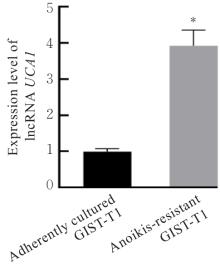
Fig. 1
Expression levels of lncRNA UCA1 in two kinds of GIST-T1 cells"

Fig. 2
Electrophoregram(A) and histograms(B,C) of expressions of autophagy-related proteins in two kinds of GIST-T1 cells"
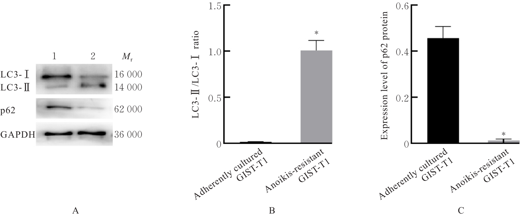
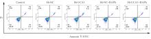
Fig. 3
Anoikis of GIST-T1 cells in various groups detected by flow cytometry"
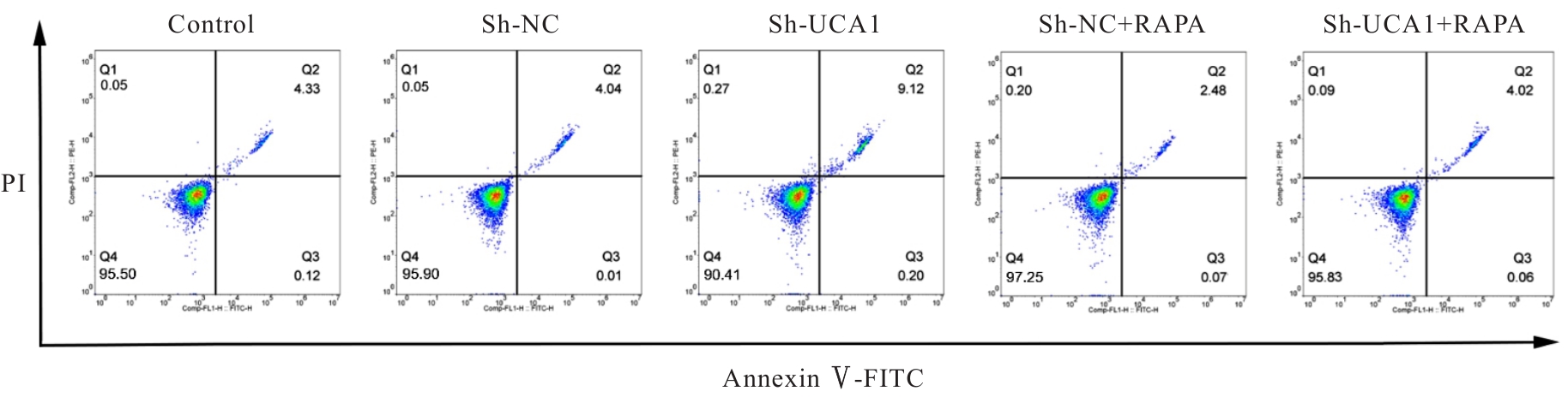
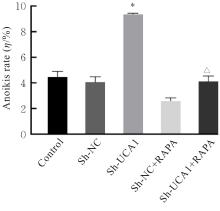
Fig. 4
Anoikis rates of GIST-T1 cells in various groups detected by flow cytometry"
Fig. 5
Electrophoregram(A) and histograms(B,C) of expressions of autophagy-related proteins in GIST-T1 cells in various groups"

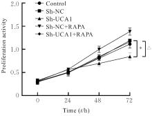
Fig. 6
Proliferation activities of GIST-T1 cells in various groups"

Fig. 7
Migration of GIST-T1 cells in various groups(×100)"

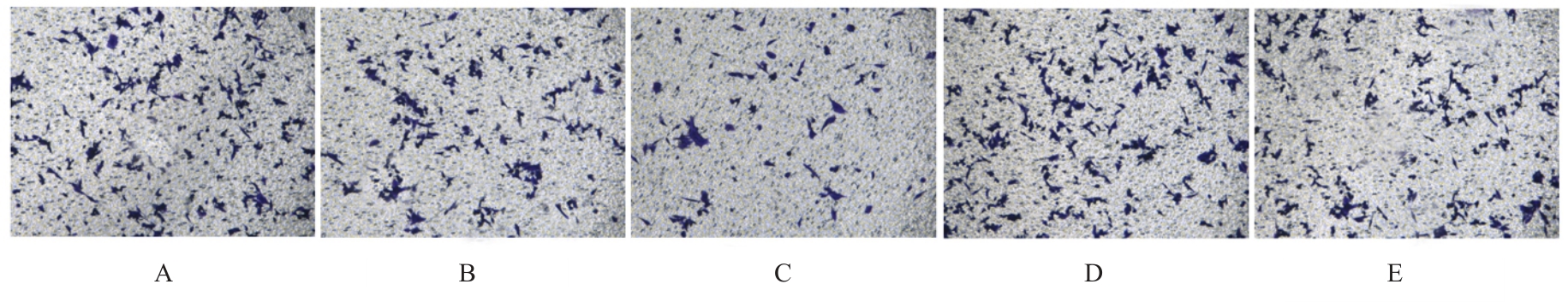
Fig. 8
Invasion of GIST-T1 cells in various groups(Crystal violet, ×100)"
Tab.1
Rates of scratch healing and numbers of invasion cells of GIST-T1 cells in various groups"
| Group | Rate of scratch healing (η/%) | Number of invasion cells |
|---|---|---|
| Control | 31.23±3.25 | 138.56±14.23 |
| Sh-NC | 32.26±3.57 | 134.92±11.80 |
| Sh-UCA1 | 10.89±1.12* | 59.50±6.04* |
| Sh-NC+RAPA | 58.75±6.32 | 166.77±17.03 |
| Sh-UCA1+RAPA | 34.51±3.49△ | 129.83±13.21△ |
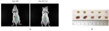
Fig. 9
Xenograft tumor growth of nude mice in two groups"
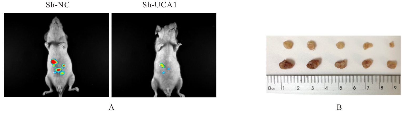
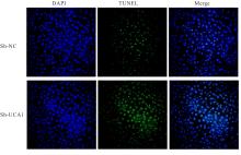
Fig. 10
Apoptosis in tumor tissue of nude mice in two groups(TUNEL,×100)"
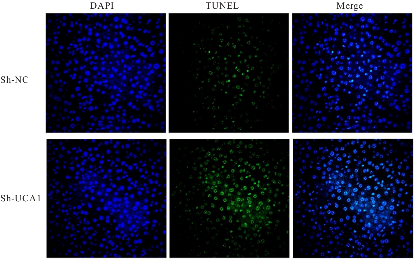
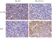
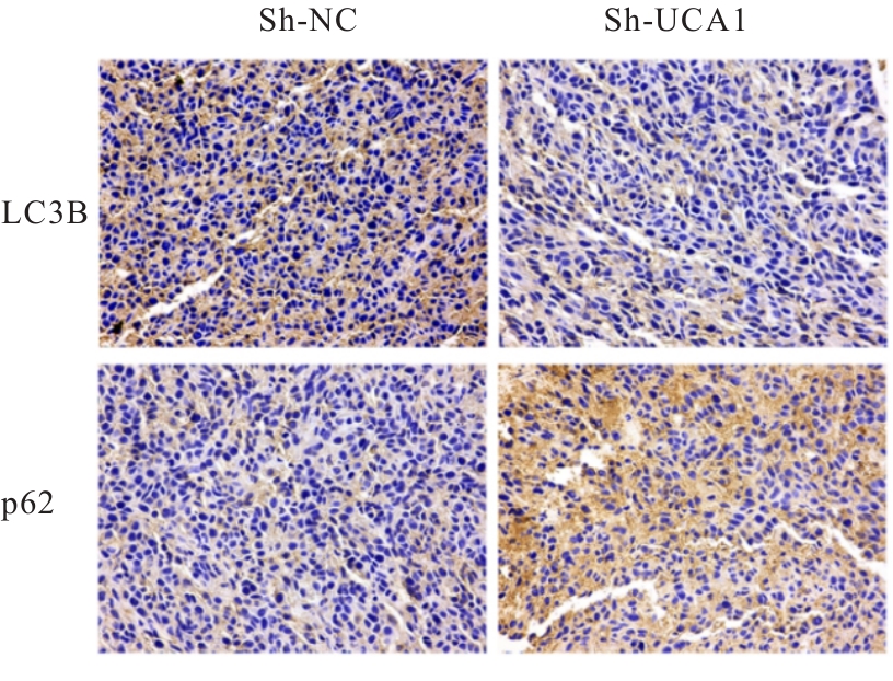
Fig. 11
Expressions of LC3B and p62 proteins in tumor tissue of nude mice in two groups(Immunohistochemistry, ×100)"
| [1] | PATIL A, HAVAL S, NICHKAODE P, et al. Gastrointestinal stromal tumor: a clinicopathological study and management[J]. Cureus, 2023, 15(11): e49469. |
| [2] | MUNTEANU A, PATRASCU S, BORDU S, et al. Clinical and morphological characteristics of gastrointestinal stromal tumor[J]. Chirurgia, 2023, 118(6): 618-623. |
| [3] | KHAN S U, FATIMA K, MALIK F. Understanding the cell survival mechanism of anoikis-resistant cancer cells during different steps of metastasis[J]. Clin Exp Metastasis, 2022, 39(5): 715-726. |
| [4] | SATTARI FARD F, JALILZADEH N, MEHDIZADEH A, et al. Understanding and targeting anoikis in metastasis for cancer therapies[J]. Cell Biol Int, 2023, 47(4): 683-698. |
| [5] | ZHANG Y G. LncRNA-encoded peptides in cancer[J]. J Hematol Oncol, 2024, 17(1): 66. |
| [6] | 李颖霞, 姜利彬, 周明霞, 等. 敲低长链非编码RNA TTTY15表达对结肠癌细胞增殖、 凋亡、 迁移和克隆形成能力的影响[J]. 郑州大学学报(医学版), 2025, 60(2): 173-176. |
| [7] | 白 雪, 伍燕平, 李忠玉, 等. lncRNA UCA1在急性髓系白血病患者中的表达及其临床意义[J]. 中国实验血液学杂志, 2024, 32(4): 999-1004. |
| [8] | ASKARI N, PIROUZ M S, MAFIKANDI V, et al. The lncRNA UCA1 enhances pancreatic cancer EMT by regulating miR-708-5p and miR-135b-5p: a bioinformatics approach[J]. Iran J Public Health, 2024, 53(7): 1659-1669. |
| [9] | RAMLI S, SIM M S, GUAD R M, et al. Long noncoding RNA UCA1 in gastrointestinal cancers: molecular regulatory roles and patterns, mechanisms, and interactions[J]. J Oncol, 2021, 2021: 5519720. |
| [10] | AN L F, DONG K J, CHI S Q, et al. lncRNA UCA1 promotes tumor progression by targeting SMARCD3 in cervical cancer[J]. Mol Carcinog, 2024, 63(3): 384-399. |
| [11] | CAO L L, LIN C, LIU Y, et al. Clinical characteristics and prognostic analysis of postoperative recurrence or metastasis of low-risk gastrointestinal stromal tumors[J]. World J Surg Oncol, 2024, 22(1): 65. |
| [12] | ADESHAKIN F O, ADESHAKIN A O, AFOLABI L O, et al. Mechanisms for modulating anoikis resistance in cancer and the relevance of metabolic reprogramming[J]. Front Oncol, 2021, 11: 626577. |
| [13] | MCCABE E M, RASMUSSEN T P. lncRNA involvement in cancer stem cell function and epithelial-mesenchymal transitions[J]. Semin Cancer Biol, 2021, 75: 38-48. |
| [14] | YANG M Y, LU H F, LIU J J, et al. lncRNAfunc: a knowledgebase of lncRNA function in human cancer[J]. Nucleic Acids Res, 2022, 50(D1): D1295-D1306. |
| [15] | LU Q, WANG L, GAO Y B, et al. lncRNA APOC1P1-3 promoting anoikis-resistance of breast cancer cells[J]. Cancer Cell Int, 2021, 21(1): 232. |
| [16] | LI Y R, FU M, SONG Y Q, et al. Long non-coding RNA MRPL23-AS1 suppresses anoikis in salivary adenoid cystic carcinoma in vitro [J]. Oral Dis, 2023, 29(4): 1588-1601. |
| [17] | MENG W J, GUO J M, HUANG L, et al. Anoikis-related long non-coding RNA signatures to predict prognosis and immune infiltration of gastric cancer[J]. Bioengineering, 2024, 11(9): 893. |
| [18] | PEÑA-MARTINEZ C, RICKMAN A D, HECKMANN B L. Beyond autophagy: LC3-associated phagocytosis and endocytosis[J]. Sci Adv, 2022, 8(43): eabn1702. |
| [19] | 金美英, 潘韦韦, 苏 婧, 等. 基于自噬调控探讨解毒通络调肝方对SD大鼠胰岛细胞保护的作用机制[J]. 中国兽医学报, 2024, 44(11): 2435-2444. |
| [20] | GULIA S, CHANDRA P, DAS A. The prognosis of cancer depends on the interplay of autophagy, apoptosis, and anoikis within the tumor microenvironment[J]. Cell Biochem Biophys, 2023, 81(4): 621-658. |
| [21] | 汪佳兵, 王石健, 魏 静, 等. 微小RNA-520e通过下调自噬调控肝癌细胞失巢凋亡抵抗[J]. 江苏医药, 2022, 48(11): 1081-1084. |
| [22] | WU Y R, CHEN Y, YAN X J, et al. Lopinavir enhances anoikis by remodeling autophagy in a circRNA-dependent manner[J]. Autophagy, 2024, 20(7): 1651-1672. |
| [1] | Ling DENG,Shouqing LI,Chunxue NIU,Xiuying LIN,Xuguang MI,Jianhua FU. Effect of bisphenol AF on senescence of human endometrial stromal cells and inhibitory role of rapamycin [J]. Journal of Jilin University(Medicine Edition), 2026, 52(2): 340-347. |
| [2] | Yao ZHENG,Mingxia FU,Weichen WANG,Weiwei CHEN,Yuchen HAN,Yu BAI,Jiajia AN. Effect of sodium selenite on biological behaviors of breast cancer doxorubicin-resistant MCF-7/ADR cells [J]. Journal of Jilin University(Medicine Edition), 2026, 52(2): 410-417. |
| [3] | Huiyan ZHU,Min CHEN,Jinxian LI,Chunli LI. Effect of enriched environment on neurofunctional damage in rats with ischemic stroke via transcription factor EB-mediated autophagy [J]. Journal of Jilin University(Medicine Edition), 2026, 52(1): 116-124. |
| [4] | Xiaohan YAO,Mingchen YAO,Zhiqing WANG,Heyang LI,Yan YAN,Ningjing LEI. Effect of silencing GPR139 gene on proliferation, apoptosis and autophagy of breast cancer cells and its mechanism [J]. Journal of Jilin University(Medicine Edition), 2026, 52(1): 1-9. |
| [5] | Zhongzheng LIU,Shubo LIAN,Wenxuan FENG,Xin WEN,Hanyu LIU,Wei HE. Prometive effect of knockdown of KIF3B gene on autophagy in mouse embryonic palatal mesenchymal cells by inhibiting Shh signaling pathway [J]. Journal of Jilin University(Medicine Edition), 2025, 51(6): 1445-1451. |
| [6] | Haidong ZHU,Changkun LYU,Wei SHI. Effect of berberine hydrochloride on autophagy of HeLa cells infected with herpes simplex virus type 1 by regulating PI3K/AKT/mTOR signaling pathway [J]. Journal of Jilin University(Medicine Edition), 2025, 51(6): 1607-1617. |
| [7] | Wenxuan LI,Minru ZONG. Research progress in role of migration of Schwann cells in repairment of peripheral nerve injury [J]. Journal of Jilin University(Medicine Edition), 2025, 51(4): 1137-1144. |
| [8] | Xiaoshuang HE,Lina XU,Mei CUI,Yu ZHAO,Bei WANG,Zheng HUANG,Yuchao WANG,Wenyan XIN,Chao WU. Effects of lncRNA DUXAP8 in lung cancer A549 cells-derived exosomes on lung cancer cell growth and its mechnism [J]. Journal of Jilin University(Medicine Edition), 2025, 51(4): 958-967. |
| [9] | Wei AN, MAIMAITITUXUN·Tuerdi,Zhitao YAO. Regulatory effect of lutein on PI3K/AKT signaling pathway and chondrocyte autophagy in mandibular joint cartilage tissue of rabbits and its mechanism [J]. Journal of Jilin University(Medicine Edition), 2025, 51(4): 976-983. |
| [10] | Guobin HE,Huan WANG. Effect of knockdown of RIP3 on autophagy, pyroptosis, and ferroptosis of hypoxia/reoxygenation-induced human renal tubular epithelial HK2 cells [J]. Journal of Jilin University(Medicine Edition), 2024, 50(6): 1644-1653. |
| [11] | Yutong WANG,Ruili RAN,Jiang BIAN,Xiaohan JIANG,Junqiu SONG,Dewei WANG,Jing YANG. Protective effect of carnosine against oxygen-glucose deprivation/reoxygenation-induced astrocyte injury through inhibition of autophagy by AMPK/mTOR signaling pathway [J]. Journal of Jilin University(Medicine Edition), 2024, 50(5): 1297-1304. |
| [12] | Yuxiao SHI,Meilan LIU,Meilin ZHU,Feng WEI. Effects of 5-Aza-CdR on autophagy and apoptosis of papillary thyroid cancer cells in subcutaneous xenograft tumor tissue of nude mice and its mechanism [J]. Journal of Jilin University(Medicine Edition), 2024, 50(5): 1330-1338. |
| [13] | Guoxing YU,Xin ZHANG,Hengwei DU,Bingjie CUI,Na GAO,Cuilan LIU,Jing DU. Effect of urolithin C on proliferation, apoptosis and autophagy of human acute myeloid leukemia HL-60 cells and its mechanism [J]. Journal of Jilin University(Medicine Edition), 2024, 50(4): 908-916. |
| [14] | Linru WANG,Jing ZHANG,Dongchan ZHAO,Jinjun WANG,Wenxian HU. Effect of silencing FOXO1 gene on autophagy and apoptosis of human aortic vascular smooth muscle cells [J]. Journal of Jilin University(Medicine Edition), 2024, 50(2): 431-441. |
| [15] | Yue WANG,Di WU,Dandan YU,Xiumei DUAN. Gastrointestinal stromal tumors with multifocal heterochronous primary resistance due to temporal gene mutations:A case report and literature review [J]. Journal of Jilin University(Medicine Edition), 2024, 50(2): 545-550. |
|