Journal of Jilin University(Medicine Edition) ›› 2026, Vol. 52 ›› Issue (2): 418-428.doi: 10.13481/j.1671-587X.20260214
• Research in basic medicine • Previous Articles Next Articles
Protective effect of silencing coagulation factor V gene on septic rats by inhibiting JNK1/2 and p38 MAPK signaling pathways
Jingyuan WANG,Fang CHEN,Yancun LIU,Shixin LI,Songtao SHOU( )
)
- Department of Emergency Medicine,General Hospital,Tianjin Medical University,Tianjin 300052,China
-
Received:2025-03-18Accepted:2025-04-30Online:2026-03-28Published:2026-04-15 -
Contact:Songtao SHOU E-mail:z2026sst02@163.com
CLC Number:
- R364.5
Cite this article
Jingyuan WANG,Fang CHEN,Yancun LIU,Shixin LI,Songtao SHOU. Protective effect of silencing coagulation factor V gene on septic rats by inhibiting JNK1/2 and p38 MAPK signaling pathways[J].Journal of Jilin University(Medicine Edition), 2026, 52(2): 418-428.
share this article
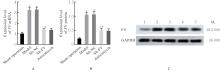
Fig. 1
Expression levels of FV mRNA and protein in lung tissue of rats in various groups"
Tab.1
Survival of rats in various groups [n=10, n(η/%)]"
| Group | Survival number | |||
|---|---|---|---|---|
| (t/d) 0 | 1 | 3 | 5 | |
Sham operation | 10(100.00) | 10(100.00) | 10(100.00) | 10(100.00) |
| Model | 10(100.00) | 8(80.00)* | 5(50.00)* | 2(20.00)* |
| Sh-NC | 10(100.00) | 9(90.00) | 5(50.00)* | 3(30.00)* |
| Sh-FV | 10(100.00) | 10(100.00)# | 8(80.00)△# | 6(60.00)△# |
| Anisomycin | 10(100.00) | 8(80.00)○ | 6(60.00)○ | 3(30.00)○ |
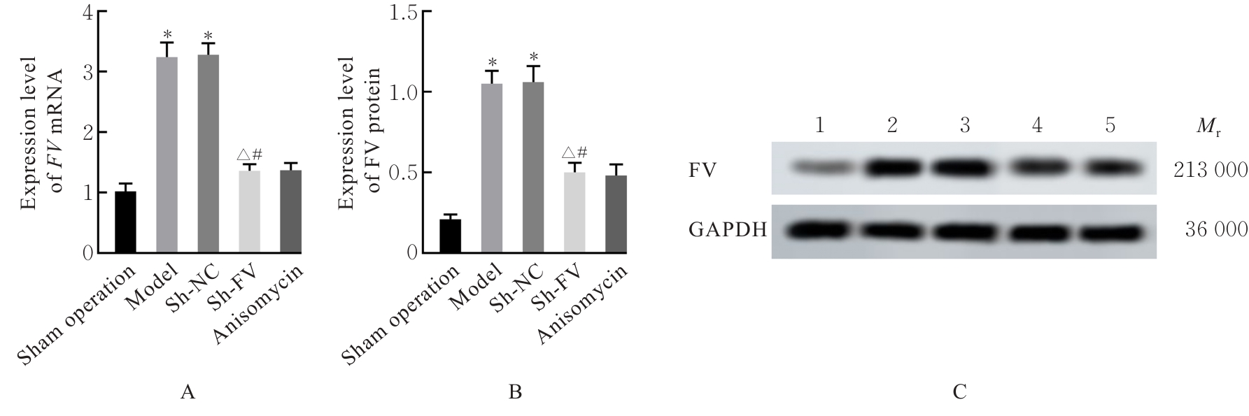
Fig. 2
Pathomorphology of various organ tissues of rats in various groups(HE,×200)"


Fig. 3
Levels of IL-1β, IL-6 and TNF-α in serum of rats in various groups"
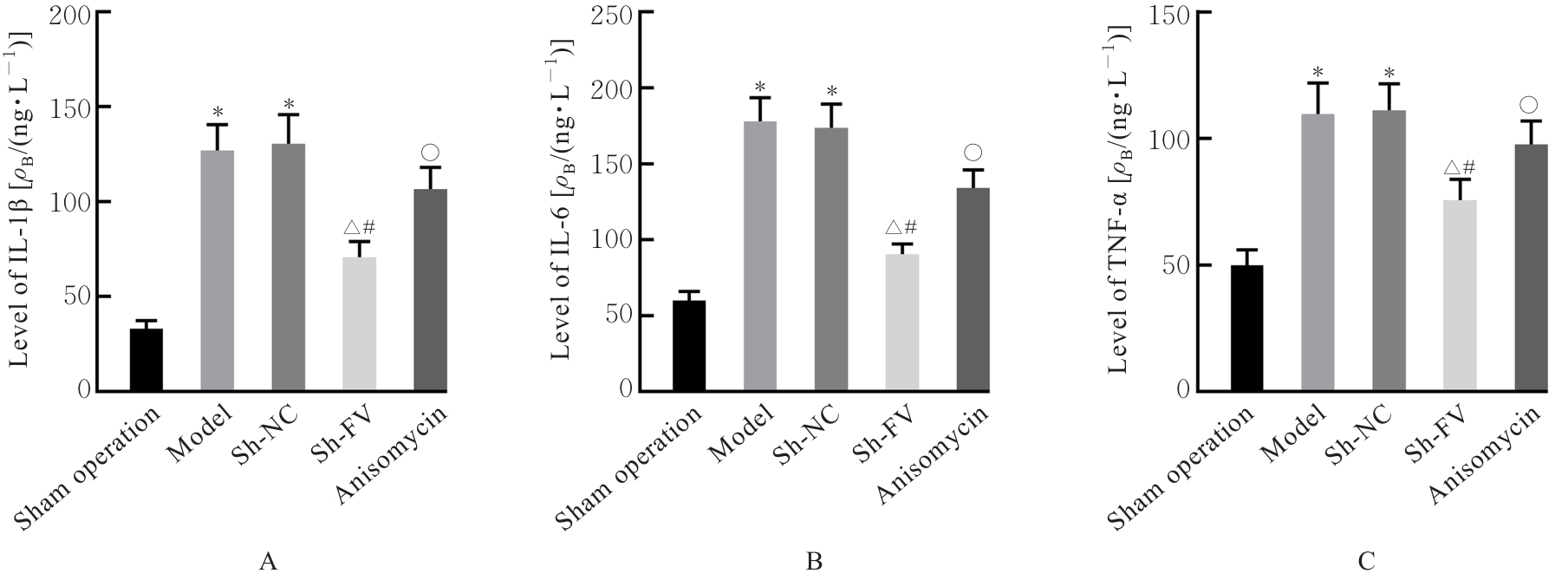
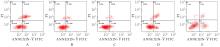
Fig. 4
Apoptosis of neutrophils in blood of rats in various groups"
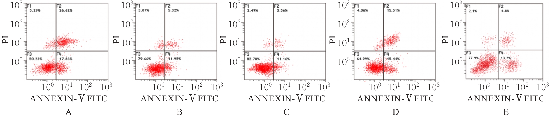
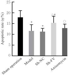
Fig. 5
Apoptotic rates of neutrophils in blood of rats in various groups"
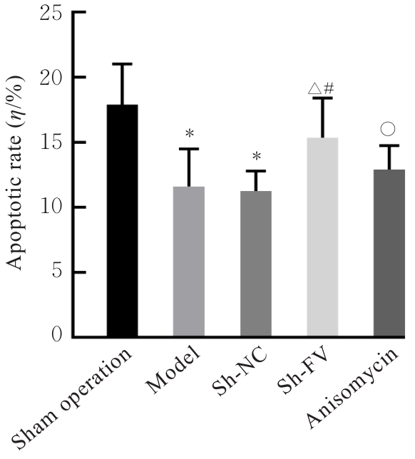
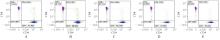
Fig. 6
Distribution of T lymphocyte subsets in blood of rats in various groups"
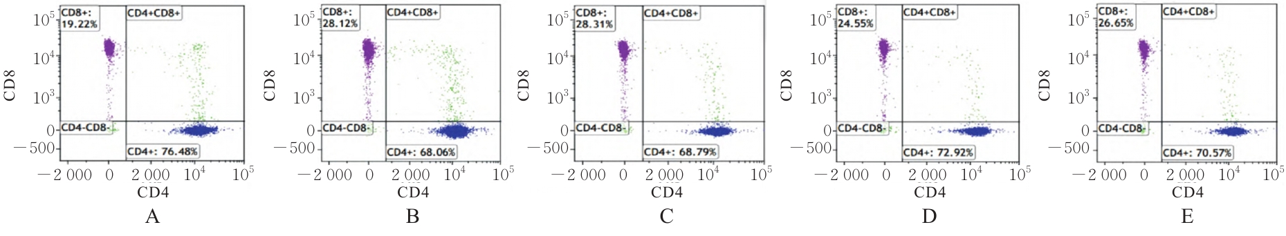
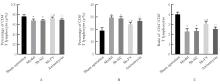
Fig. 7
Percentages of CD4+ and CD8+ T lymphocytes and ratios of CD4+/CD8+ T lymphocytes in blood of rats in various groups"
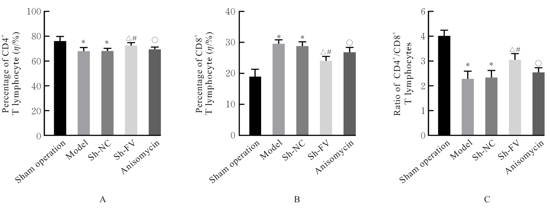
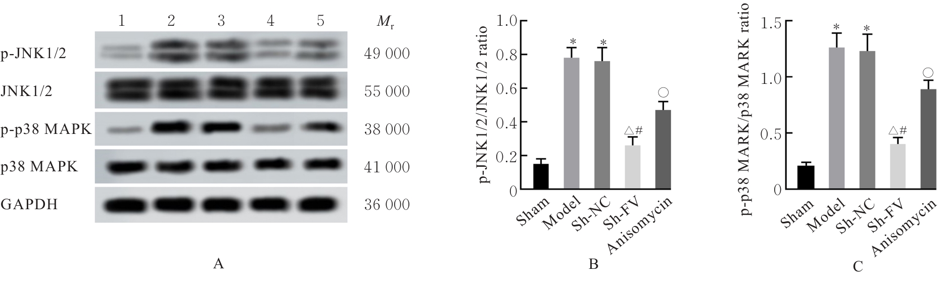
Fig. 8
Electrophoregram(A) and histograms(B,C) of expressions of JNK1/2 and p38 MAPK signaling pathway related proteins in lung tissue of rats in various groups"
| [1] | SRZIĆ I, NESEK ADAM V, TUNJIĆ PEJAK D. Sepsis definition: what’s new in the treatment guidelines[J]. Acta Clin Croat, 2022, 61(): 67-72. |
| [2] | RUDD K E, JOHNSON S C, AGESA K M, et al. Global, regional, and national sepsis incidence and mortality, 1990-2017: Analysis for the Global Burden of Disease Study[J]. Lancet, 2020, 395(10219): 200-211. |
| [3] | WANG B B, ZHU L L, JIA B, et al. Sepsis induces non-classic innate immune memory in granulocytes[J]. Cell Rep, 2023, 42(9): 113044. |
| [4] | UNAR A, BERTOLINO L, PATAUNER F, et al. Pathophysiology of disseminated intravascular coagulation in sepsis: A clinically focused overview[J]. Cells, 2023, 12(17): 2120. |
| [5] | LIND S M, SLETTEN M, HELLENES M, et al. Coagulation factor V in breast cancer: A p53-regulated tumor suppressor and predictive marker for treatment response to chemotherapy[J]. J Thromb Haemost, 2024, 22(6): 1569-1582. |
| [6] | 文柯力. 脓毒症合并弥散性血管内凝血的机制及抗凝治疗研究进展[J]. 现代医药卫生, 2019, 35(9): 1370-1374. |
| [7] | 张 铭, 雷 娇, 龚 禹, 等. 芍药苷通过JNK/NEK7/NLRP3通路缓解脓毒症相关急性肾损伤[J]. 中国病理生理杂志, 2024, 40(9): 1711-1717. |
| [8] | ZHANG Y R, CHEN Z R, GUO J Y, et al. Factor XII and prekallikrein promote microvascular inflammation and psoriasis in mice[J]. Br J Pharmacol, 2024, 181(19): 3760-3778. |
| [9] | 沈磊, 董林, 贺福义, 等. 三七总皂苷调节RAGE/MAPK/NF-κB信号通路对糖尿病骨质疏松大鼠骨密度及骨代谢的影响[J]. 中国老年学杂志, 2024, 44(23): 5768-5772. |
| [10] | 温 亚, 李 燕, 白思怡, 等. 瑞香素调节STING/TBK1/IRF3信号通路对脓毒症大鼠免疫炎症反应的影响[J]. 西安交通大学学报(医学版), 2024, 45(5): 822-827. |
| [11] | ZENG Y C, CAO G D, LIN L, et al. Resveratrol attenuates sepsis-induced cardiomyopathy in rats through anti-ferroptosis via the Sirt1/Nrf2 pathway[J]. J Invest Surg, 2023, 36(1): 2157521. |
| [12] | NEDEVA C. Inflammation and cell death of the innate and adaptive immune system during sepsis[J]. Biomolecules, 2021, 11(7): 1011. |
| [13] | CHEN W J, PAN J Y. Anatomical and pathological observation and analysis of SARS and COVID-19: Microthrombosis is the main cause of death[J]. Biol Proced Online, 2021, 23(1): 4. |
| [14] | FANG Z D, ZHANG X W, HUANG Y Y, et al. Piperlongumin improves survival in the mouse model of sepsis: Effect on coagulation factors and lung inflammation[J]. Inflammation, 2022, 45(6): 2513-2528. |
| [15] | MOHAMMED B M, PELC L A, RAU M J, et al. Cryo-EM structure of coagulation factor V short[J]. Blood, 2023, 141(26): 3215-3225. |
| [16] | XUAN W X, WU X, ZHENG L C, et al. Gut microbiota-derived acetic acids promoted sepsis-induced acute respiratory distress syndrome by delaying neutrophil apoptosis through FABP4[J]. Cell Mol Life Sci, 2024, 81(1): 438. |
| [17] | 罗玉娇, 郑小莉, 罗 波, 等. PD-L1通过调控PKC-α-NF-κB信号通路延迟脓毒血症中性粒细胞凋亡[J]. 中国免疫学杂志, 2020, 36(17): 2049-2052. |
| [18] | GENDRE B, MARTINEZ-PEREZ A, KLEBER M E, et al. Genome-wide search for nonadditive allele effects identifies PSKH2 as involved in the variability of factor V activity[J]. J Am Heart Assoc, 2024, 13(21): e034943. |
| [19] | 郑涵雪, 张连生, 李莉娟. 微移植治疗急性髓系白血病不同预处理方案精细免疫指标的差异性研究[J]. 中国实用内科杂志, 2025, 45(1): 54-61. |
| [20] | 谢小丽, 张予晋, 谭 瑶, 等. JAK/STAT通路介导免疫缺陷大鼠CD4+T细胞分化的研究[J]. 湖南中医药大学学报, 2023, 43(7): 1206-1214. |
| [21] | DE GROOT A S, ROSENBERG A S, SHAHJAHAN MIAH S M, et al. Identification of a potent regulatory T cell epitope in factor V that modulates CD4+ and CD8+ memory T cell responses[J]. Clin Immunol, 2021, 224: 108661. |
| [22] | 王 静, 辛绍斌. 脓毒症亚型的研究进展[J]. 中国实用内科杂志, 2025, 45(9): 785-792. |
| [23] | FAWZY M A, MAHER S A, BAKKAR S M, et al. Pantoprazole attenuates MAPK (ERK1/2, JNK, p38)- NF-κB and apoptosis signaling pathways after renal ischemia/reperfusion injury in rats[J]. Int J Mol Sci, 2021, 22(19): 10669. |
| [24] | MOHYELDIN R H, ALAAELDIN R, SHARATA E E, et al. LCZ696 attenuates sepsis-induced liver dysfunction in rats; the role of oxidative stress, apoptosis, and JNK1/2-P38 signaling pathways[J]. Life Sci, 2023, 334: 122210. |
| [25] | ZHANG W, CHEN H Z, XU Z Y, et al. Liensinine pretreatment reduces inflammation, oxidative stress, apoptosis, and autophagy to alleviate sepsis acute kidney injury[J]. Int Immunopharmacol, 2023, 122: 110563. |
| [26] | CUI Y N, TIAN N, LUO Y H, et al. High-dose Vitamin C injection ameliorates against sepsis-induced myocardial injury by anti-apoptosis, anti-inflammatory and pro-autophagy through regulating MAPK, NF-κB and PI3K/AKT/mTOR signaling pathways in rats[J]. Aging, 2024, 16(8): 6937-6953. |
| [27] | ISHIKURA S, OGAWA M, DOI K, et al. Zfat-deficient CD4⁺ CD8⁺ double-positive thymocytes are susceptible to apoptosis with deregulated activation of p38 and JNK[J]. J Cell Biochem, 2015, 116(1): 149-157. |
| [1] | Jinyu LI,Zhonghui LI,Aibin CHENG,Xuan BU,Jing BAI,Jianjun WANG. Improvement effect of TREM-1 inhibitory peptide LR12 on myocardial injury in septic mice and its mechanism [J]. Journal of Jilin University(Medicine Edition), 2026, 52(2): 366-374. |
| [2] | Jingyuan WANG,Fang CHEN,Yancun LIU,Shixin LI,Songtao SHOU. Effect of USF2 knockdown on coagulation dysfunction in septic rats and its mechanism [J]. Journal of Jilin University(Medicine Edition), 2026, 52(1): 171-181. |
| [3] | Yaping LI,Chunyan TAN,Lujie ZHAO,Jiayi ZHAO,Ting LI,Xiao YANG,Xiaoyun YANG. Inductive effect of lysophosphatidic acid combined with 6-hydroxydopamine on apoptosis of SH-SY5Y cells and its mechanism [J]. Journal of Jilin University(Medicine Edition), 2026, 52(1): 152-161. |
| [4] | Tao SUN,Zhiyin DAI,Xuan LI,Chaopu ZHANG,Shu DING,Jianwei ZHAO. Predictive value of pan-immune-inflammation index for major adverse cardiovascular events within 1 year after PCI in elderly patients with coronary heart disease [J]. Journal of Jilin University(Medicine Edition), 2025, 51(6): 1655-1660. |
| [5] | Kun YANG,Qianyao FU,Yongqiang SUN,Kun YANG,Jun MENG. Protective effect of dexmedetomidine on intestinal mucosal injury in rats with enterogenous sepsis and its mechanism [J]. Journal of Jilin University(Medicine Edition), 2025, 51(4): 855-865. |
| [6] | Ming xing YANG,Wen DONG,Ji LI. Inductive effect of peiminine on apoptosis of lung cancer A549 cells and its mechanism [J]. Journal of Jilin University(Medicine Edition), 2022, 48(3): 711-717. |
| [7] | ZHANG Xiaoxuan, MA Zheng, YU Ning, JIAO Guangmei, DOU Zhijie, ZHAO Liang, GAO Yanjun. Inhibitory effect of butylphthalide on hippocampal neuron apoptosis in ischemic stroke rats and itsinfluence in p38 MAPK signaling pathway [J]. Journal of Jilin University(Medicine Edition), 2019, 45(05): 1086-1091. |
| [8] | ZHENG Guilang, GUO Yuxiong, LYU Juanjuan, HUANG Jinda, LIU Cui, ZENG Qiyi. Protective effect of overexpression of uncoupling protein 2 on mitochondria in cardiomyocytes of rats with sepsis [J]. Journal of Jilin University Medicine Edition, 2018, 44(04): 698-703. |
| [9] | QIU Lijie, ZHANG Ye, ZHANG Wenjing, QIAN Donghua. Meta-analysis on curative effects of statins in treatment of pneumoniaor or sepsis [J]. Journal of Jilin University Medicine Edition, 2016, 42(02): 336-344. |
| [10] | LU Feiping, CHEN Wei, ZHEN Jie, MA Li. Changes of lymphocyte subsets in elderly patients with sepsis before and after immune modulation therapy [J]. Journal of Jilin University Medicine Edition, 2015, 41(05): 994-997. |
| [11] | LIU Hua-Wen, XU Xiang-Hong, LANG Sen-Yang, CAi Ai-Min. Expression of p38MAPK in cultured cortical neurons in vitro and intervention effect of W-7 [J]. J4, 2011, 37(1): 47-50. |
|