Journal of Jilin University(Medicine Edition)
Ameliorating effect of epicatechin on liver injury induced by acetaminophen in mice and its mechanism
Huiyuan YU1,2,Ling JIN3,Ying YU4,Xue WANG5,Bing WANG6( )
)
- 1.Department of Radiotherapy,First Hospital,Jilin University,Changchun 130021,China
2.Department of Clinical Veterinary Medicine,College of Veterinary Medicine,Jilin University,Changchun 130062,China
3.Department of Clinical Laboratory,First Hospital,Jilin University,Changchun 130021,China
4.Reproductive Center,Prenatal Diagnosis Center,First Hospital,Jilin University,Changchun 130021,China
5.Department of Clinical Laboratory,Songyuan Jilin Oilfield Hospital,Songyuan 138000,China
6.Department of Radiotherapy,Tumor Hospital,Jilin Province,Changchun 130012,China
-
Received:2024-12-09Accepted:2025-01-16Online:2025-01-26Published:2025-01-26 -
Contact:Bing WANG E-mail:zisu8688@163.com
CLC Number:
- R575
Cite this article
Huiyuan YU, Ling JIN, Ying YU, Xue WANG, Bing WANG. Ameliorating effect of epicatechin on liver injury induced by acetaminophen in mice and its mechanism[J].Journal of Jilin University(Medicine Edition), 2025, (): 1-10.
share this article
Add to citation manager EndNote|Reference Manager|ProCite|BibTeX|RefWorks
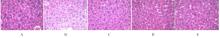
Fig. 1
Pathomorphology of liver tissue of mice in various groups (HE, ×400)"
Tab.1
Levels of ALT and AST in serum of mice in varrous groups"
| Group | Serum ALT level [λB/(U·L-1)] | Serum AST level [λB/(U·L-1)] |
|---|---|---|
| Blank control | 32.75±17.52 | 47.12±22.56 |
| APAP model | 582.63±36.32* | 509.22±15.97* |
| EC | ||
| Low dose | 425.33±21.47△ | 396.55±10.29△ |
| Medium dose | 215.69±19.59△△ | 189.96±30.21△△ |
| High dose | 153.92±24.91△△ | 109.85±26.91△△ |
Tab.2
Activities of MPO and levels of TNF-α and IL-1β in liver tissue of mice in various groups"
| Group | MPO activity[λB/(U·L-1)] | TNF-α level [ωB/(ng·g-1)] | IL-1β level [ωB/(ng·g-1)] |
|---|---|---|---|
| Blank control | 1.023±0.352 | 119.36±10.32 | 47.12±22.56 |
| APAP model | 5.336±1.096* | 756.83±63.98* | 509.22±15.97* |
| EC | |||
| Low dose | 4.589±0.966△ | 572.41±98.25△ | 396.55±10.29△ |
| Medium dose | 2.158±0.765△△ | 276.85±50.67△△ | 189.96±30.21△△ |
| High dose | 1.864±0.919△△ | 159.72±56.91△△ | 109.85±26.91△△ |
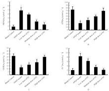
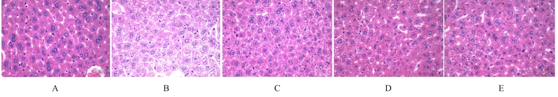
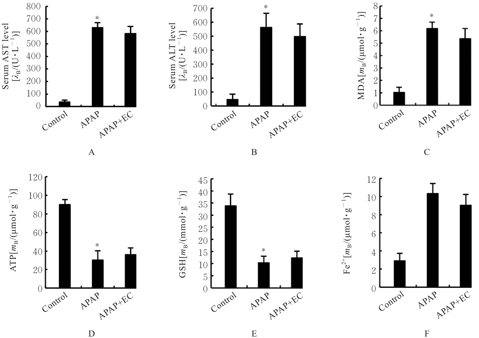
Fig. 2
Levels of MDA,ATP, GSH, and Fe2? in liver tissue homogenates of mice in various groups"

Fig. 3
Electrophoregram(A) and histograms(B,C) of expressions of GPX4 and xCT proteins in liver tissue of mice in various groups"

Fig.4
Electrophoregram(A) and histograms(B, C) of expressions of NF-κB p-p65 and p-IκBα,proteins in liver tissue of mice in various groups"
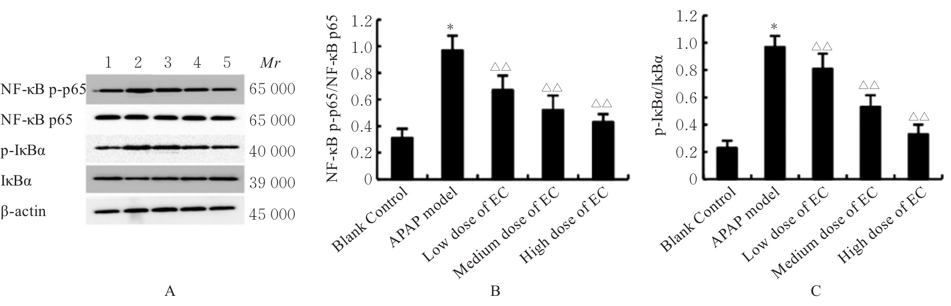
Fig. 5
Electrophoregram(A) and histograms(B, C) of Nrf2 and HO-1 in liver tissue of mice in various groups"

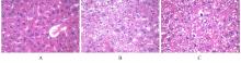
Fig. 6
Pathomorphology of liver tissue of Nrf2-/- mice in varions groups (HE, ×400)"
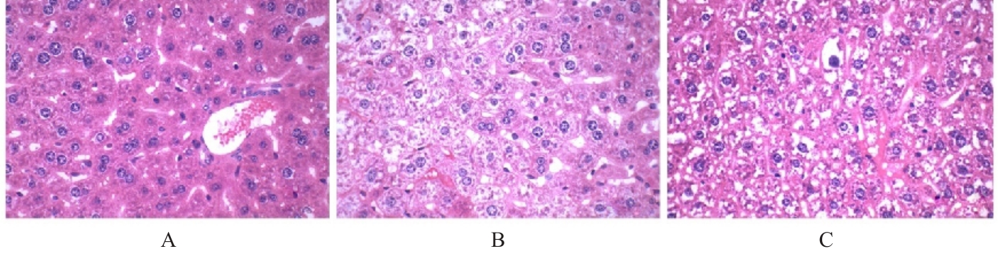
Fig. 7
Levels of AST and ALT in serum and levels of MDA, ATP, GSH, and Fe2+ in liver tissue of Nrf2-/- mice in various groups"
| 1 | SIEMIONOW K, TEUL J, DRAGOWSKI P, et al. New potential biomarkers of acetaminophen-induced hepatotoxicity[J]. Adv Med Sci, 2016, 61(2): 325-330. |
| 2 | WOODHEAD J L, HOWELL B A, YANG Y, et al. An analysis of N-acetylcysteine treatment for acetaminophen overdose using a systems model of drug-induced liver injury[J]. J Pharmacol Exp Ther, 2012, 342(2): 529-540. |
| 3 | MORRIS G, ANDERSON G, DEAN O, et al. The glutathione system: a new drug target in neuroimmune disorders[J]. Mol Neurobiol, 2014, 50(3): 1059-1084. |
| 4 | WALLACE K P, CENTER S A, HICKFORD F H, et al. S-adenosyl-L-methionine (SAMe) for the treatment of acetaminophen toxicity in a dog[J]. J Am Anim Hosp Assoc, 2002, 38(3): 246-254. |
| 5 | YU H T, GUO P Y, XIE X Z, et al. Ferroptosis, a new form of cell death, and its relationships with tumourous diseases[J]. J Cell Mol Med, 2017, 21(4): 648-657. |
| 6 | DIXON S J, LEMBERG K M, LAMPRECHT M R, et al. Ferroptosis: an iron-dependent form of nonapoptotic cell death[J]. Cell, 2012, 149(5): 1060-1072. |
| 7 | CAPELLETTI M M, MANCEAU H, PUY H, et al. Ferroptosis in liver diseases: an overview[J]. Int J Mol Sci, 2020, 21(14): 4908. |
| 8 | CHEN J Y, LI X P, GE C D, et al. The multifaceted role of ferroptosis in liver disease[J]. Cell Death Differ, 2022, 29(3): 467-480. |
| 9 | WANG C, LIU T, TONG Y M, et al. Ulinastatin protects against acetaminophen-induced liver injury by alleviating ferroptosis via the SIRT1/NRF2/HO-1 pathway[J]. Am J Transl Res, 2021, 13(6): 6031-6042. |
| 10 | LAN X, HAN X N, LI Q, et al. (-)-Epicatechin, a natural flavonoid compound, protects astrocytes against hemoglobin toxicity via Nrf2 and AP-1 signaling pathways[J]. Mol Neurobiol, 2017, 54(10): 7898-7907. |
| 11 | TIAN X, XUE Y S, XIE G G, et al. (-)-Epicatechin ameliorates cigarette smoke-induced lung inflammation via inhibiting ROS/NLRP3 inflammasome pathway in rats with COPD[J]. Toxicol Appl Pharmacol, 2021, 429: 115674. |
| 12 | XING J, YU Z L, ZHANG X Y, et al. Epicatechin alleviates inflammation in lipopolysaccharide-induced acute lung injury in mice by inhibiting the p38 MAPK signaling pathway[J]. Int Immunopharmacol, 2019, 66: 146-153. |
| 13 | WU C X, LI F F, ZHANG X X, et al. (-)-epicatechin ameliorates monosodium urate-induced acute gouty arthritis through inhibiting NLRP3 inflammasome and the NF-κB signaling pathway[J]. Front Pharmacol, 2022, 13: 799552. |
| 14 | MA X, LI M M, LU G C, et al. Anti-inflammation of epicatechin mediated by TMEM35A and TMPO in bovine mammary epithelial cell line cells and mouse mammary gland[J]. J Dairy Sci, 2021, 104(12): 12925-12938. |
| 15 | TACKE F, LUEDDE T, TRAUTWEIN C. Inflammatory pathways in liver homeostasis and liver injury[J]. Clin Rev Allergy Immunol, 2009, 36(1): 4-12. |
| 16 | YOU Q, HOLT M, YIN H, et al. Role of hepatic resident and infiltrating macrophages in liver repair after acute injury[J]. Biochem Pharmacol, 2013, 86(6): 836-843. |
| 17 | HOLT M P, CHENG L L, JU C. Identification and characterization of infiltrating macrophages in acetaminophen-induced liver injury[J]. J Leukoc Biol, 2008, 84(6): 1410-1421. |
| 18 | KRENKEL O, MOSSANEN J C, TACKE F. Immune mechanisms in acetaminophen-induced acute liver failure[J]. Hepatobiliary Surg Nutr, 2014, 3(6): 331-343. |
| 19 | JAESCHKE H, DAVID WILLIAMS C, RAMACHANDRAN A, et al. Acetaminophen hepatotoxicity and repair: the role of sterile inflammation and innate immunity[J]. Liver Int, 2012, 32(1): 8-20. |
| 20 | HAYDEN M S, GHOSH S. Regulation of NF-κB by TNF family cytokines[J]. Semin Immunol, 2014, 26(3): 253-266. |
| 21 | LIU A M, TANAKA N, SUN L, et al. Saikosaponin d protects against acetaminophen-induced hepatotoxicity by inhibiting NF-κB and STAT3 signaling[J]. Chem Biol Interact, 2014, 223: 80-86. |
| 22 | LAI Y F, DONG J, WU Y, et al. Lipid peroxides mediated ferroptosis in electromagnetic pulse-induced hippocampal neuronal damage via inhibition of GSH/GPX4 axis[J]. Int J Mol Sci, 2022, 23(16): 9277. |
| 23 | YAMADA N, KARASAWA T, TAKAHASHI M. Role of ferroptosis in acetaminophen-induced hepatotoxicity[J]. Arch Toxicol, 2020, 94(5): 1769-1770. |
| 24 | FORCINA G C, DIXON S J. GPX4 at the crossroads of lipid homeostasis and ferroptosis[J]. Proteomics, 2019, 19(18): e1800311. |
| 25 | ZHU L J, CHEN D Z, ZHU Y, et al. GPX4-regulated ferroptosis mediates S100-induced experimental autoimmune hepatitis associated with the Nrf2/HO-1 signaling pathway[J]. Oxid Med Cell Longev, 2021, 2021(1): 6551069. |
| 26 | PANIERI E, PINHO S A, AFONSO G J M, et al. NRF2 and mitochondrial function in cancer and cancer stem cells[J]. Cells, 2022, 11(15): 2401. |
| 27 | PANG C, ZHENG Z Y, SHI L, et al. Caffeic acid prevents acetaminophen-induced liver injury by activating the Keap1-Nrf2 antioxidative defense system[J]. Free Radic Biol Med, 2016, 91: 236-246. |
| 28 | LI H Y, WENG Q Q, GONG S, et al. Kaempferol prevents acetaminophen-induced liver injury by suppressing hepatocyte ferroptosis via Nrf2 pathway activation[J]. Food Funct, 2023, 14(4): 1884-1896. |
| 29 | CAI X P, HUA S Y, DENG J W, et al. Astaxanthin activated the Nrf2/HO-1 pathway to enhance autophagy and inhibit ferroptosis, ameliorating acetaminophen-induced liver injury [J]. ACS Appl Mater Interfaces, 2022, 14(38): 42887-42903. |
| 30 | LI L, HUANG W X, WANG S K, et al. Astragaloside Ⅳ attenuates acetaminophen-induced liver injuries in mice by activating the Nrf2 signaling pathway[J]. Molecules, 2018, 23(8): 2032. |
| 31 | PARK T J, PARK J H, LEE G S, et al. Quantitative proteomic analyses reveal that GPX4 downregulation during myocardial infarction contributes to ferroptosis in cardiomyocytes[J]. Cell Death Dis, 2019, 10(11): 835. |
| [1] | Baolian MA,Xiaoxue HU,Xiaowen AI,Yonglan ZHANG. Inhibitory effect of diosmetin on ferroptosis of GC-2 spermatocytes induced by RSL3 in mice and its mechanism [J]. Journal of Jilin University(Medicine Edition), 2024, 50(6): 1481-1490. |
| [2] | Meng QU,Rui HUANG,Xinda JU,Yuxin LIU,Jichen XIA,Jiaxin HUANG,Chunyan YU,Zhiheng DONG. Ameliorative effect of ginsenoside Rh1 on kidney injury in diabetic mice through activation of Nrf2/HO-1 signaling pathway [J]. Journal of Jilin University(Medicine Edition), 2024, 50(6): 1565-1571. |
| [3] | Yi LONG,Ziyi YOU,Xiuying TAN,Rou ZHANG,Yuhan ZHANG,Lina YANG. Protective effect of sodium butyrate on acute liver injury in mice induced by lipopolysaccharide combined with D-galactosamine and its mechanism [J]. Journal of Jilin University(Medicine Edition), 2024, 50(6): 1614-1620. |
| [4] | Guobin HE,Huan WANG. Effect of knockdown of RIP3 on autophagy, pyroptosis, and ferroptosis of hypoxia/reoxygenation-induced human renal tubular epithelial HK2 cells [J]. Journal of Jilin University(Medicine Edition), 2024, 50(6): 1644-1653. |
| [5] | Fangyang JIANG,Jing XIAO,He CHANG,Mingyang SUN,Wenjing ZHANG,Guangfu LYU,He LIN,Zhe LIN,Xiaowei HUANG,Yuchen WANG. Effect of polygonatum odoratum polysaccharide on acute kidney injury in mice induced by cisplatin and its ferroptosis mechanism [J]. Journal of Jilin University(Medicine Edition), 2024, 50(5): 1235-1242. |
| [6] | Yanjue YE,Ziyi TANG,Yupei TAN,Shiying YANG,Yong LIU,Li YIN. Effect of azathioprine on ferroptosis in spermatocytes of mice induced by RSL3 [J]. Journal of Jilin University(Medicine Edition), 2024, 50(5): 1217-1226. |
| [7] | Xiaoyong PENG,Yu ZHU,Shuangbo ZHANG,Yingguo ZHU,Tao LI,Liangming LIU,Jianmin WANG,Guangming YANG. Alleviative effect of fluid resuscitation on damage of structure injury of vascular cells after blast injury complicated with hemorrhagic shock in rats by inhibiting ferroptosis of vascular tissue [J]. Journal of Jilin University(Medicine Edition), 2024, 50(5): 1227-1234. |
| [8] | Junping WEI,Dajia FU,Qingwen MENG,Daofei LIN,Yanzai LIN. Effect of bone marrow mesenchymal stem cell-derived exosomes on myocardial fibrosis in rats induced by isoproterenol and its mechanism [J]. Journal of Jilin University(Medicine Edition), 2024, 50(5): 1348-1357. |
| [9] | Yingqun NI,Mao YANG,Di YANG,Chenglin GUO,Wenjun ZHU,Yaqin YU,Qin LU,Jinzhi LUO,Chunqin WU,Zhaohui FANG. Screening of key differentially expressed genes involved in osteogenic differentiation of lower limb vascular smooth muscle cells and validation [J]. Journal of Jilin University(Medicine Edition), 2024, 50(3): 620-627. |
| [10] | Yuexin LIU,Yongwei LAI,Yanchun WANG,Bo XU,Qian LU,Ying AN,Kuang REN,Hongyan FAN. Improvement effect of total flavonoids of Sophora flavescens on oxidative stress induced by lead acetate in leydig cells in mice and its in effect on Keap1/Nrf2 signaling pathway [J]. Journal of Jilin University(Medicine Edition), 2024, 50(2): 320-325. |
| [11] | Ruipeng ZHANG,Jie LI. Resistance and regeneration effects of lncRNA GPRC5D-AS1 on muscle atrophy of myocytes in mice induced by dexamethasone and its mechanism [J]. Journal of Jilin University(Medicine Edition), 2023, 49(6): 1457-1465. |
| [12] | Jing GUAN,Shen HA,Hao YUAN,Ying CHEN,Pengju LIU,Zhi LIU,Shuang JIANG. Protective effect of Modified Xiao-Xian-Xiong Decoction on liver injury in rats with type 2 diabete mellitus and its mechanism [J]. Journal of Jilin University(Medicine Edition), 2023, 49(3): 608-616. |
| [13] | Qiaoling YANG,Lu FU,Yu SHI,Yanjue YE,Rifeng LU,Yong LIU,Li YIN. Inhibitory effect of rosiglitazone on ferroptosis of renal tubular epithelial cells in mice with acute renal injury induced by lipopolysaccharide and its mechanism [J]. Journal of Jilin University(Medicine Edition), 2023, 49(2): 351-359. |
| [14] | Renyi YANG,Shuwang PENG,Yongheng WANG,Yuxuan DONG,Shanshan DUAN. Construction of ferroptosis prognostic risk model of thyroid cancer and bioinformatics analysis on its potential mechanism [J]. Journal of Jilin University(Medicine Edition), 2023, 49(2): 402-413. |
| [15] | Xuechun DU,Baosheng LI,Shuwei QIAO,Yanzhen OU,Zhen LI,Weiyan MENG. Effect of Porphyromonas gingivalis-LPS on expression levels of ferroptosis-related factors in macrophages [J]. Journal of Jilin University(Medicine Edition), 2022, 48(5): 1148-1155. |
|
||

