Journal of Jilin University(Medicine Edition) ›› 2026, Vol. 52 ›› Issue (2): 398-409.doi: 10.13481/j.1671-587X.20260212
• Research in basic medicine • Previous Articles Next Articles
Inhibitory effect of Chaiqi Yigan formula on malignant biological behaviors of liver cancer HepG2 cells by regulating macrophage polarization
Xiqian ZHANG1,Zhibo DANG2( ),Yunan DU1,Pei WU1,Hang XIE3,Gaofeng TAN1
- 1.Department of Geriatrics,Henan Provincial Hospital of Traditional Chinese Medicine,Second Affiliated Hospital,Henan University of Chinese Medicine,Zhengzhou 450053,China
2.Department of Hepatobiliary,Spleen and Stomach Diseases,Henan Provincial Hospital of Traditional Chinese Medicine,Second Affiliated Hospital,Henan University of Chinese Medicine,Zhengzhou 450053,China
3.Department of Critical Care Medicine,Henan Provincial Hospital of Traditional Chinese Medicine,Second Affiliated Hospital,Henan University of Chinese Medicine,Zhengzhou 450053,China
-
Received:2025-05-31Accepted:2025-07-06Online:2026-03-28Published:2026-04-15 -
Contact:Zhibo DANG E-mail:1622097861@qq.com
CLC Number:
- R273
Cite this article
Xiqian ZHANG,Zhibo DANG,Yunan DU,Pei WU,Hang XIE,Gaofeng TAN. Inhibitory effect of Chaiqi Yigan formula on malignant biological behaviors of liver cancer HepG2 cells by regulating macrophage polarization[J].Journal of Jilin University(Medicine Edition), 2026, 52(2): 398-409.
share this article
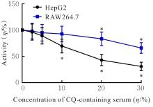
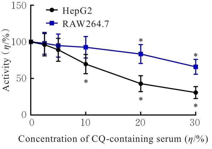
Fig. 1
Activities of HepG2 and RAW264.7 cells after treated with different concentrations of CQ-containing serum"
Tab.1
Levels of IL-6 and IL-10 in supernant of RAW264.7 cells after treated with different concentrations of CQ-containing serum [n=6, x±s, ρB/(ng·L-1)]"
| Group | IL-6 | IL-10 |
|---|---|---|
| Blank | 79.04±13.35 | 23.01±3.92 |
| Co-culture | 96.15±12.10 | 82.75±5.13* |
| 2.5% CQ-containing serum | 220.28±13.64*△ | 63.52±2.87△ |
| 5.0% CQ-containing serum | 345.76±14.15*△ | 45.02±3.24△ |
| 10.0% CQ-containing serum | 402.85±15.86*△ | 27.93±2.90△ |
| 20.0% CQ-containing serum | 420.23±17.12*△ | 25.80±3.45△ |
| 30.0% CQ-containing serum | 435.71±16.53*△ | 24.14±4.16△ |
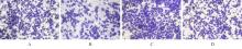
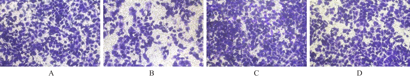
Fig. 2
Migration of HepG2 cells in various groups (Crystal violet, ×200)"
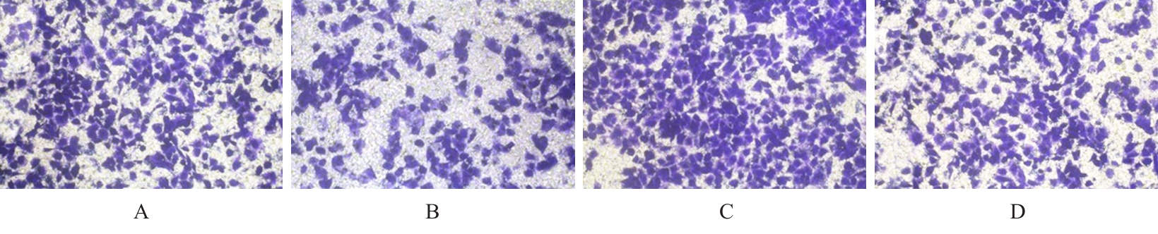
Fig. 3
Invasion of HepG2 cells in various groups (Crystal violet, ×200)"
Tab.2
Cell activities and numbers of migration and invasion HepG2 cells in various groups"
| Group | Cell activity (η/%) | Number of migration cells | Number of invasion cells |
|---|---|---|---|
| Control | 100.00±0.00 | 232.50±19.23 | 329.00±21.36 |
| CQ | 48.90±9.62* | 119.00±14.15* | 132.50±16.22* |
| PDTC | 205.45±13.25* | 343.50±22.46* | 471.00±27.40* |
| CQ+PDTC | 92.70±10.84△ | 215.00±20.14△ | 314.50±23.05△ |
Fig. 4
Polarization phenotypes of macrophages in RAW264.7 cells in various groups detected by flow cytometry"
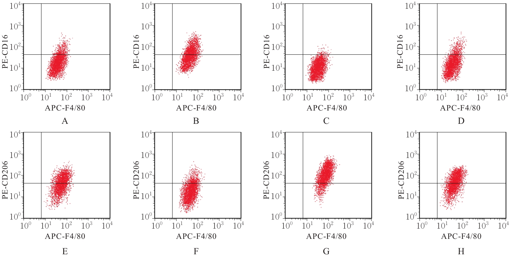
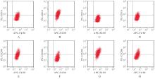
Fig. 5
Polarization phenotypes of macrophages in tumor tissue of nude mice in various groups detected by flow cytometry"
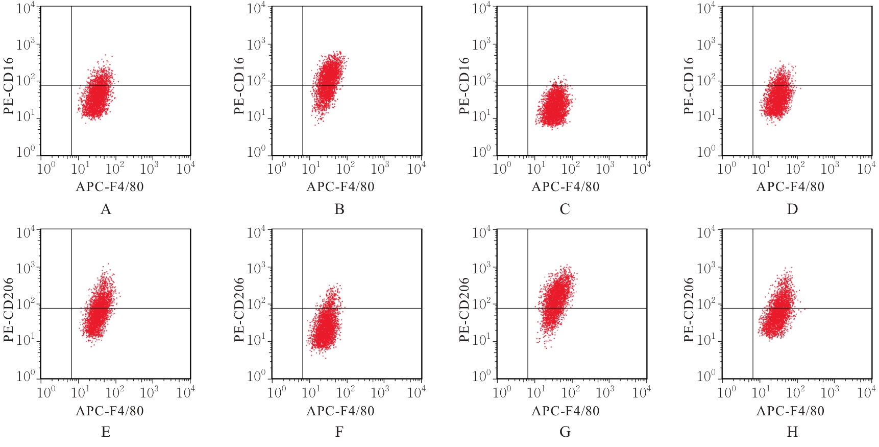
Tab.3
Percentages of M1 type and M2 type macrophages in RAW264.7 cells and tumor tissue of nude mice in various groups"
| Group | M1 type macrophage | M2 type macrophage | ||
|---|---|---|---|---|
| RAW264.7 | Tumor tissue | RAW264.7 | Tumor tissue | |
| Control | 18.05±3.30 | 20.84±2.62 | 52.43±4.73 | 45.31±4.12 |
| CQ | 52.32±4.25* | 63.75±4.80* | 20.36±3.15* | 16.70±2.90* |
| PDTC | 1.84±0.60* | 2.56±0.79* | 81.90±5.64* | 71.89±4.53* |
| CQ+PDTC | 20.13±2.72△ | 22.13±2.45△ | 49.86±3.92△ | 43.25±3.64△ |
Tab.4
Levels of M1/M2 type macrophage secretion factors in supernatant of RAW264.7 cells in various groups [n=6, x±s, ρB/(ng·L-1)]"
| Group | IL-6 | TNF-α | iNOS | TGF-β | Arg-1 | IL-10 |
|---|---|---|---|---|---|---|
| Control | 93.54±9.37 | 42.71±5.02 | 11.74±1.15 | 31.15±3.76 | 126.21±9.16 | 82.75±6.13 |
| CQ | 234.76±15.72* | 184.69±9.20* | 29.83±2.04* | 14.49±2.53* | 49.86±7.95* | 29.10±4.51* |
| PDTC | 40.13±7.51* | 13.45±3.62* | 3.67±0.92* | 47.62±4.14* | 198.74±8.78* | 131.82±8.36* |
| CQ+PDTC | 101.08±11.13△ | 47.93±4.84△ | 12.95±1.48△ | 29.05±3.12△ | 118.54±8.20△ | 76.91±6.42△ |
Tab.5
Levels of M1/M2 type macrophage secretion factors in serum of nude mice in various groups [n=6, x±s, ρB/(ng·L-1)]"
| Group | IL-6 | TNF-α | iNOS | TGF-β | Arg-1 | IL-10 |
|---|---|---|---|---|---|---|
| Control | 60.92±6.73 | 29.62±4.14 | 5.98±0.37 | 15.56±2.63 | 71.64±7.13 | 59.12±5.20 |
| CQ | 156.45±13.62* | 130.51±6.97* | 19.32±0.64* | 7.47±1.82* | 23.60±6.04* | 18.63±3.57* |
| PDTC | 27.63±5.21* | 8.75±2.70* | 0.83±0.25* | 24.06±3.15* | 117.95±8.20* | 105.82±7.69* |
| CQ+PDTC | 67.10±7.24△ | 33.56±3.92△ | 5.13±0.46△ | 13.91±2.81△ | 66.17±7.92△ | 54.96±4.84△ |
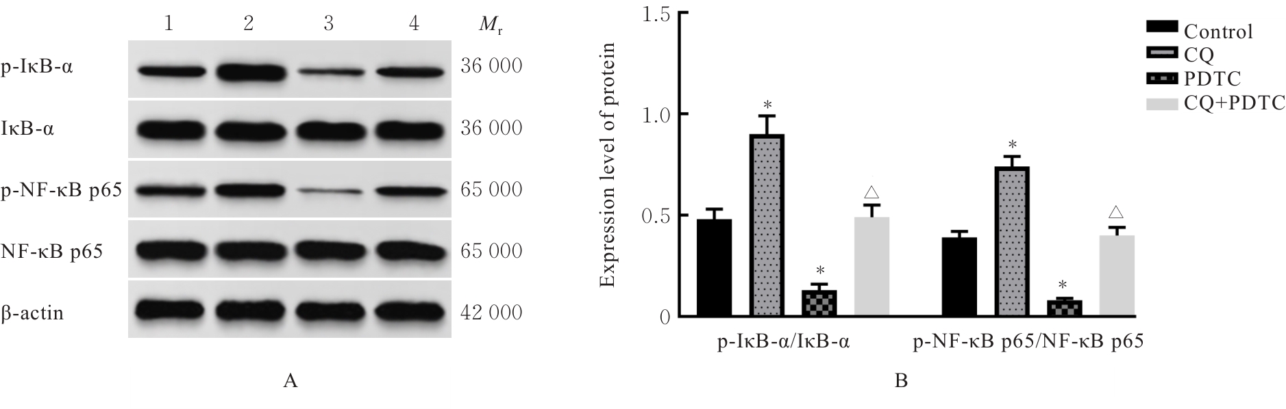
Fig. 6
Electrophoregram (A) and histogram (B) of expressions of NF-κB pathway related proteins in RAW264.7 cells in various groups"
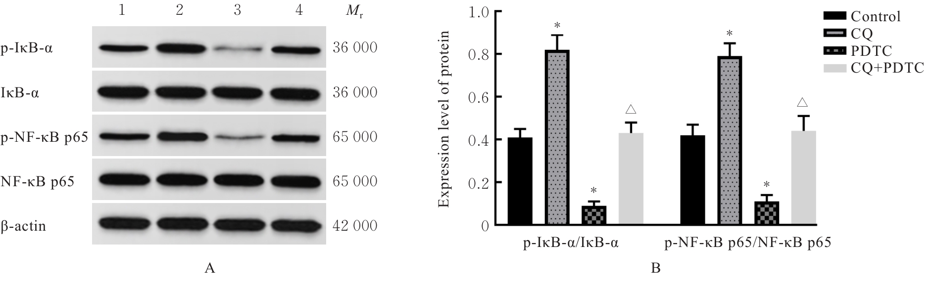
Fig. 7
Electrophoregram(A) and histogram(B) of expressions of NF-κB pathway related proteins in tumor tissue of nude mice in various groups"
| [1] | LV S M, WANG J H, LI L. Extracellular vesicular lncRNA FAL1 promotes hepatocellular carcinoma cell proliferation and invasion by inducing macrophage M2 polarization[J]. J Physiol Biochem, 2023, 79(3): 669-682. |
| [2] | CHEN S P, ZHANG P L, ZHU G Q, et al. Targeting GSDME-mediated macrophage polarization for enhanced antitumor immunity in hepatocellular carcinoma[J]. Cell Mol Immunol, 2024, 21(12): 1505-1521. |
| [3] | CAI J L, SONG L N, ZHANG F, et al. Targeting SRSF10 might inhibit M2 macrophage polarization and potentiate anti-PD-1 therapy in hepatocellular carcinoma[J]. Cancer Commun, 2024, 44(11): 1231-1260. |
| [4] | HAN S L, BAO X Y, ZOU Y F, et al. D-lactate modulates M2 tumor-associated macrophages and remodels immunosuppressive tumor microenvironment for hepatocellular carcinoma[J]. Sci Adv, 2023, 9(29): eadg2697. |
| [5] | TAN S Y, WANG Z H, LI N, et al. Transcription factor Zhx2 is a checkpoint that programs macrophage polarization and antitumor response[J]. Cell Death Differ, 2023, 30(9): 2104-2119. |
| [6] | LI L, ZUO W T, LIU H, et al. Oxoaporphine Pr(Ⅲ) complex inhibits hepatocellular carcinoma progression and metastasis by disrupting tumor cell-macrophage crosstalk[J]. Biomed Pharmacother, 2023, 169: 115849. |
| [7] | 游敏玲, 罗满芳, 廖蔚茜, 等. 柴芪益肝颗粒联合紫杉醇抑制裸鼠肝癌生长的在体成像研究[J]. 南方医科大学学报, 2012, 32(7): 1042-1045. |
| [8] | 周怡驰, 晏 军, 胡世平, 等. 基于生物分子网络调控研究柴芪益肝方治疗肝纤维化的作用机制[J]. 世界中西医结合杂志, 2021(3): 393-400, 445. |
| [9] | 吕景娣, 王永辉, 冷茹冰, 等. 比卡鲁胺对乳腺癌细胞周期、巨噬细胞极化及p38/p-STAT1水平的影响[J]. 现代药物与临床, 2023, 38(7): 1553-1559. |
| [10] | 张春焕, 阚士锋, 魏守娟, 等. LPS通过宫颈癌细胞来源的IL-6调节巨噬细胞分泌和趋化活性[J]. 现代妇产科进展, 2021, 30(12): 905-909. |
| [11] | 罗江萍, 刘永红, 马全庆, 等. 柴芪益肝颗粒对小鼠肝移植瘤的作用[J]. 广东医学, 2011, 32(3): 284-286. |
| [12] | 袁兆林, 刘 娣, 朱绪锋. HMGB1/RAGE轴在结直肠癌肥大细胞浸润中的作用机制研究[J]. 中国免疫学杂志, 2022, 38(15): 1861-1865. |
| [13] | COFFIN P, HE A W. Hepatocellular carcinoma: past and present challenges and progress in molecular classification and precision oncology[J]. Int J Mol Sci, 2023, 24(17): 13274. |
| [14] | SINGH S P, ARORA V, MADKE T, et al. Hepatocellular carcinoma-Southeast Asia updates[J]. Cancer J, 2023, 29(5): 259-265. |
| [15] | WU D Q, LI Y J. Application of adoptive cell therapy in hepatocellular carcinoma[J]. Immunology, 2023, 170(4): 453-469. |
| [16] | PONVILAWAN B, ROTH M T. Sequencing systemic therapy in hepatocellular carcinoma[J]. Curr Treat Options Oncol, 2023, 24(11): 1580-1597. |
| [17] | 王 妮, 贺屹巍, 陈燕京, 等. 柴芪疏肝调脾方联合生物反馈对腹腔镜直肠癌低位保肛术后胃肠、肛门功能指标恢复的影响[J]. 临床和实验医学杂志, 2023, 22(5): 491-495. |
| [18] | 马贵萍, 刘博文, 李晓斌, 等. 基于mTORC1/SREBP1/SCD1信号通路探讨柴芪益肝颗粒含药血清对肝癌HepG2细胞铁死亡的影响[J]. 中成药, 2025, 47(4): 1305-1309. |
| [19] | LU Y J, SUN Q K, GUAN Q F, et al. The XOR-IDH3α axis controls macrophage polarization in hepatocellular carcinoma[J]. J Hepatol, 2023, 79(5): 1172-1184. |
| [20] | LIU Y Y, TANG Y F, JIANG H L, et al. Exosome-related FTCD facilitates M1 macrophage polarization and impacts the prognosis of hepatocellular carcinoma[J]. Biomolecules, 2023, 14(1): 41. |
| [21] | LI G L, TANG J F, TAN W L, et al. The anti-hepatocellular carcinoma effects of polysaccharides from Ganoderma lucidum by regulating macrophage polarization via the MAPK/NF-κB signaling pathway[J]. Food Funct, 2023, 14(7): 3155-3168. |
| [22] | 冯雯倩, 杜 洋, 毛德文, 等. 核因子κB信号通路在肝脏疾病中的作用机制及潜在治疗靶点[J]. 临床肝胆病杂志, 2025, 41(9): 1949-1955. |
| [23] | YU Z, LI Y Y, LI Y, et al. Bufalin stimulates antitumor immune response by driving tumor-infiltrating macrophage toward M1 phenotype in hepatocellular carcinoma[J]. J Immunother Cancer, 2022, 10(5): e004297. |
| [24] | XU G L, FENG D J, YAO Y, et al. Listeria-based hepatocellular carcinoma vaccine facilitates anti-PD-1 therapy by regulating macrophage polarization[J]. Oncogene, 2020, 39(7): 1429-1444. |
| [25] | LEI C H, GAO Z H, LV X Z, et al. Saikosaponin-b2 inhibits primary liver cancer by regulating the STK4/IRAK1/NF-κB pathway[J]. Biomedicines, 2023, 11(10): 2859. |
| [1] | Ruihan GE,Chen LI,Shengpeng WANG,Yang LU,Caixia TAN,Haotian CUI,Xinmin WANG,Le ZHANG. Bioinformatic analysis on regulatory mechanism of MAPK-Mcl-1 signaling pathway and macrophage polarization during Bacillus Calmette-Guérin infection and its experimental validation [J]. Journal of Jilin University(Medicine Edition), 2026, 52(2): 440-450. |
| [2] | Jiaxin WANG,Junwen MAO,Xuan ZHANG. Expression of natural autoantibodies against apoptosis inhibitory genes in plasma of patients with hepatocellular carcinoma and its clinical significance [J]. Journal of Jilin University(Medicine Edition), 2026, 52(1): 228-235. |
| [3] | Limei WEN,Yali GUO,Wenmei MA,Taotao XUE,Ruoyu GENG,Chong MA,Xinhong ZHANG,Jianhua YANG. Bioinformatic analysis of TCGA database based on INPP4B gene expression in hepatocellular carcinoma and its experimental validation [J]. Journal of Jilin University(Medicine Edition), 2025, 51(6): 1618-1629. |
| [4] | Kun YANG,Qianyao FU,Yongqiang SUN,Kun YANG,Jun MENG. Protective effect of dexmedetomidine on intestinal mucosal injury in rats with enterogenous sepsis and its mechanism [J]. Journal of Jilin University(Medicine Edition), 2025, 51(4): 855-865. |
| [5] | Han LIN,Qiuyan YANG,Jieyue ZHONG,Bolun CHEN,Wangxia TONG. Improvement effect of cordycepin on ferroptosis in HepG2 cells induced by RSL3 and its mechanism [J]. Journal of Jilin University(Medicine Edition), 2025, 51(3): 576-589. |
| [6] | Xiaoxia HU,Yalong LI,Dongliang YANG,Bazeren LA,Xinyue LIU. Effect of high glucose on polarization of Raw264.7 macrophages in vitro [J]. Journal of Jilin University(Medicine Edition), 2025, 51(2): 403-411. |
| [7] | Xiaoyan WANG,Xuelian LI,Bin LIANG,Wenfei TIAN,Hailin MA,Zhijing MO. Analysis on relationship between CALU and prognosis of hepatocellular carcinoma patients and its mechanism based on transcriptome and single cell sequencing data [J]. Journal of Jilin University(Medicine Edition), 2025, 51(2): 447-459. |
| [8] | Tan CHEN,Yan CHEN. Research progress in mechanism of fibrosis regulated by macrophage polarization [J]. Journal of Jilin University(Medicine Edition), 2024, 50(5): 1465-1473. |
| [9] | Yongjing YANG,Tianyang KE,Shixin LIU,Xue WANG,Dequan XU,Tingting LIU,Ling ZHAO. Synergistic sensitization of apatinib mesylate and radiotherapy on hepatocarcinoma cells invitro [J]. Journal of Jilin University(Medicine Edition), 2024, 50(4): 1009-1015. |
| [10] | Jinlian LI,Lanzhen HUANG,Xishi HUANG,Kangzhi LI,Jiali JIANG,Miaomiao ZHANG,Qunying WU. Bioinformatics analysis on key genes related to prognosis, diagnosis, and immune cell infiltration of hepatocellular carcinoma and their potential therapeutic drugs [J]. Journal of Jilin University(Medicine Edition), 2024, 50(4): 1062-1075. |
| [11] | Shilei GAO,Jiaqiang WANG,Weitao YAO,Zhichao TIAN,Chao LI,Xiaoxiao LIANG,Xin WANG. Effect of miR-761 on epithelial-mesenchymal transition in osteosarcoma MG63 cells by regulating tumor-associated macrophage polarization [J]. Journal of Jilin University(Medicine Edition), 2024, 50(4): 978-988. |
| [12] | Yiyan YU,Zhimin ZHANG,Jiawen CHEN,Xin LIU,Yan LI,Hongyan ZHAO. Research progress in relationship between macrophage polarization and oral diseases [J]. Journal of Jilin University(Medicine Edition), 2024, 50(3): 864-871. |
| [13] | Xiaoyan WANG,Hao ZHANG,Zehao GUO,Jun CAO,Zhijing MO. Screening of UBE2S interacting protein and construction of prognostic model in hepatocellular carcinoma [J]. Journal of Jilin University(Medicine Edition), 2024, 50(1): 168-177. |
| [14] | Xiaopeng YU,Renyi YANG,Zuomei HE,Puhua ZENG. Establishment and validation of nomogram of cancer specific survival of patients with hepatocellular carcinoma with negative alpha fetoprotein based on SEER Database [J]. Journal of Jilin University(Medicine Edition), 2024, 50(1): 188-197. |
| [15] | Yong DONG,Lingyao XU,Jing HUA,Han LIANG,Dongya LIU,Junbo ZHAO,Zhenglu SUN,Cheng CHENG,Shutang WEI. Effect of macrophage exosomal lncRNA HULC on migration, invasion,and metastasis of hepatocellular carcinoma cells and its mechanism [J]. Journal of Jilin University(Medicine Edition), 2023, 49(5): 1217-1226. |
|