吉林大学学报(医学版) ›› 2021, Vol. 47 ›› Issue (6): 1455-1461.doi: 10.13481/j.1671-587X.20210615

• 基础研究 • 上一篇    下一篇

外源性神经生长因子对形觉剥夺性近视豚鼠巩膜组织的保护作用及其机制

张新,巨朝娟(),金鑫,熊朝晖,赵燕   

  1. 河北医科大学第一医院眼科,河北 石家庄 050031
  • 收稿日期:2020-09-04 出版日期:2021-11-28 发布日期:2021-12-14
  • 通讯作者: 巨朝娟 E-mail:xinzh1978@163.com
  • 作者简介:张 新(1978-),女,河北省衡水市人,副教授,副主任医师,医学硕士,主要从事眼视光方面的研究。
  • 基金资助:
    河北省卫健委医学科研项目(20190422)

Protective effect of exogenous nerve growth factor on scleral tissue of guinea pigs with form-deprived myopia and its mechanism

Xin ZHANG,Chaojuan JU(),Xin JIN,Chaohui XIONG,Yan ZHAO   

  1. Department of Ophthalmology,First Hospital,Hebei Medical University,Shijiazhuang 050031,China
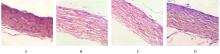
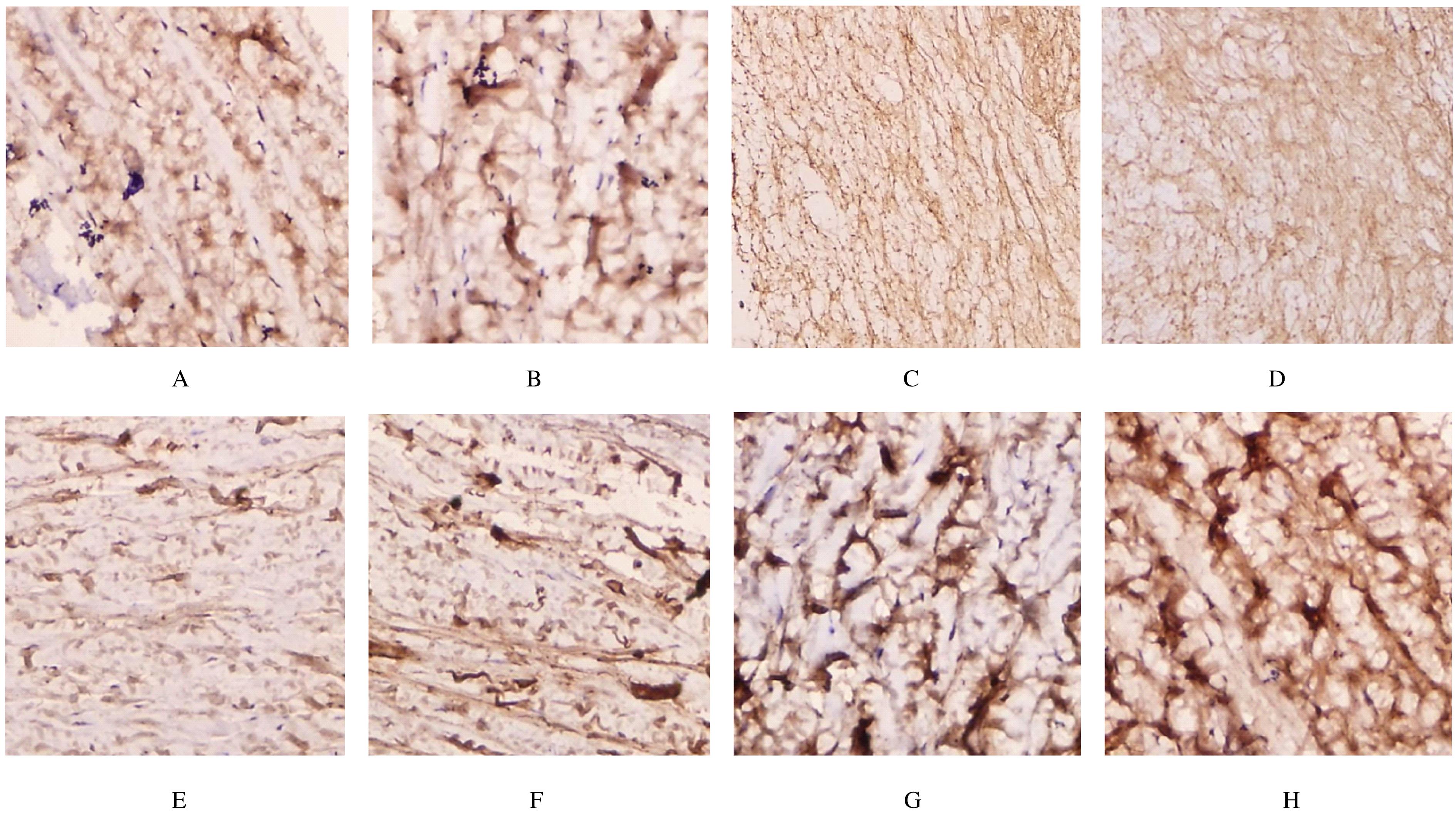
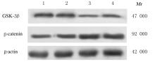
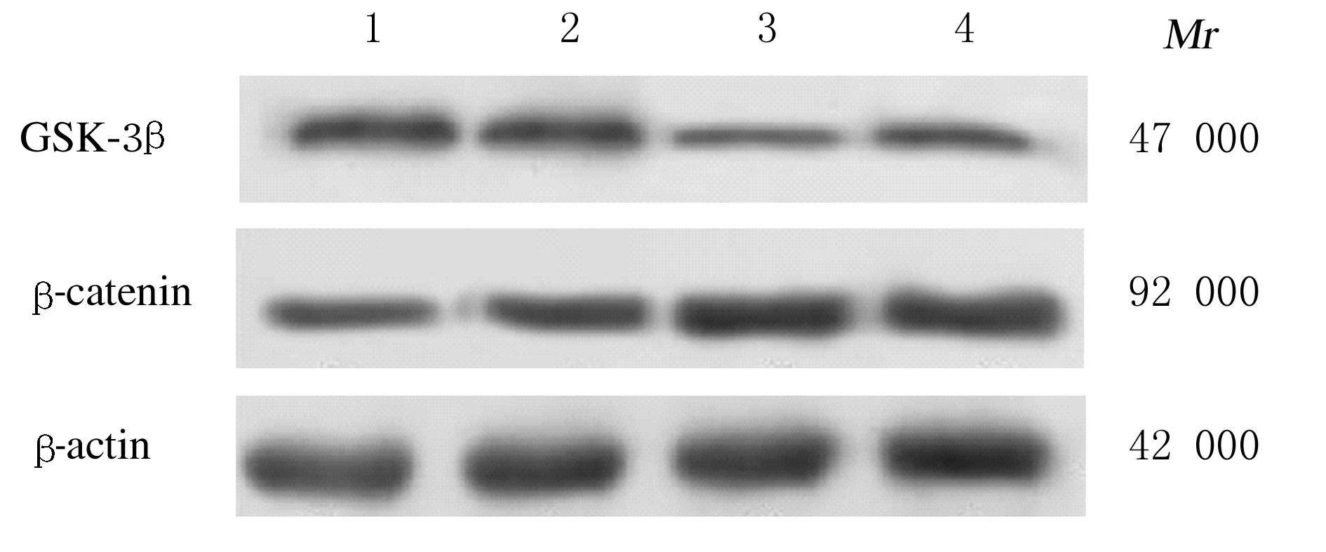
  • Received:2020-09-04 Online:2021-11-28 Published:2021-12-14
  • Contact: Chaojuan JU E-mail:xinzh1978@163.com

摘要: 目的

探讨外源性神经生长因子(NGF)对形觉剥夺性近视(FDM)豚鼠的保护作用,并阐明其机制。

方法

48只豚鼠随机分为对照组、模型组、低浓度NGF组和高浓度NGF组(n=12)。除对照组外,其余各组豚鼠采用右眼戴-10.0屈光度(D)透镜的方法建立FDM模型,左眼不进行处理。低和高浓度NGF组豚鼠分别右眼注射500和1 000 BU NGF,模型组和对照组豚鼠给予等体积生理盐水,连续给药21 d。分别采用红外偏心验光仪和眼科A/B超声诊断仪检测各组豚鼠双眼D和眼轴长度, HE染色观察各组豚鼠巩膜组织病理形态表现,实时荧光定量PCR(RT-qPCR)法检测各组豚鼠巩膜组织中糖原合成酶激酶 3β(GSK-3β)、β-连环蛋白(β-catenin)和血管内皮生长因子(VEGF) mRNA表达水平,免疫组织化学法检测各组豚鼠巩膜组织中GSK-3β和β-catenin阳性细胞率, Western blotting法检测各组豚鼠巩膜组织中GSK-3β和β-catenin蛋白表达水平。

结果

与模型组比较,低和高浓度NGF组豚鼠右眼D明显升高(P<0.05),眼轴长度明显减少(P<0.05)。HE染色,对照组豚鼠巩膜组织结构正常,模型组豚鼠巩膜组织厚度减少,胶原纤维排列紊乱,纤维之间出现明显空隙;低和高浓度NGF组豚鼠巩膜组织厚度增加,结构相对清晰。与模型组比较,低和高浓度NGF组豚鼠右眼巩膜组织中GSK-3β mRNA和蛋白表达水平明显降低(P<0.05),VEGF 和β-catenin mRNA及蛋白表达水平明显升高(P<0.05)。

结论

外源性NGF能够通过调控Wnt/β-catenin信号通路,抑制GSK-3β表达,增加β-catenin表达,对FDM豚鼠巩膜组织起保护作用。

关键词: 神经生长因子, 形觉剥夺性近视, Wnt/β-catenin信号通路, 血管内皮生长因子, 糖原合成酶激酶3β, β-连环蛋白

Abstract: Objective

To discuss the protective effect of exogenous nerve growth factor (NGF) in the guinea pigs with form-deprived myopia(FDM),and to charify its mechanism.

Methods

A total of 48 guinea pigs were randomly divided into control group, model group, low concentration of NGF group and high concentration of NGF group(n=12). In addition to control group, the FDM models of guinea pigs in other groups were established by wearing lens with 10.0 diopter (D) in the right eyes, and the left eyes were not treated. The guinea pigs in low and high concentrations of NGF groups were respectively injected with 500 and 1 000 BU NGF in the right eyes for 21 d. The D and axial lengths of eyes of the guinea pigs in various groups were measured by infrared eccentric optometer and ophthalmic A/B ultrasound diagnostic apparatus. HE staining was used to observe the pathomophology of scleral tissue of the guinea pigs in various groups. Real-time fluorescence quantificative PCR (RT-qPCR) method was used to detect the expression levels of glycogen synthase kinase-3β(GSK-3β),β-catenin, and vascular endothelial growth factor (VEGF) mRNA in scleral tissue of the guinea pigs in various groups. Immunohistochemistry was used to detect the rates of GSK-3β and β-catenin positive cells in scleral tissue of the guinea pigs in various groups.Western blotting method was used to detect the expression levels of GSK-3β and β-catenin proteins in scleral tissue of the guinea pigs in various groups.

Results

Compared with model group, the D of right eyes of the guinea pigs in low and high concentrations of NGF groups were increased significantly (P<0.05), while the axial lengths of eyes were decreased significantly (P<0.05).The HE staining results showed that the thickness of scleral tissue of the guinea pigs in model group were decreased, the collagen fibers were disordered, and the obvious interstitial spaces appeared between the fibers; the thickness of sclera tissue of the guinea pigs in low and high concentrations of NGF groups were increased, and the structures were relatively clear. Compared with model group, the expression levels of GSK-3β mRNA and protein in scleral tissue of right eyes of the guinea pigs in low and high concentrations of NGF groups were significantly decreased (P<0.05), while the expression levels of VEGF and β-catenin mRNA and protein were significantly increased (P<0.05).

Conclusion

The exogenous NGF can protect the scleral tissue of the guinea pigs with FDM by regulating the Wnt/β-catenin signaling pathway, inhibiting the expression of GSK-3β and increasing the expression of β-catenin.

Key words: nerve growth factor, form-deprived myopia, Wnt/β-catenin signal pathway, vascular endothelial growth factor, glycogen synthase kinase 3β, β-catenin

中图分类号: 

  • R788.1