吉林大学学报(医学版) ›› 2022, Vol. 48 ›› Issue (5): 1139-1147.doi: 10.13481/j.1671-587X.20220506

• 基础研究 • 上一篇    

当归红芪超滤物对X射线引起人脐静脉内皮细胞损伤的保护作用及其机制

武兵兵1,张爱平1,赵信科2,李应东2,刘凯1,2()   

  1. 1.甘肃中医药大学中西医结合学院 中西医结合临床重点实验中心,甘肃 兰州 730000
    2.甘肃中医药大学附属医院心血管内科,甘肃 兰州 730000
  • 收稿日期:2021-11-18 出版日期:2022-09-28 发布日期:2022-11-15
  • 通讯作者: 刘凯 E-mail:xubo_l@163.com
  • 作者简介:武兵兵(1986-),男,甘肃省会宁县人,在读硕士研究生,主要从事中西医结合防治心脑血管疾病方面的研究。
  • 基金资助:
    国家中医药管理局国家中医药循证能力建设项目(2019XZZX-XXG002);甘肃省科技厅自然科学基金项目(21JR7RA562);甘肃省中医药研究中心开放课题(ZYZX-2020-16)

Protective effect of ultra-filtration extract from Angelica Sinensis Radix and Hedysari Radix on human umbilical vein endothelial cell injury induced by X-ray and its mechanism

Bingbing WU1,Aiping ZHANG1,Xinke ZHAO2,Yingdong LI2,Kai LIU1,2()   

  1. 1.Key Clinical Experimental Center of Integrated Traditional Chinese and Western Medicine,College of Integrated Chinese and Western Medicine,Gansu University of Traditional Chinese Medicine,Lanzhou 730000,China
    2.Department of Cardiovascular Medicine,Affiliated Hospital,Gansu University of Traditional Chinese Medicine,Lanzhou 730000,China
  • Received:2021-11-18 Online:2022-09-28 Published:2022-11-15
  • Contact: Kai LIU E-mail:xubo_l@163.com

摘要:

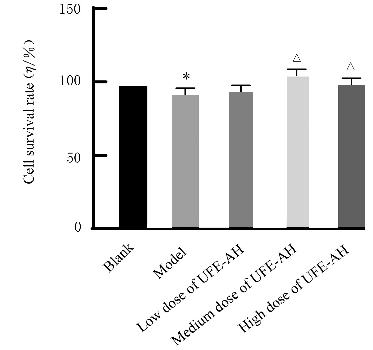
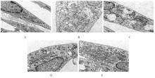
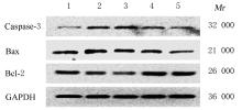
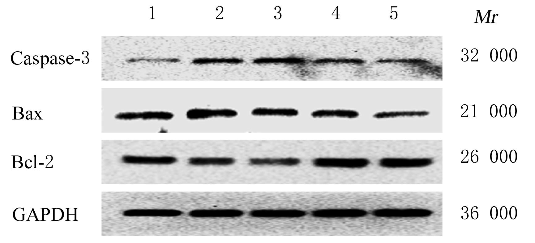
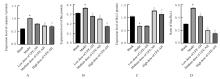
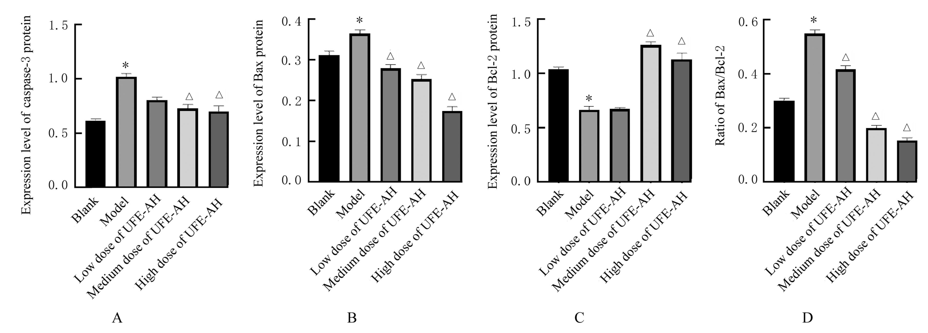
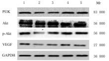
目的 探讨当归红芪超滤物(UFE-AH)对X射线引起人脐静脉内皮细胞(HUVECs)损伤的保护作用,阐明其可能的机制。 方法 体外培养 HUVECs,采用不同剂量(0、2、4、6、8和10 Gy)X射线辐射,筛选出最佳辐射剂量(6 Gy);用不同浓度(0、50、100、200、400、600、800和1 000 μg·L-1)UFE-AH干预HUVECs,筛选出最佳促增殖浓度(100、200和400 μg·L-1)。实验分为空白组、模型组(6 Gy X射线)、低剂量UFE-AH组(6 Gy X射线+100 μg·L-1 UFE-AH)、中剂量UFE-AH组(6 Gy X射线+200 μg·L-1 UFE-AH)和高剂量UFE-AH组(6 Gy X射线+400 μg·L-1 UFE-AH)。CCK-8法检测各组细胞存活率,流式细胞术检测各组细胞凋亡率,透射电子显微镜下观察各组细胞超微结构,Western blotting法检测各组细胞中磷脂酰肌醇3-激酶(PI3K)、蛋白激酶B(Akt)、磷酸化Akt(p-Akt)、血管内皮生长因子(VEGF)、B细胞淋巴瘤2(Bcl-2)、Bcl-2相关 X 蛋白(Bax)和含半胱氨酸的天冬氨酸蛋白水解酶3(caspase-3)蛋白表达水平。 结果 CCK-8法检测,与0 Gy比较,当辐射剂量大于2 Gy时,辐射后48和72 h X射线对HUVECs的抑制率随辐射剂量增加而升高(P<0.05或P<0.01),6 Gy及6 Gy以上辐射剂量细胞抑制率升高更明显(P<0.01)。与0 μg·L-1 UFE-AH比较,100、200、400和600 μg·L-1UFE-AH作用时细胞存活率升高(P<0.05或P<0.01),且100、200和400 μg·L-1UFE-AH作用24、48和72 h时细胞存活率明显升高(P<0.01)。与空白组比较,模型组细胞存活率明显降低(P<0.01);与模型组比较,中和高剂量UFE-AH 组细胞存活率明显升高(P<0.01)。流式细胞术检测,与空白组比较,模型组细胞凋亡率明显升高(P<0.01);与模型组比较,低、中和高剂量UFE-AH组细胞凋亡率均明显降低(P<0.01)。透射电子显微镜观察,空白组细胞膜完整,胞质较均匀,线粒体呈卵圆形,未见明显肿胀,胞内可见少量溶酶体;与空白组比较,模型组细胞重度肿胀,细胞膜多处破损,胞质稀松且出现多个空泡区,细胞核呈轻度不规则形,线粒体数量明显减少,呈轻度或中度肿胀,基质变浅且不均,线粒体少部分嵴局部断裂、变短,胞内可见较大量自噬溶酶体(ASS);与模型组比较,低、中和高剂量UFE-AH组细胞肿胀减轻,胞内细胞器空泡逐渐减少,线粒体肿胀减轻,数量增多,胞内可见一定量ASS,但仍未恢复正常。Western blotting 法检测,与空白组比较,模型组细胞中PI3K、p-Akt、Bcl-2和VEGF蛋白表达水平明显降低(P<0.01),Bax和caspase-3蛋白表达水平及Bax/Bcl-2比值明显升高(P<0.01);与模型组比较,中和高剂量 UFE-AH组细胞中PI3K、p-Akt、Bcl-2和VEGF蛋白表达水平明显升高(P<0.01),Bax和caspase-3蛋白表达水平及Bax/Bcl-2比值明显降低(P<0.01)。 结论 一定剂量的UFE-AH对X射线引起的HUVECs 损伤具有保护作用,其机制可能与UFE-AH影响HUVECs中PI3K、Akt、p-Akt、VEGF、Bcl-2、Bax和caspase-3蛋白表达有关。

关键词: 当归红芪超滤物, 血管内皮细胞, 细胞凋亡, X射线, 辐射, 放射治疗

Abstract:

Objective: To investigate the protective effect of ultra-filtration extract from Angelica Sinensis Radix and Hedysari Radix (UFE-AH) on the injury of human umbilical vein endothelial cells (HUVECs) induced by X-ray,and to clarify its possible mechanism. Methods The HUVECs were cultured in vitro and irradiated with different doses (0,2,4,6,8 and 10 Gy) of X-ray; the optimal radiation dose (6 Gy) was selected. The HUVECs were intervened with different concentrations (0,50,100,200,400,600,800 and 1 000 μg·L-1) of UFE-AH, and the optimal proliferation promoting concentrations (100,200 and 400 μg·L-1) were selected.The experiment was divided into blank group, model group (6 Gy X-ray), low dose of UFE-AH group (6 Gy X-ray + 100 μg·L-1 UFE-AH), medium dose of UFE-AH group (6 Gy X-ray+200 μg·L-1 UFE-AH) and high dose of UFE-AH group (6 Gy X-ray + 400 μg·L-1 UFE-AH).The survival rates of cells in various groups were measured by CCK-8 assay, the apoptotic rates of cells in various groups were measured by flow cytometry,the ultrastructures were observed by transmission electron microscope, and the expression levels of phosphatidylinositol 3-kinase (PI3K), protein kinase B (Akt), phosphorylated Akt(p-Akt),vascular endothelial growth factor (VEGF),B-cell lymphoma-2(Bcl-2),Bcl-2 associated X protein (Bax) and cysteinyl aspartate specific proteinase-3(caspase-3) proteins in the cells in various groups were measured by Western blotting method. Results The CCK-8 assay results showed that compared with 0 Gy X-ray,the inhibitory rates of HUVECs irradiated by X-ray were increased with the increase of radiation dose when the indiation dose was greater than 2 Gy at 48 and 72 h after radiation (P<0.05 or P<0.01), and the increasing of inhibition rates at 6 Gy X-ray and above were more significant(P<0.01). Compared with 0 μg·L-1 UFE-AH, the survival rates of cells after treated with 100,200,400 and 600 μg·L-1UFE-AH were increased(P<0.05 or P<0.01),especially treated with 100,200 and 400 μg·L-1 UFE-AH for 24,48 and 72 h (P<0.01). Compared with blank group,the survival rate of the cells in model group was decreased(P<0.01);compared with model group, the cell survival rates in medium and high doses of UFE-AH groups were significantly increased(P<0.01). Compared with blank group,the apoptotic rate of the cells in model group was significantly increased(P<0.01);compared with model group, the apoptotic rates of the cells in different doses of UFE-AH groups were significantly decreased(P<0.01).The transmission electron microscope results showed that the cell membrane in blank group was intact and the cytoplasm was more uniform, the mitochondria were oval; no obvious swelling was observed, and a small amount of lysosomes were observed in the cells;compared with blank group, the cells in model group had severe swelling, multiple damage of the cell membrane, loose cytoplasm and many vacuolar areas, mildly irregular nuclei, the significantly reduced mitochondria, mild or moderate swelling, shallower and uneven matrix, local breakage and shortening of a small part of the cristae of mitochondria, and a large amount of autolysosomes (ASS) in the cells; compared with model group, the cell swelling in low, medium, and high doses of UFE-AH groups was alleviated,intracellular organelle vacuoles were gradually reduced, mitochondrial swelling was alleviated, the mitochondrial number was increased, and a certain amount of ASS were observed, but they still did not return to normal. Compared with blank group,the expression levels of PI3K, p-Akt, Bcl-2 and VEGF proteins in the cells in model group were significantly decreased(P<0.01), and the expression levels of Bax and caspase-3 proteins and the Bax/Bcl-2 ratio were significantly increased(P<0.01); compared with model group, the expression levels of PI3K, p-Akt, Bcl-2 and VEGF proteins in the cells in medium and high doses of UFE-AH groups were significantly increased(P<0.01),and the expression levels of Bax and caspase-3 proteins and the Bax/Bcl-2 ratio were significantly decreased(P<0.01). Conclusion UFE-AH at a certain dose has a protective effect on the X-ray-induced injury of HUVECs, and its mechanism may be related to the effect of UFE-AH on the PI3K, Akt, p-Akt, VEGF, Bcl-2, Bax and caspase-3 protein expressions in the HUVECs.

Key words: Ultra-filtration extract from Angelica Sinensis Radix and Hedysari Radix, Vascular endothelial cells, Apoptosis, X-ray, Radiation, Radiation therapy

中图分类号: 

  • R815.2