吉林大学学报(医学版) ›› 2022, Vol. 48 ›› Issue (5): 1314-1323.doi: 10.13481/j.1671-587X.20220527

• 临床研究 • 上一篇    

妊娠期糖尿病患者胎盘组织中Apelin和APJ的表达及其对滋养层细胞胰岛素抵抗和行为的影响

刘莎莎1,赵云1,孙国强1,陈菲菲2()   

  1. 1.湖北省妇幼保健院产科,湖北 武汉 430070
    2.湖北省妇幼保健院妇产科,湖北 武汉 430070
  • 收稿日期:2021-10-29 出版日期:2022-09-28 发布日期:2022-11-15
  • 通讯作者: 陈菲菲 E-mail:49381389@qq.com
  • 作者简介:刘莎莎(1985-),女,湖北省荆州市人,主治医师,医学学士,主要从事妇产科疾病诊治方面的研究。
  • 基金资助:
    湖北省卫健委科研项目(WJ2019M227)

Expressions of Apelin and APJ in placental tissue of gestational diabetes mellitus patients and its effect on insulin resistance and behavior of trophoblast cells

Shasha LIU1,Yun ZHAO1,Guoqiang SUN1,Feifei CHEN2()   

  1. 1.Department of Obstetrics,Hubei Provincial Maternal and Child Health Hospital,Wuhan 430070,China
    2.Department of Obstetrics and Gynecology,Hubei Maternal and Child Health Hospital,Wuhan 430070,China
  • Received:2021-10-29 Online:2022-09-28 Published:2022-11-15
  • Contact: Feifei CHEN E-mail:49381389@qq.com

摘要:

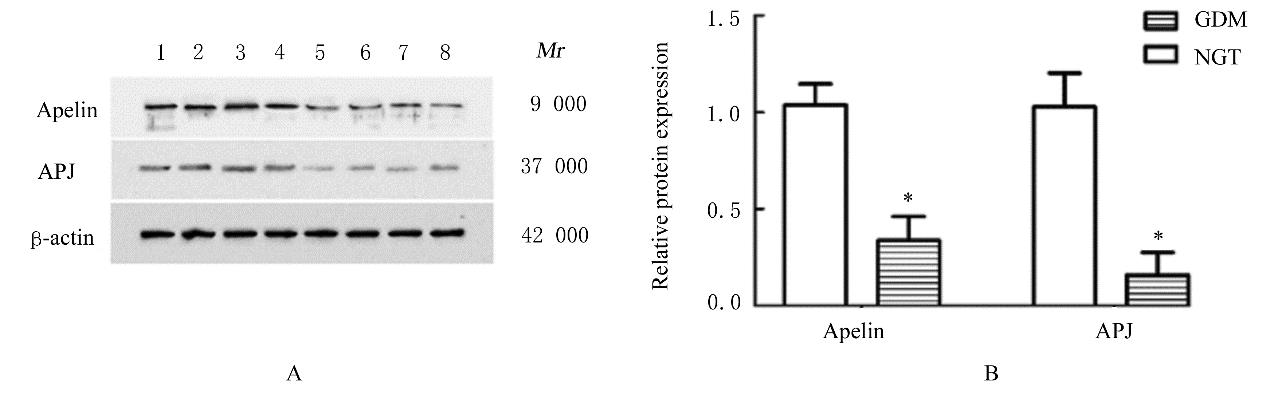
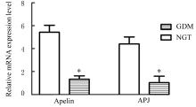
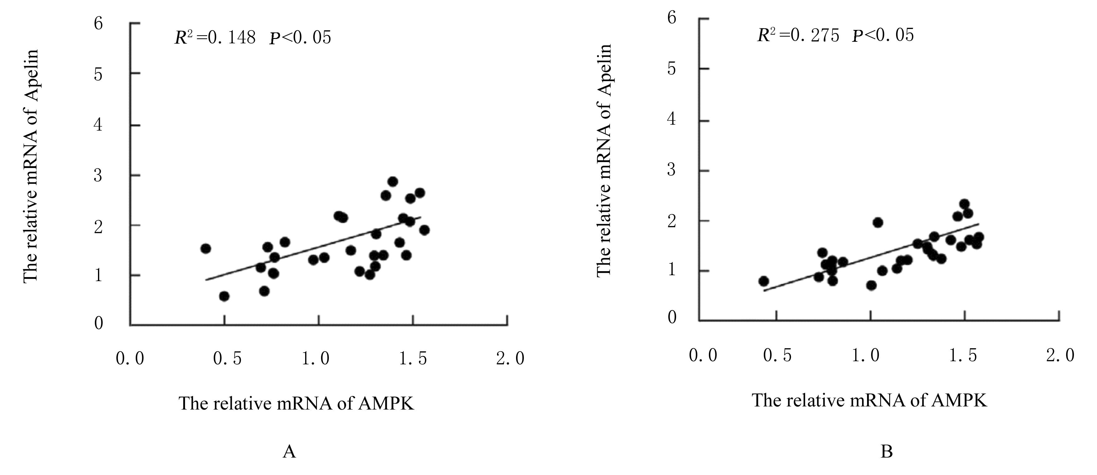
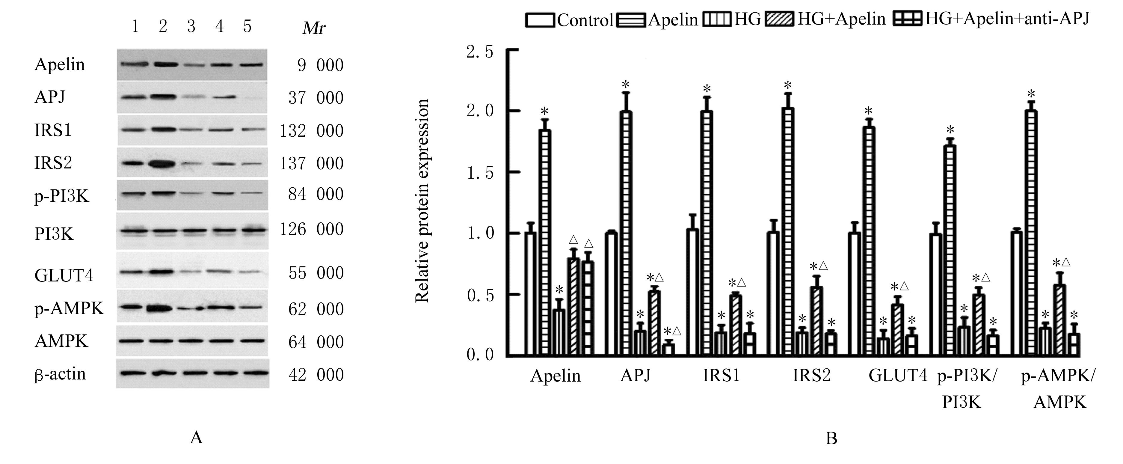
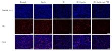
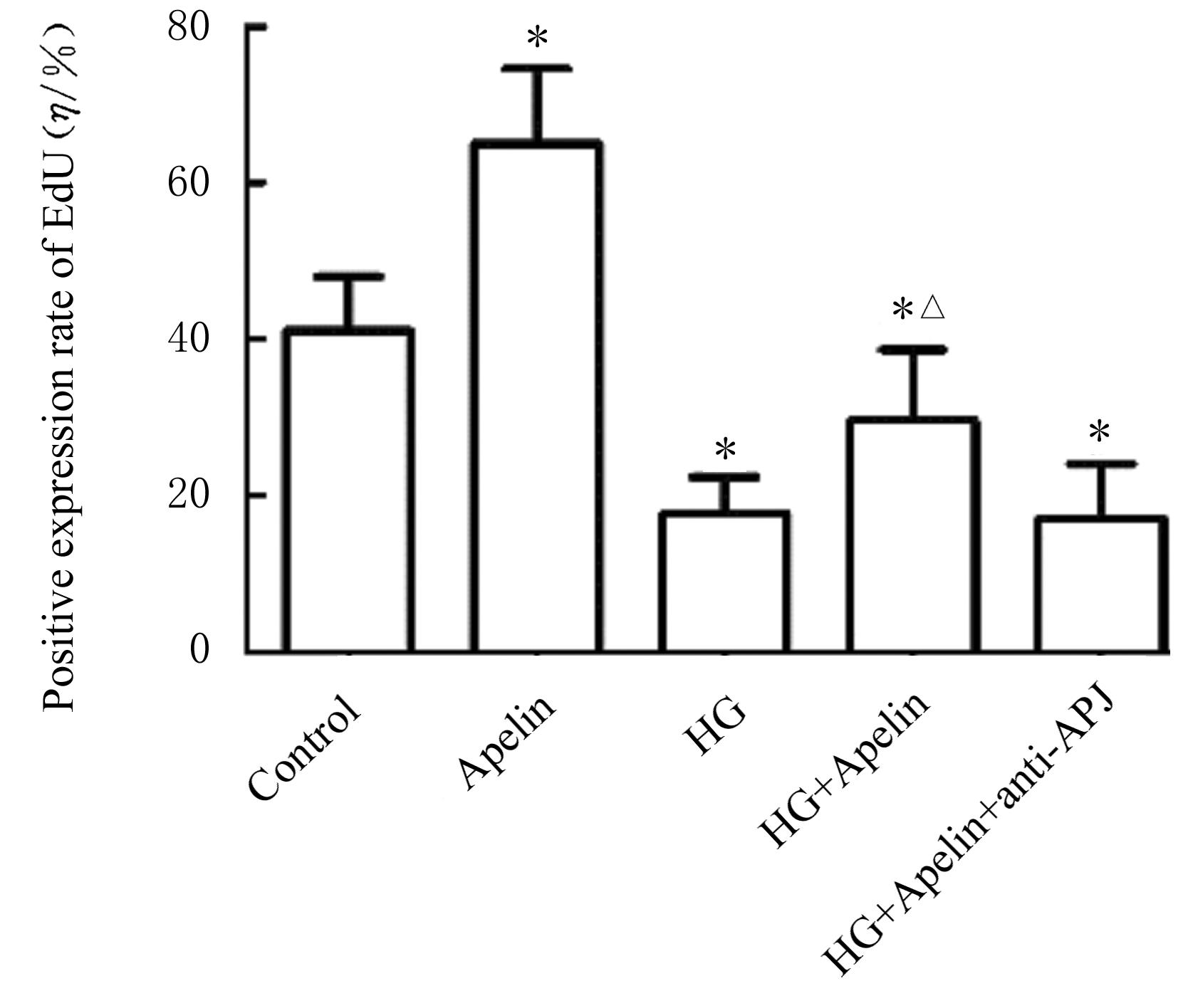
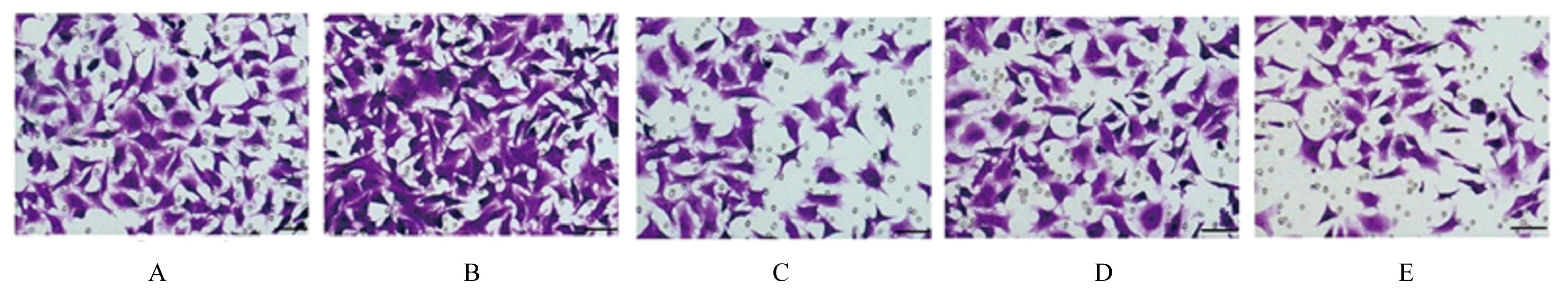
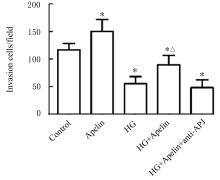
目的 观察妊娠期糖尿病(GDM)患者胎盘组织中Apelin和其受体血管紧张素Ⅱ1型受体相关蛋白(APJ)表达情况,探讨其对滋养层细胞胰岛素抵抗(IR)、增殖和侵袭能力的影响。 方法 收集GDM患者(GDM组,30例)和糖耐量正常(NGT)孕产妇(NGT组,30例)胎盘组织。采用免疫组织化学法检测2组研究对象胎盘组织中Apelin和APJ阳性表达率,Western blotting法检测胎盘组织中Apelin和APJ蛋白表达水平,实时荧光定量PCR(RT-qPCR)法检测胎盘组织中Apelin、APJ和腺苷酸活化蛋白激酶(AMPK)mRNA表达水平,Pearson相关分析法分析Apelin和APJ mRNA表达水平与AMPK mRNA表达水平的相关性。体外培养滋养层细胞HTR-8/SVneo(HTR-8),分为对照组、Apelin组、高糖组(HG组)、HG+Apelin组和HG+Apelin+anti-APJ组;采用Western blotting法检测各组细胞中Apelin、APJ和胰岛素信号传递中胰岛素受体底物1(IRS1)、胰岛素受体底物2(IRS2)和葡萄糖转运蛋白4(GLUT4)蛋白表达水平及磷脂酰肌醇3-激酶蛋白磷酸化(p-PI3K/PI3K)和AMPK蛋白磷酸化(p-AMPK/AMPK)水平,5-溴-2-脱氧尿嘧啶(EdU)试剂盒检测各组细胞EdU阳性表达率,Transwell小室实验检测各组侵袭细胞数。 结果 Apelin和APJ在胎盘绒毛组织中广泛表达;与NGT组比较,GDM组患者胎盘组织中Apelin和APJ mRNA及蛋白表达水平明显降低(P<0.05)。相关性分析,GDM组患者胎盘组织中Apelin和APJ mRNA表达水平与AMPK mRNA表达水平呈正相关关系(R2=0.148,P<0.05;R2=0.275, P<0.05)。Western blooting法检测,与对照组比较,Apelin组细胞中Apelin、APJ、IRS1、IRS2和GLUT4蛋白表达水平及p-PI3K/PI3K和p-AMPK/AMPK比值明显升高(P<0.05),HG组、HG+Apelin组和HG+Apelin+anti-APJ组细胞中Apelin、APJ、IRS1、IRS2和GLUT4蛋白表达水平及p-PI3K/PI3K和p-AMPK/AMPK比值均明显降低(P<0.05);与HG组比较,HG+Apelin组细胞中IRS1、IRS2和GLUT4蛋白表达水平及p-PI3K/PI3K和p-AMPK/AMPK比值明显升高(P<0.05),HG+Apelin+anti-APJ组细胞中上述指标差异均无统计学意义(P>0.05)。EdU和Transwell小室实验,与对照组比较,Apelin组细胞EdU阳性表达率和侵袭细胞数明显升高(P<0.05),HG组、HG+Apelin组和HG+Apelin+anti-APJ组细胞EdU阳性表达率和侵袭细胞数均明显降低(P<0.05);与HG组比较,HG+Apelin组细胞EdU阳性表达率和侵袭细胞数均明显升高(P<0.05),HG+Apelin+anti-APJ组上述指标差异均无统计学意义(P>0.05)。 结论 Apelin和APJ在GDM患者胎盘组织中表达降低,外源性给予Apelin后可改善滋养层细胞IR,并促进HG环境下的滋养层细胞增殖和侵袭能力,其机制可能与Apelin上调AMPK信号通路蛋白表达有关。

关键词: 妊娠期糖尿病, Apelin, 血管紧张素Ⅱ1型受体相关蛋白, 胰岛素抵抗, 滋养层细胞

Abstract:

Objective: To investigate the expressions of Apelin and its receptor angtiotensin Ⅱ type 1 receptor associated protein (APJ) in placenta tissue of the patients with gestational diabetes mellitus (GDM),and to explore its effect on the insulin resistance(IR), proliferation and invasion of trophoblast cells. Methods The placenta tissue of patients with GDM (GDM group,30 cases) and pregnant women with normal glucose tolerance (NGT) (NGT group,30 cases) were collected. Immunohistochemistry was used to detect the rates of positive cells of Apelin and APJ in placenta tissue of the subjects in two groups; Western blotting method was used to detect the expression levels of Apelin and APJ proteins in placenta tissue; real-time fluorescence quantitative PCR(RT-qPCR) method was used to detect the expression levels of Apelin, APJ and adenosine monophosphate-activated protein kinase (AMPK)mRNA; Pearson correlation analysis method was used to analyze the correlation between the expression levels of Apelin and APJ mRNA and the expression level of AMPK mRNA. The trophoblast cells HTR-8/SVneo (HTR-8) were cultured in vitro and divided into control group, Apelin group, high glucose group(HG group), HG+Apelin group,and HG+Apelin+anti-APJ group.The expression levels of Apelin, APJ, insulin receptor substrate 1 (IRS1), insulin receptor substrate 2 (IRS2),and glucose transporter 4 (GLUT4) proteins and the phosphorylated levels of phosphatidylinositol 3-kinase (PI3K)(p-PI3K/PI3K) and AMPK (p-AMPK/AMPK) in the cells in various groups were detected by Western blotting method; 5-bromo-2-deoxyuracil(EdU)method was used to detect EdU positive expression rates in the cells in various groups; the number of invasion cells in various groups was detected by Transwell chamber assay. Results Apelin and APJ were widely expressed in placental villus tissue; compared with NGT group, the expression levels of Apelin and APJ mRNA and protein in placenta tissue of the patients in GDM group were significantly decreased (P<0.05). The correlation analysis results showed that the expression levels of Apelin and APJ mRNA in placenta tissue of the patients in GDM group was positively correlated with the expression level of AMPK mRNA (R2=0.148, P<0.05;R2=0.275, P<0.05).The Western blotting results showed that compared with control group, the expression levels of Apelin, APJ, IRS1, IRS2,and GLUT4 proteins and the ratios of p-PI3K/PI3K and p-AMPK/AMPK in Apelin group were significantly increased (P<0.05), the expression levels of Apelin,APJ,IRS1,IRS2,and GLUT4 proteins and the ratios of p-PI3K/PI3K and p-AMPK/AMPK in HG group, HG+Apelin group and HG+Apelin+anti-APJ group were significantly decreased (P<0.05); compared with HG group, the expression levels of IRS1, IRS2, and GLUT4 proteins and the ratios of p-PI3K/PI3K and p-AMPK/AMPK in HG+Apelin group were significantly increased(P<0.05), and the differences in the indicators mentioned above in HG+Apelin+anti-APJ group were not statistically significant (P>0.05).The EdU and Transwell assay results showed compared with control group, the EdU positive expression rate and the number of invasion cells in Apelin group were significantly increased (P<0.05), while those in HG group, HG+Apelin group and HG+Apelin+anti-APJ group were significantly decreased (P<0.05); compared with HG group, the EdU positve expression rate and the number of invasion cells in HG+Apelin group were significantly increased (P<0.05), while the differences between HG group and HG+Apelin+anti-APJ group were not statistically significant (P>0.05). Conclusion The expressions of Apelin and APJ in placenta tissue of the GDM patients are decreased, and exogenous administration of Apelin can improve IR of trophoblast cells and promote the abilities of proliferation and invasion of trophoblast cells under HG environment,and its mechanism may be related to the up-regulation of AMPK signaling pathway proteins by Apelin.

Key words: Gestational diabetes mellitus, Apelin, Angtiotensin Ⅱ type 1 receptor associated protein, Insulin resistance, Trophoblast cells

中图分类号: 

  • R714.256