吉林大学学报(医学版) ›› 2022, Vol. 48 ›› Issue (6): 1375-1381.doi: 10.13481/j.1671-587X.20220601

• 基础研究 •    下一篇

大鼠臂旁外侧核化学毁损对脂多糖致热反应的影响

高文敏,何田慧,姚兰,吴沆烘,陈圳炜,赖玉培,胥建辉,张洁()   

  1. 成都医学院体温与炎症四川省高校重点实验室,四川 成都 610500
  • 收稿日期:2022-03-04 出版日期:2022-11-28 发布日期:2022-12-07
  • 通讯作者: 张洁 E-mail:zhangjiefa8888@126.com
  • 作者简介:高文敏(1995-),女,山西省朔州市人,在读硕士研究生,主要从事体温调节和发热的发病机制方面的研究。
  • 基金资助:
    国家自然科学基金项目(31771289);成都医学院校基金项目(CYTD19-04);成都医学院研究生创新项目(YCX2021-08)

Effect of lateral parabrachial nucleus chemical lession on fever induced by lipopolysaccharide in rats

Wenmin GAO,Tianhui HE,Lan YAO,Hanghong WU,Zhenwei CHEN,Yupei LAI,Jianhui XU,Jie ZHANG()   

  1. Key Laboratory of Thermoregulation and Inflammation of Sichuan Higher Education Institutes,Chengdu Medical College,Chengdu 610500,China
  • Received:2022-03-04 Online:2022-11-28 Published:2022-12-07
  • Contact: Jie ZHANG E-mail:zhangjiefa8888@126.com

摘要:

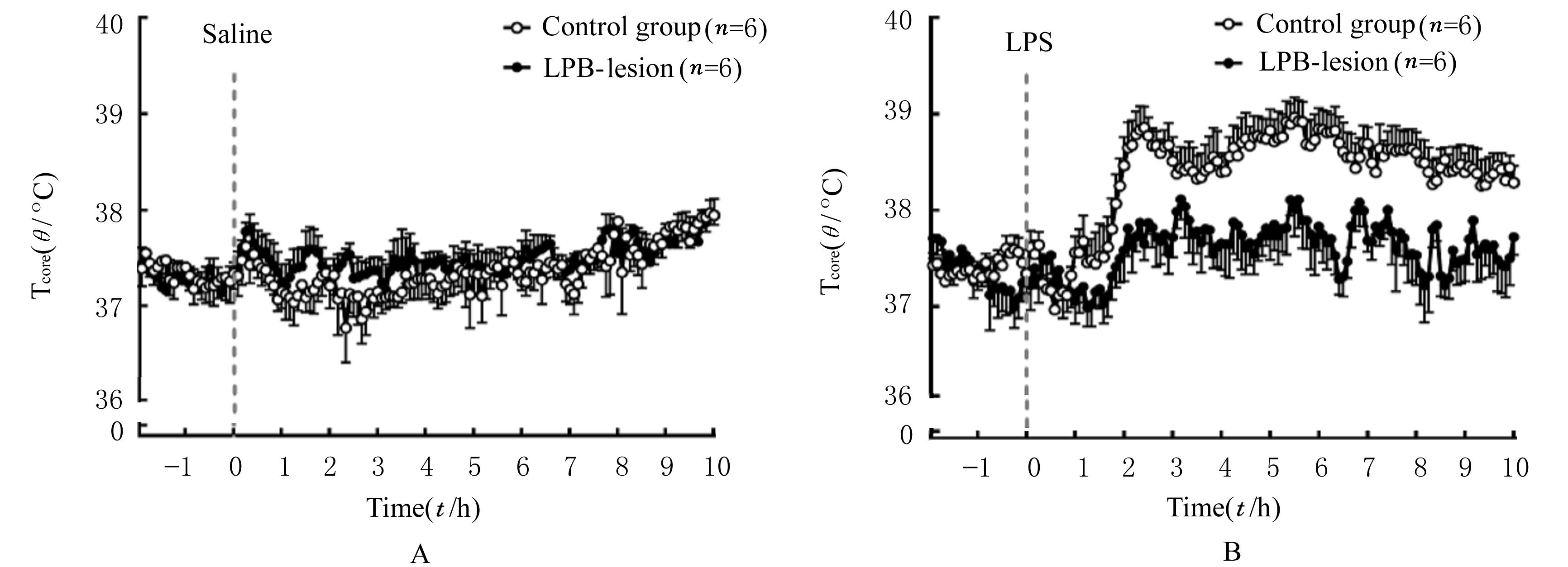
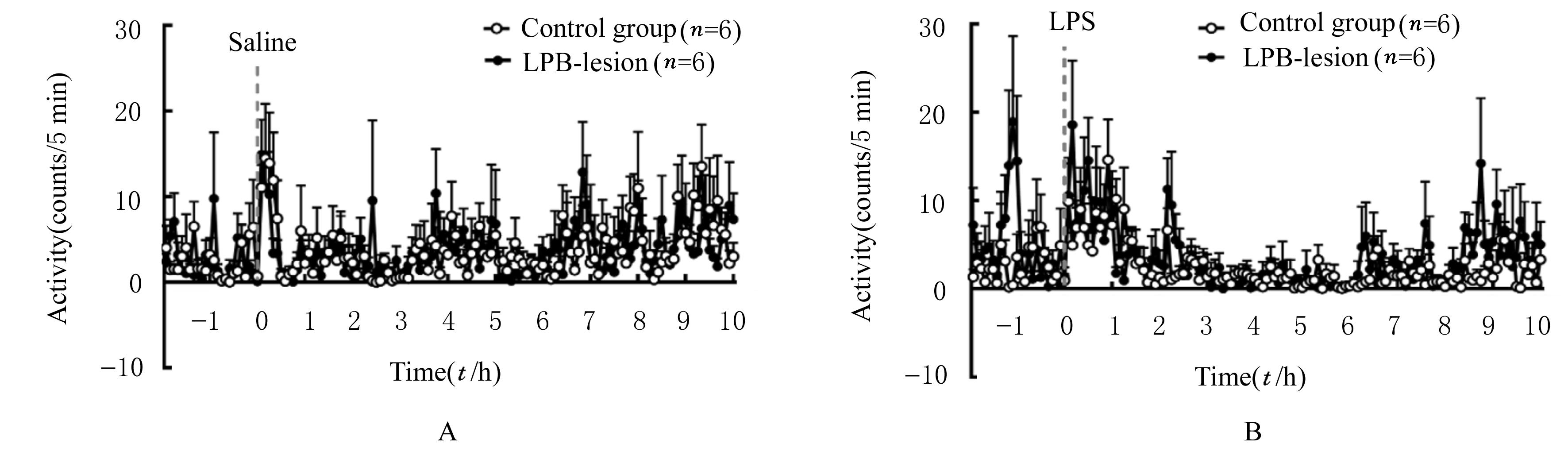
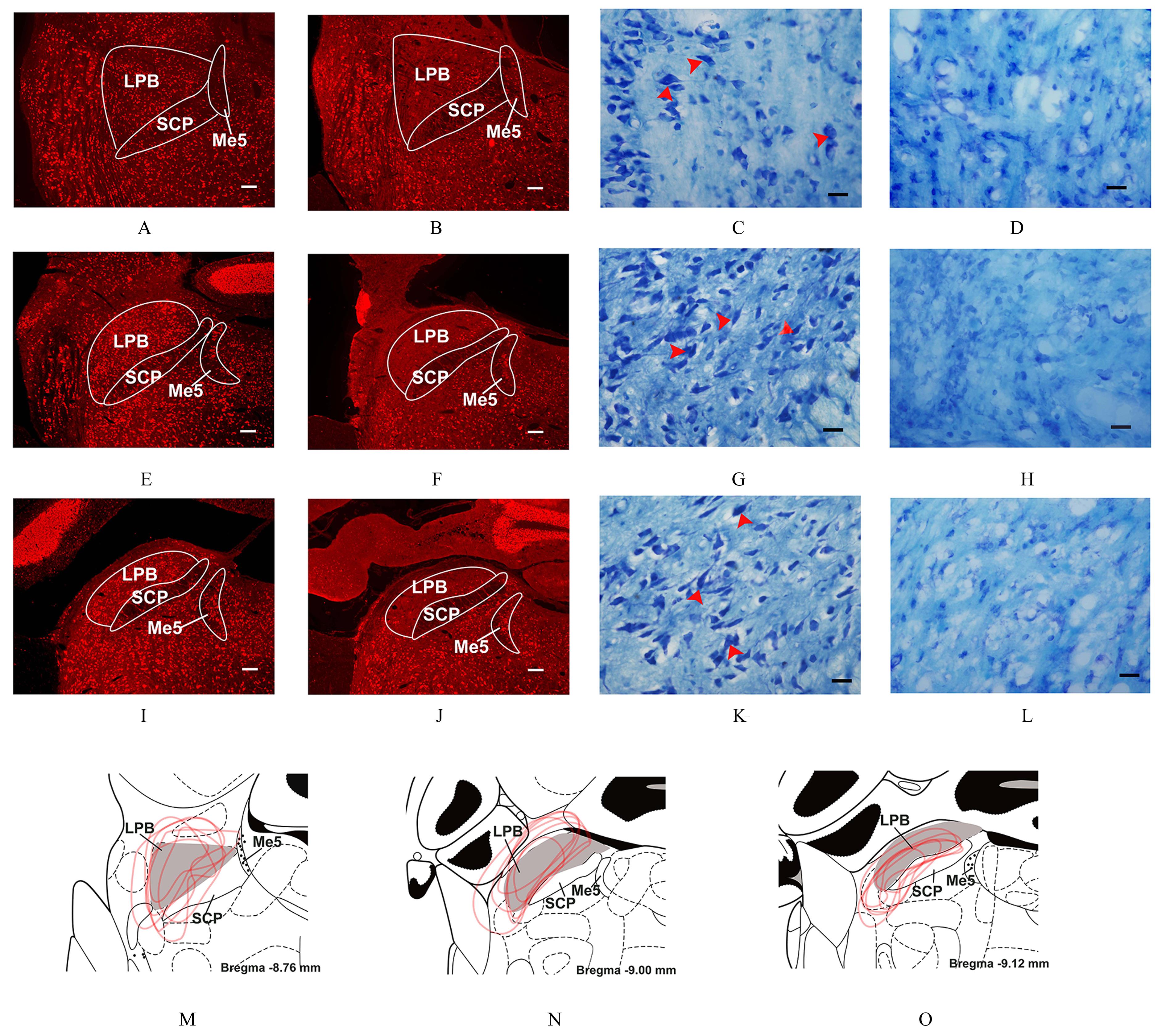
目的 探讨毁损臂旁外侧核(LPB)对大鼠脂多糖(LPS)致热反应的影响,阐明LPB是否参与LPS致热反应过程。 方法 将12只雄性SD大鼠随机分为对照组和LPB毁损组,每组6只。采用核团注射的方法在LPB毁损组大鼠LPB局部注射鹅膏氨酸进行化学毁损,对照组大鼠注入等量生理盐水,恢复至少1周,腹腔植入监测体核温度(Tcore)所需的无线遥测发射子。采用无线遥控测温的方法监测2组大鼠腹腔注射生理盐水或LPS后不同时间点Tcore和活动度,采用免疫荧光染色和尼氏染色法检测2组大鼠LPB中的NeuN和尼氏小体的分布。 结果 对照组大鼠腹腔注射LPS(100 μg·kg-1)后诱导出典型的双相发热,在腹腔注射LPS 后1.5 h开始升温,2.5 h(1相)Tcore首次达到峰值,3.5 h再次升温且5.5 h(2相)Tcore第2次达到峰值;LPS毁损组大鼠腹腔注射LPS后也出现发热反应,5.5 h时Tcore较基础Tcore明显升高(P<0.05);与对照组比较,LPB毁损组大鼠发热反应明显减弱,腹腔注射LPS后2.5和5.5 h时Tcore升高幅度均低于对照组(P<0.05)。对照组和LPS毁损组大鼠腹腔注射生理盐水或LPS后活动度均较注射前增加,但组间比较差异无统计学意义(P>0.05)。对照组大鼠LPB中分布大量NeuN免疫阳性神经元,尼氏小体大而数量多,细胞形态完整,边缘清晰;LPB毁损组大鼠头尾方向前、中和后3个水平的LPB毁损区域几乎未见NeuN免疫阳性神经元,神经元胞体缺失,尼氏小体大量消失。 结论 LPB化学毁损模型部分消除了LPS的致热反应,表明LPB在LPS致热反应过程中起一定作用。

关键词: 臂旁外侧核, 化学毁损, 脂多糖, 双相发热, 尼氏体

Abstract:

Objective: To investigate the effect of lateral parabrachial (LPB) chemical lesion on the fever induced by lipopolysaccharide (LPS),and to clarify whether LPB was involved in the fever induced by LPS. Methods Twelve adult male SD rats were randomly divided into control group and LPB-lesion group(n=6).The rats in LPB-lesion group were bilaterally injected with ibotenic acid into LPB by nuclear injection to induce chemical lession, and the rats in control group were injected with the equal amount of saline into LPB. The rats were survived for at least 1 week and then were implantated with telemetry transmitter for monitoring the body core temperature (Tcore) in the abdominal cavity. The Tcore and activities of the rats in two groups at different time points after intraperitoneal injection of normal saline or LPS were monitored by radiotelemetry,and the distributions of NeuN and Nissl bodies in LPB of the rats in two groups were detected by immunofluorescence staining and Nissl staining. Results The rats in control group were induced the typically biphasic fever after intraperitoneal injection of LPS (100 μg·kg-1),and the Tcore started to rise 1.5 h after intraperitoneal injection of LPS, firstly reached its peak at 2.5 h (the first phase), started to rise again at 3.5 h and reached its peak again at 5.5 h (the second phase) after injection of LPS; the rats in LPB-lesion group also had fever reaction after intraperitoneal injection of LPS.At 5.5 h, the Tcore was significantly higher than the basic Tcore P<0.05);compared with control group, the fever reaction of the rats in LPB-lesion group was significantly weakened, and the increasing of Tcore at 2.5 and 5.5 h after intraperitoneal injection of LPS was lower than that in control group (P<0.05).After intraperitoneal injection of normal saline or LPS, the activities of the rats in control group and LPB-lesion group were increased temporarily compared with those before injection, but there were no significant differences between two groups (P>0.05).A large number of NeuN immunopositive neurons were distributed in LPB of the rats in control group, with large number of Nissl bodies and the shape was big,and the morphology of the cells was complete and the margin was clear; in LPB-lesion group, there were almost no NeuN immunoreactive neurons in the LPB lesion area at the rostral, middle,and caudal levels in the rostrocaudal directions,the neuronal soma was missing, and the Nissl bodies were largely disappeared. Conclusion LPB chemical lession model partially eliminate the LPS-induced fever,indicating that LPB plays a role in the LPS-induced fever.

Key words: Lateral parabrachial nucleus, Chemical lesion, Lipopolysaccharide, Biphasic fever, Nissl body

中图分类号: 

  • R364.6