吉林大学学报(医学版) ›› 2023, Vol. 49 ›› Issue (4): 850-857.doi: 10.13481/j.1671-587X.20230404

• 基础研究 • 上一篇    下一篇

黄芩苷对腹主动脉结扎诱导的大鼠心肌肥厚和细胞凋亡的作用及其机制

刘冠1,陶贵周1(),王洪新2()   

  1. 1.锦州医科大学附属第一医院心内科, 辽宁 锦州 121001
    2.锦州医科大学药理教研室,辽宁 锦州 121001
  • 收稿日期:2022-06-17 出版日期:2023-07-28 发布日期:2023-07-26
  • 通讯作者: 陶贵周,王洪新 E-mail:tgz56789@163.com;hongxinwang@jzmu.edu.cn
  • 作者简介:刘 冠(1988-),男,辽宁省锦州市人,主治医师,医学硕士,主要从事心血管系统疾病临床方面的研究。
  • 基金资助:
    国家自然科学基金项目(81973553)

Effect of baicalin on myocardial hypertrophy and apoptosis induced by abdominal aorta ligation in rats and its mechanism

Guan LIU1,Guizhou TAO1(),Hongxin WANG2()   

  1. 1.Department of Cardiology,First Affiliated Hospital,Jinzhou Medical University,Jinzhou 121001,China
    2.Department of Pharmacology,Jinzhou Medical University,Jinzhou 121001,China
  • Received:2022-06-17 Online:2023-07-28 Published:2023-07-26
  • Contact: Guizhou TAO,Hongxin WANG E-mail:tgz56789@163.com;hongxinwang@jzmu.edu.cn

摘要:

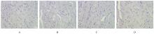
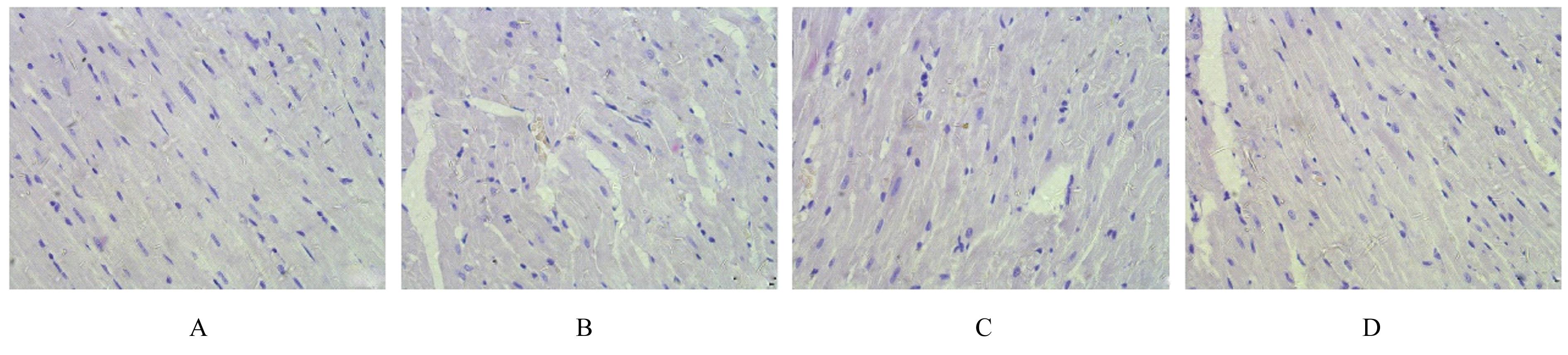
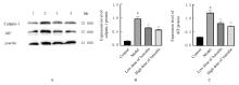
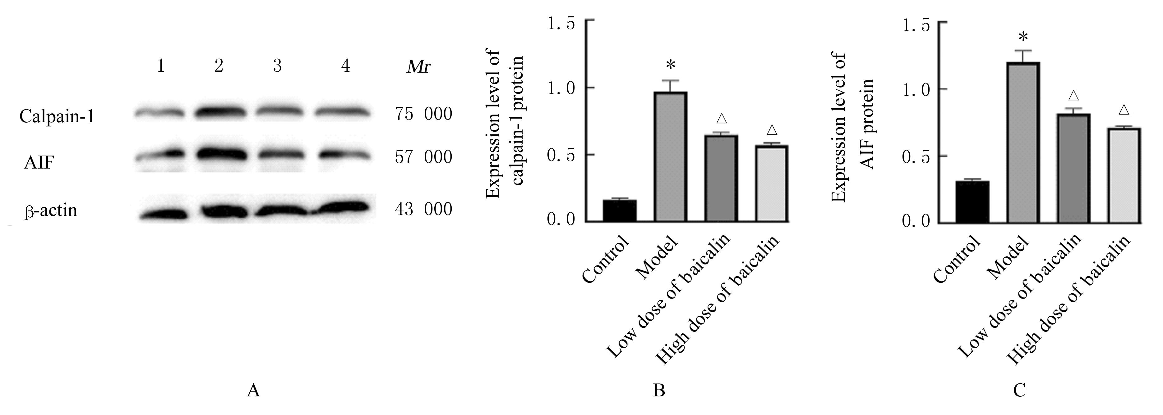
目的 探讨黄芩苷对腹主动脉结扎诱导的大鼠心肌肥厚和细胞凋亡的改善作用,并阐明其潜在的作用机制。 方法 SD大鼠随机分为对照组、模型组、低剂量黄芩苷组和高剂量黄芩苷组,采用结扎腹主动脉的方法构建心肌肥厚大鼠模型,低和高剂量黄芩苷组大鼠分别给予50和100 mg·kg-1黄芩苷6周后,采用超声心动图检测各组大鼠左心室射血分数(LVEF)、左心室后壁舒张末期厚度(LVPWd)、左心室后壁收缩末期厚度(LVPWs)、室间隔舒张末期厚度(IVSd)和室间隔收缩末期厚度(IVSs),计算各组大鼠心脏质量指数(HWI)和左心室质量指数(LVWI),HE染色观察各组大鼠心肌组织病理形态表现,酶联免疫吸附试验(ELISA)法检测各组大鼠血清中肿瘤坏死因子α(TNF-α)、白细胞介素6(IL-6)、白细胞介素8(IL-8)和白细胞介素1β(IL-1β)水平,Western blotting法检测各组大鼠心肌组织中B细胞淋巴瘤2(Bcl-2)、Bcl-2相关X蛋白(Bax)、凋亡诱导因子(AIF)和钙蛋白酶1(calpain-1)蛋白表达水平。 结果 与对照组比较,模型组大鼠LVEF明显降低(P<0.01),LVPWd、LVPWs、IVSd、IVSs、HWI和LVWI明显升高(P<0.01),心肌组织中心肌细胞排列紊乱,血清中TNF-α、IL-6、IL-8和IL-1β水平明显升高(P<0.01),心肌组织中Bcl-2蛋白表达水平明显降低(P<0.01),Bax、AIF和calpain-1蛋白表达水平及Bax/Bcl-2比值明显升高(P<0.01)。与模型组比较,低和高剂量黄芩苷组大鼠LVEF明显升高(P<0.01),LVPWd、LVPWs、IVSd、IVSs、HWI和LVWI明显降低(P<0.05或P<0.01),心肌组织中心肌细胞排列整齐,血清中TNF-α、IL-6、IL-8和IL-1β水平明显降低(P<0.01),心肌组织中Bcl-2蛋白表达水平升高(P<0.01),Bax、AIF和calpain-1蛋白表达水平及Bax/Bcl-2比值明显降低(P<0.05或P<0.01)。与低剂量黄芩苷组比较,高剂量黄芩苷组大鼠LVPWs和HWI明显降低(P<0.05),血清中TNF-α、IL-6和IL-1β水平及心肌组织中Bax蛋白表达水平明显降低(P<0.05),其他指标差异均无统计学意义(P>0.05)。 结论 黄芩苷可改善腹主动脉结扎诱导的大鼠心肌肥厚并抑制炎症反应和细胞凋亡,其作用机制可能与调控calpain-1/AIF信号通路有关。

关键词: 黄芩苷, 细胞凋亡, 钙蛋白酶1, 凋亡诱导因子, 心肌肥厚

Abstract:

Objective To discuss the improvement effect of baicalin on the myocardial hypertrophy and apoptosis induced by abdominal aorta ligation of the rats, and to clarify the potential mechanism. Methods The SD rats were randomly divided into control group, model group, low dose of baicalin (50 mg·kg-1) group, and high dose of baicalin (100 mg·kg-1) group.The rat model of myocardial hypertrophy was established by abdominal aorta ligation method. The rats in low and high doses of baicalin groups were given 50 and 100 mg·kg-1 baicalin.After six weeks, the left ventricular ejection fraction (LVEF), left ventricular posterior wall thickness at end-diastolic (LVPWd), left ventricular posterior wall thickness at end-systolic (LVPWs),interventricular septal thickness at end-diastolic (IVSd) and interventricular septal thickness at end-systolic (IVSs) of rats in various groups were measured by echocardiography; the heart weight index(HWI) and left ventricular weight index (LVWI) of rats in various groups were calculated; HE staining was used to observe the pathomorphology of myocardium tissue of the rats in various groups; the serum levels of interleukin-8 (IL-8),interleukin-1β (IL-1β),tumor necrosis factor-α (TNF-α), and interleukin-6 (IL-6) of the rats in various groups were detected by enzyme-linked immunosorbnent assay(ELISA) method;the expression levels of B-cell lymphoma-2 (Bcl-2), Bcl-2 associated X protein (Bax),apoptosis-inducing factor (AIF),and calpain-1 proteins in myocardium tissue of the rats in various groups were detected by Western blotting method. Results Compared with control group, the LVEF of the rats in model group was significantly decreased(P<0.01),and the LVPWd, LVPWs, IVSd, IVSs, HWI, and LVWI were significantly increased(P<0.01), the myocardium tissue were disordered arrangement; the serum levels of TNF-α,IL-6,IL-8, and IL-1β were increased(P<0.01); the expression levels of Bcl-2 protein in myocardium tissue of the rats were decreased(P<0.01), while the expression levels of Bax, AIF, and calpain-1 proteins were increased(P<0.01),and the ratio of Bax/Bcl-2 was increased(P<0.01). Compared with model group, the LVEF of the rats in low and high doses of baicalin groups was increased(P<0.01),and the LVPWd, LVPWs, IVSd, IVSs, HWI, and LVWI were decreased(P<0.05 or P<0.01);the cardiomyocytes in myocardium tissue were arranged in order;the serum levels of TNF-α,IL-6,IL-8, and IL-1β were decreased(P<0.05);the expression levels of Bcl-2 protein in myocardium tissue of the rats were increased(P<0.01), while the expression levels of Bax, AIF, and calpain-1 proteins were decreased(P<0.05), and the ratios of Bax/Bcl-2 were decreased(P<0.05 or P<0.01). Compared with low dose of baicalin group, the LVPWS and HWI of the rats in high dose of baicalin group were decreased(P<0.05);the levels of TNF-α,IL-6,IL-8,and IL-1β in serum and the expression level of Bax protein were decreased(P>0.05),but there were no statistically significant differences in the other indexes(P>0.05). Conclusion Baicalin can improve the myocardial hypertrophy and inhibit the inflammation and apoptosis induced by abdominal aorta ligation of the rats, and its mechanism may be related to the regulation of calpain-1/AIF signaling pathway.

Key words: Baicalin, Apoptosis, Calpain-1, Apoptosis-inducing factor, Myocardial hypertrophy

中图分类号: 

  • R285.5