吉林大学学报(医学版) ›› 2024, Vol. 50 ›› Issue (3): 602-611.doi: 10.13481/j.1671-587X.20240303

• 基础研究 • 上一篇    

C57BL/6小鼠大脑皮层区与基底神经节隆起区神经元突触发育过程比较

赵艳1,2,卢广泉2,杜金乐2,潘雨绮2,董子意2,康鑫2,高弋婷2,高方2,杨加周1()   

  1. 1.延安大学医学院组织学与胚胎学教研室,陕西 延安 716099
    2.空军军医大学基础医学院神经 生物学教研室,陕西 西安 710032
  • 收稿日期:2023-12-18 出版日期:2024-05-28 发布日期:2024-07-01
  • 通讯作者: 杨加周 E-mail:jiazhouy@163.com
  • 作者简介:赵 艳(1993-),女,云南省威信县人,在读硕士研究生,主要从事神经发育基础方面的研究。
  • 基金资助:
    国家自然科学基金面上项目(82071529)

Comparison of development process of neuronal synapse between cerebral cortex and basal ganglia eminence regions in C57BL/6 mice

Yan ZHAO1,2,Guangquan LU2,Jinle DU2,Yuqi PAN2,Ziyi DONG2,Xin KANG2,Yiting GAO2,Fang GAO2,Jiazhou YANG1()   

  1. 1.Department of Histology and Embryology,School of Medical Sciences,Yan’an University,Yan’an 716099,China
    2.Department of Neurobiology,School of Basic Medical Sciences,Air Force Medical University,Xi’an 710032,China
  • Received:2023-12-18 Online:2024-05-28 Published:2024-07-01
  • Contact: Jiazhou YANG E-mail:jiazhouy@163.com

摘要:

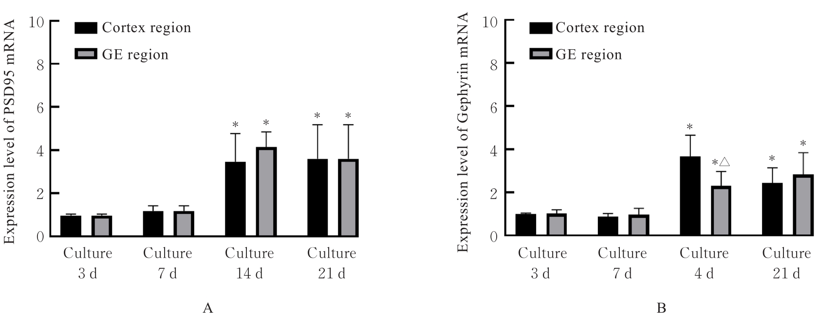
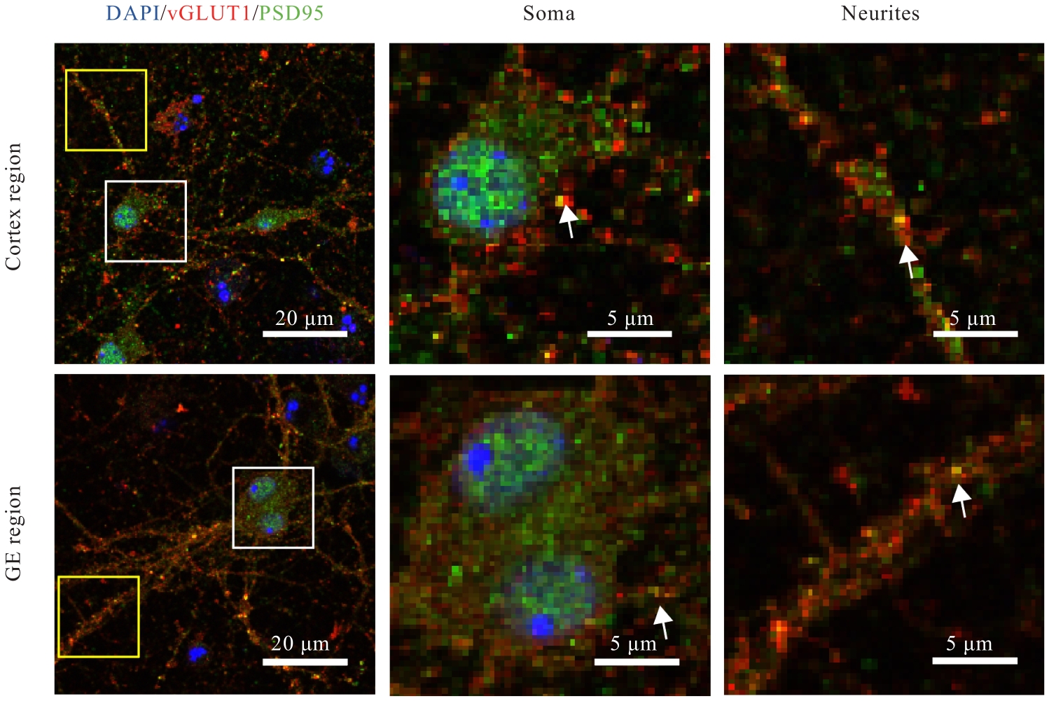
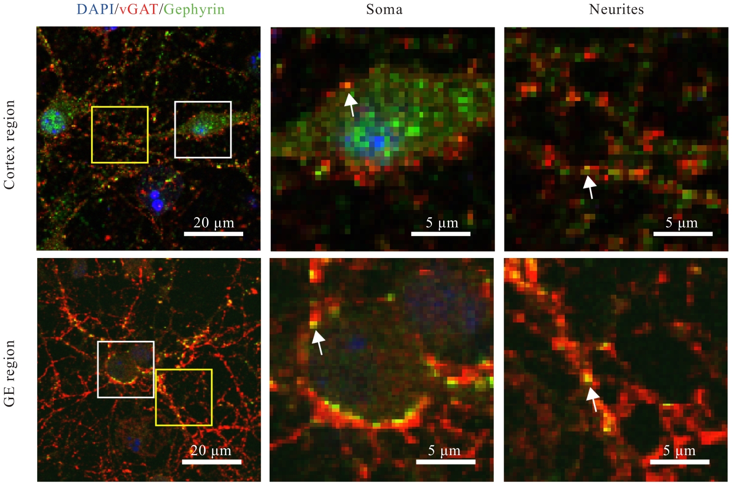
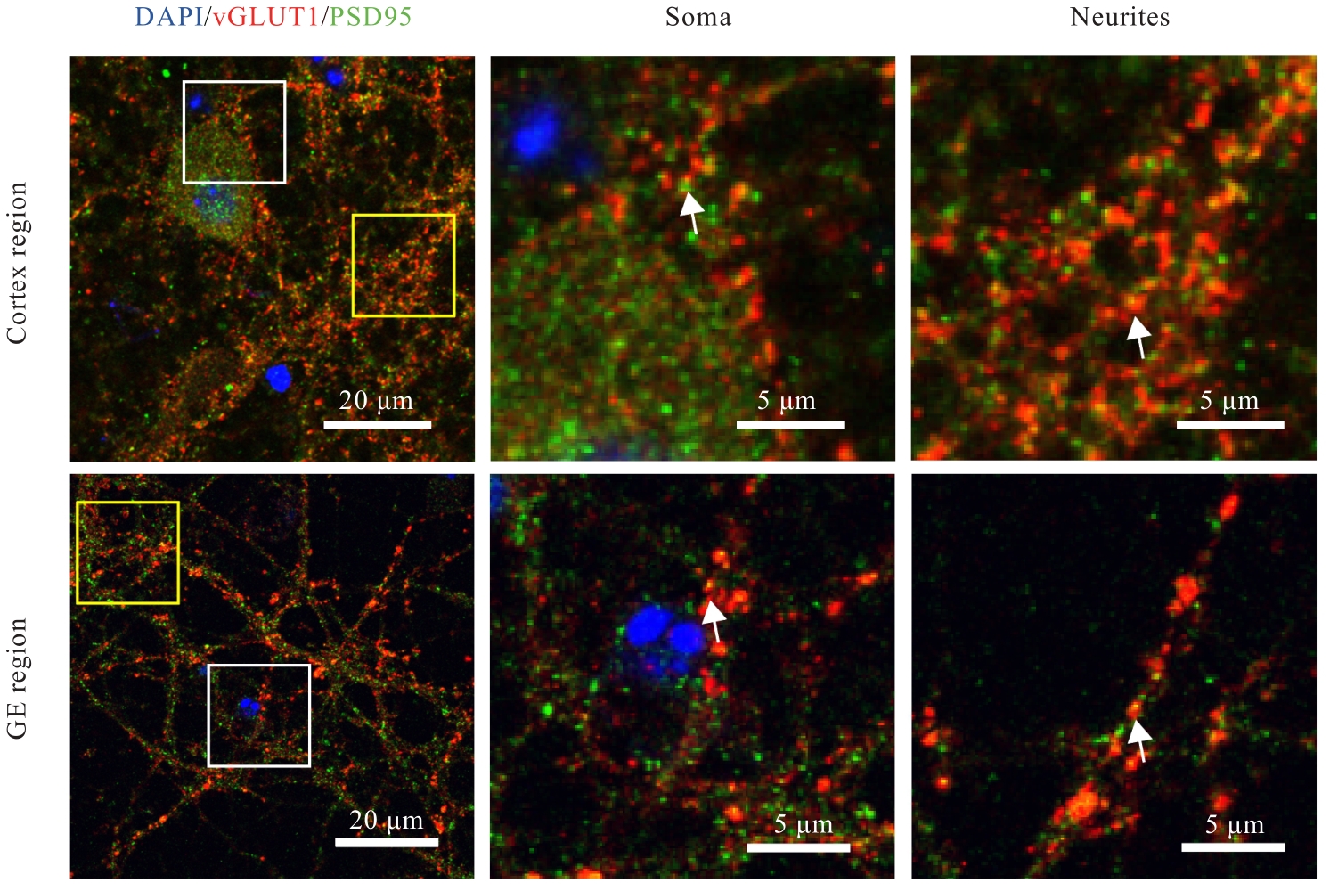
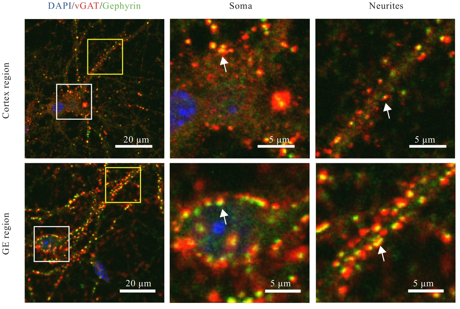
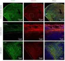
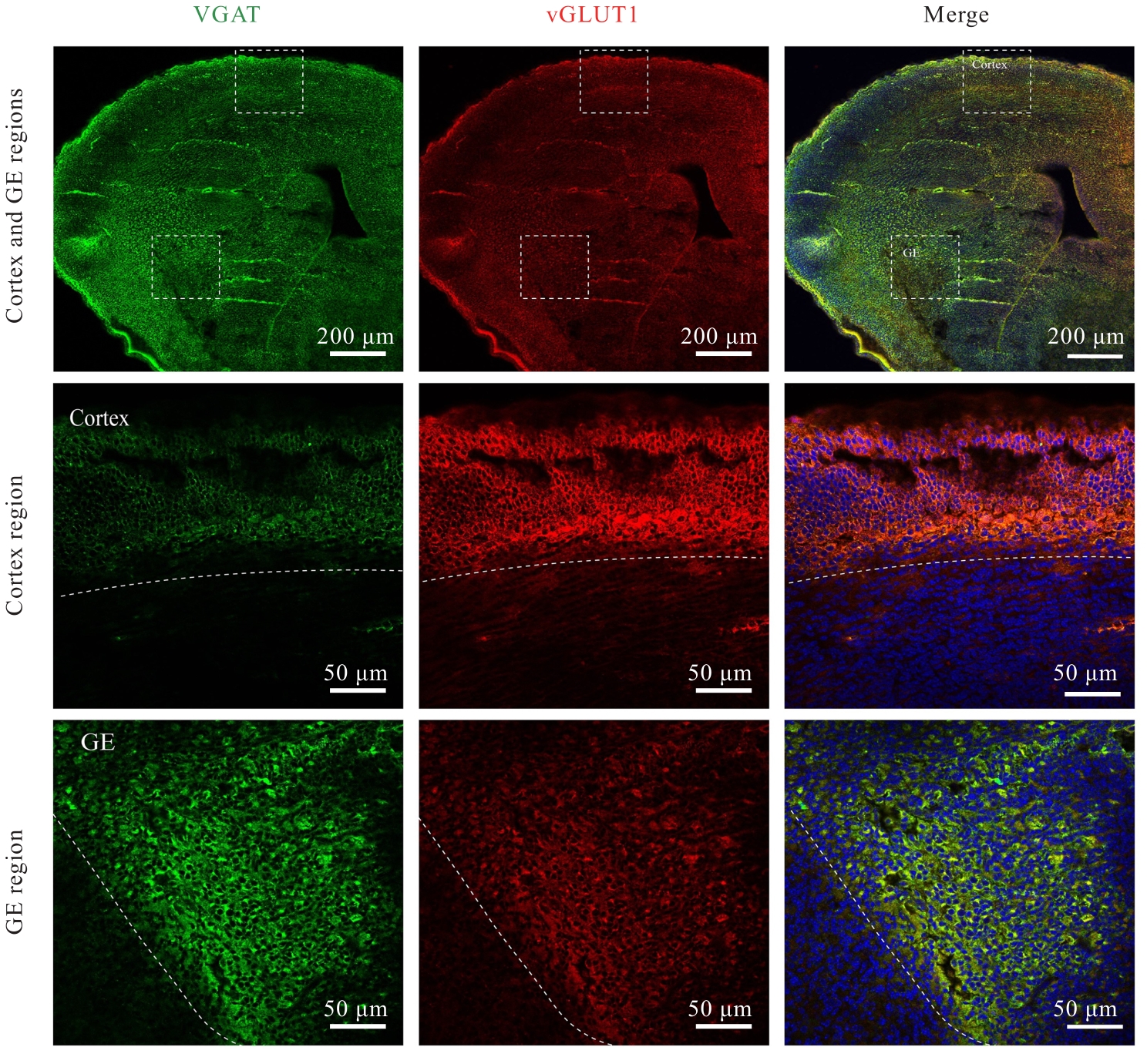
目的 观察小鼠皮层区和基底神经节隆起(GE)区神经元突触发育过程,阐明兴奋性突触和抑制性突触在不同脑区的体内外发育差异。 方法 C57BL/6雌鼠于妊娠第13.5~15.5天断颈处死后,经无菌操作取胚胎小鼠,显微镜下逐步分离获取胚胎小鼠脑组织皮层区和GE区。体外原代培养胚胎小鼠神经元,于培养3、7、14和21 d分别收集细胞样品,并将其作为培养3、7、14和21 d组。采用实时荧光定量PCR(RT-qPCR)法检测各组小鼠皮层区和GE区原代培养神经元中突触后表达蛋白突触后密度蛋白95(PSD95)及桥尾蛋白(Gephyrin)mRNA表达水平。免疫荧光法检测各组小鼠皮层区和GE区原代培养神经元中囊泡谷氨酸转运蛋白1(vGLUT1)、PSD95、囊泡γ-氨基丁酸(GABA)转运蛋白(vGAT)及Gephyrin蛋白表达水平。免疫荧光法检测胚胎小鼠脑组织皮层区和GE区神经元中vGLUT1及vGAT蛋白表达水平。 结果 与培养3 d组比较,培养14和21 d组小鼠皮层区和GE区原代培养神经元中PSD95及Gephyrin mRNA表达水平明显升高(P<0.01);与皮层区比较,培养14 d组小鼠GE区原代培养神经元中Gephyrin mRNA表达水平明显降低(P<0.01)。显微镜下观察,培养14 d组小鼠皮层区和GE区原代培养神经元中兴奋性突触及抑制性突触均初步发育,相关蛋白呈阳性表达;其中兴奋性突触相关蛋白阳性表达在皮层区神经元中更为明显,且突触前分子vGLUT1和突触后分子PSD95在皮层区神经元的胞体及突起部位均呈现共定位的特征;抑制性突触前分子vGAT蛋白和突触后分子Gephyrin蛋白在GE区神经元胞体及突起中也呈现共定位的特征,且突触前分子较相应的突触后分子蛋白阳性表达更明显。与皮层区比较,培养14 d组小鼠GE区原代培养神经元中vGLUT1和PSD95蛋白表达水平明显降低(P<0.01),vGAT和Gephyrin蛋白表达水平明显升高(P<0.01)。培养21 d组小鼠皮层区和GE区原代培养神经元突触相关蛋白阳性表达增加,兴奋性突触和抑制性突触进一步成熟并完善。皮层区和GE区原代培养神经元的胞体及突起部位均形成了丰富的突触前后对应的表达模式,突触结构逐步发育良好,且突触前分子较相应的突触后分子蛋白阳性表达更明显。与皮层区比较,培养21 d组小鼠GE区原代培养神经元中vGLUT1和PSD95蛋白表达水平均明显降低(P<0.01),vGAT和Gephyrin蛋白表达水平均明显升高(P<0.01)。与皮层区比较,胚胎小鼠脑组织GE区神经元中vGLUT1蛋白表达水平明显降低(P<0.01),vGAT蛋白表达水平明显升高(P<0.05)。 结论 皮层区和GE区神经元的突触发育具有明显的差异性,皮层区兴奋性突触发育较早,GE区抑制性突触发育较早。突触的脑区特异性发育提示不同细胞类型的神经疾病可能具有不同的发育来源。

关键词: 神经元发育, 兴奋性突触, 抑制性突触, 皮层区, 基底神经节隆起区

Abstract:

Objective To observe the development process of the neuronal synapse in cerebral cortex and basal ganglionic eminence (GE) regions of the mice, and to clarify the differences in the development of excitatory and inhibitory synapses in different brain regions in vivo and in vitro. Methods The female C57BL/6 mice were euthanized by cervical dislocation from the 13.5th day to the 15.5th day during the pregnancy, and the embryos were collected under the sterile conditions. The cortex and GE regions of brain tissue of the embryonic mice were gradually isolated under microscope. The primary neurons from the embryonic mice were cultured in vitro, and the cell samples were collected on the 3rd, 7th, 14th, and 21th days, respectively, and regarded as culture 3 d, 7 d, 14 d, and 21 d groups. The expression levels of postsynaptic density 95 (PSD95) and Gephyrin mRNA in the primary neurons from the cortex and GE regions of the mice in various groups were detected by real-time fluorescence quantitative PCR (RT-qPCR)method. Immunofluorescence method was used to detect the expression levels of vesicular glutamate transporter 1 (vGLUT1), PSD95, vesicular GABA transporter (vGAT), and Gephyrin proteins in the neurons from the cortex and GE regions of the mice in various groups. Immunofluorescence method was also used to detect the expression levels of vGLUT1 and vGAT proteins in the neurons from the cortical and GE regions in brain tissue of the embryonic mice. Results Compared with culture 3 d group, the expression levels of PSD95 and Gephyrin mRNA in cortex and GE regions of the mice in culture 14 d and 21 d groups were significantly increased (P<0.01). Compared with cortex area, the expression level of Gephyrin mRNA in the neurons from GE region of the mice in culture 14 d group was significantly decreased (P<0.01). The microscope observation results showed that the excitatory and inhibitory synapses in the neurons from cortex and GE regions of the mice in culture 14 d group showed preliminary development, with positive expression of relevant proteins; among them, the excitatory synaptic proteins showed more distinct positive expression in the cortex neurons, and the presynaptic vGLUT1 and postsynaptic PSD95 molecules exhibited co-localization in the cell bodies and protrusions of the cortical neurons; the inhibitory presynaptic vGAT protein and postsynaptic Gephyrin protein in the neurons from GE region also exhibited co-localization in the cell bodies and protrusions, and there were more distinct expressions of the presynaptic molecule proteins than postsynaptic molecule proteins. Compared with cortex region, the levels of vGLUT1 and PSD95 proteins in the neurons from GE region of the mice in culture 14 d group were significantly decreased (P<0.01), while the levels of vGAT and gephyrin proteins were significantly increased (P<0.01). In culture 21 d group, the positive expressions of synaptic protein in the neurons from cortex and GE regions were increased, and the excitatory and inhibitory synapses further matured and enhanced. In the neurons from cortex and GE regions, rich patterns of corresponding pre- and postsynaptic expression were formed in the cell bodies and protrusions, and synapse structures showed gradual, positive development, with more apparent expression of presynaptic molecules compared wih postsynaptic proteins. Compared with cortex region, the levels of vGLUT1 and PSD95 proteins in the neurons from GE region of the mice in culture 21 d group were significantly decreased (P<0.01), and the levels of vGAT and Gephyrin proteins were significantly increased (P<0.01). Compared with cortex region, the expression level of vGLUT1 protein in the neurons from GE region in brain tissue of the embryonic mice was significantly decreased (P<0.01), while the expression level of vGAT protein was significantly increased (P<0.05). Conclusion There are distinct differences in synaptic development between the neurons from cortex and GE regions, the excitatory synapses develope earlier in the cortical region and the inhibitory synapses develope earlier in the GE region. The region-specific development of synapses suggests that different types of neural diseases with different cell types might originate from different developmental processes.

Key words: Neuronal development, Excitatory synapses, Inhibitory synapses, Cortex, Ganglionic eminence

中图分类号: 

  • R394