吉林大学学报(医学版) ›› 2023, Vol. 49 ›› Issue (3): 549-556.doi: 10.13481/j.1671-587X.20230301

• 基础研究 •    下一篇

芹菜素对小鼠RAW264.7巨噬细胞极化和炎症反应的作用及其机制

李海涛,李沁,蔡飞,胡国富,滕云飞()   

  1. 华中科技大学同济医学院附属协和医院血管外科,湖北 武汉 430000
  • 收稿日期:2022-08-17 出版日期:2023-05-28 发布日期:2023-06-20
  • 通讯作者: 滕云飞 E-mail:cf241cf@126.com
  • 作者简介:李海涛(1984-),男,湖北省武汉市人,讲师,医学博士,主要从事血管瘤和动脉硬化方面的研究。
  • 基金资助:
    国家自然科学基金项目(81900432)

Effect of apigenin on polarization and inflammation of mouse RAW264.7 macrophages and its mechanism

Haitao LI,Qin LI,Fei CAI,Guofu HU,Yunfei TENG()   

  1. Department of Vascular Surgery,Affiliated Union Hospital,Tongji Medical College,Huazhong University of Science and Technology,Wuhan 430000,China
  • Received:2022-08-17 Online:2023-05-28 Published:2023-06-20
  • Contact: Yunfei TENG E-mail:cf241cf@126.com

摘要:

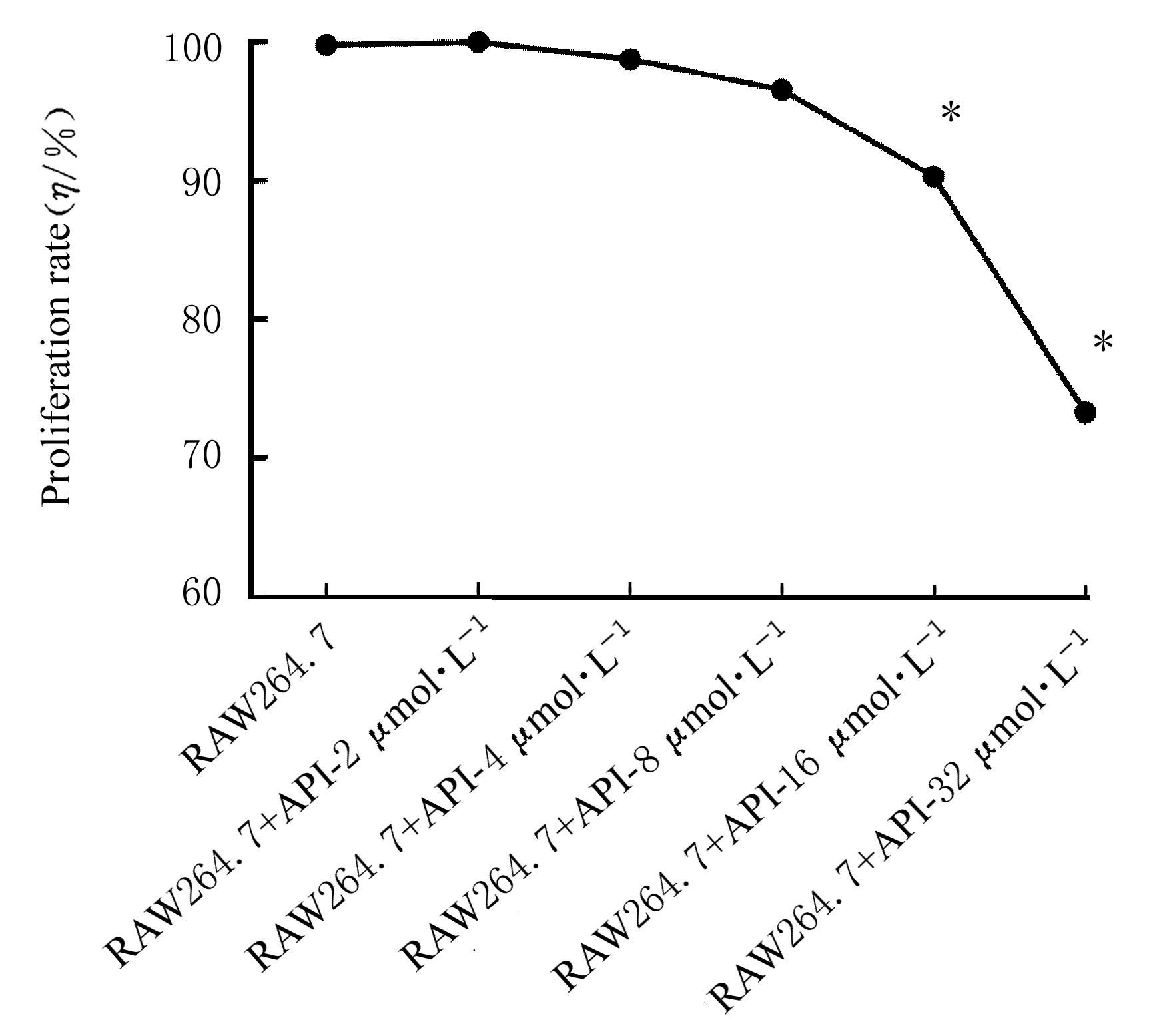
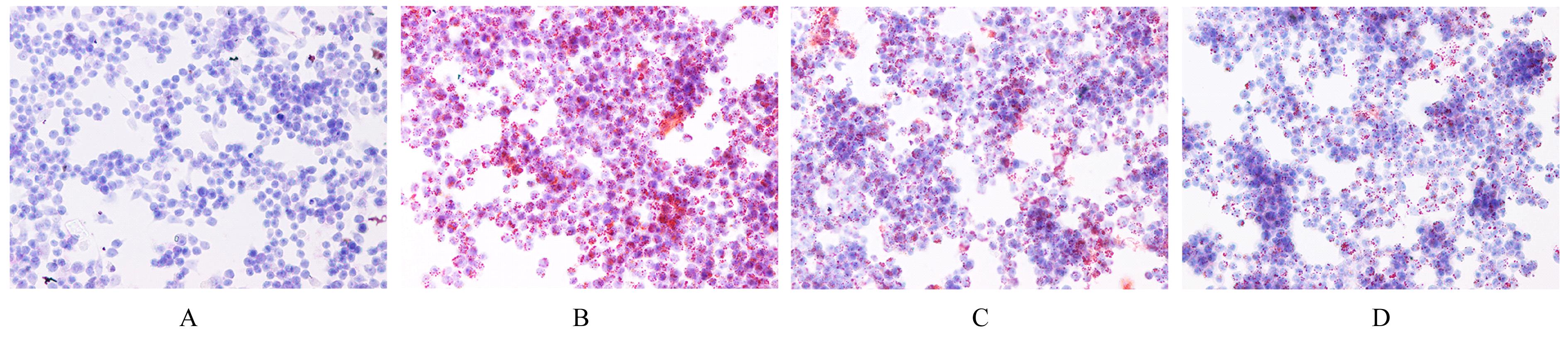
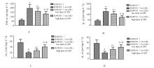
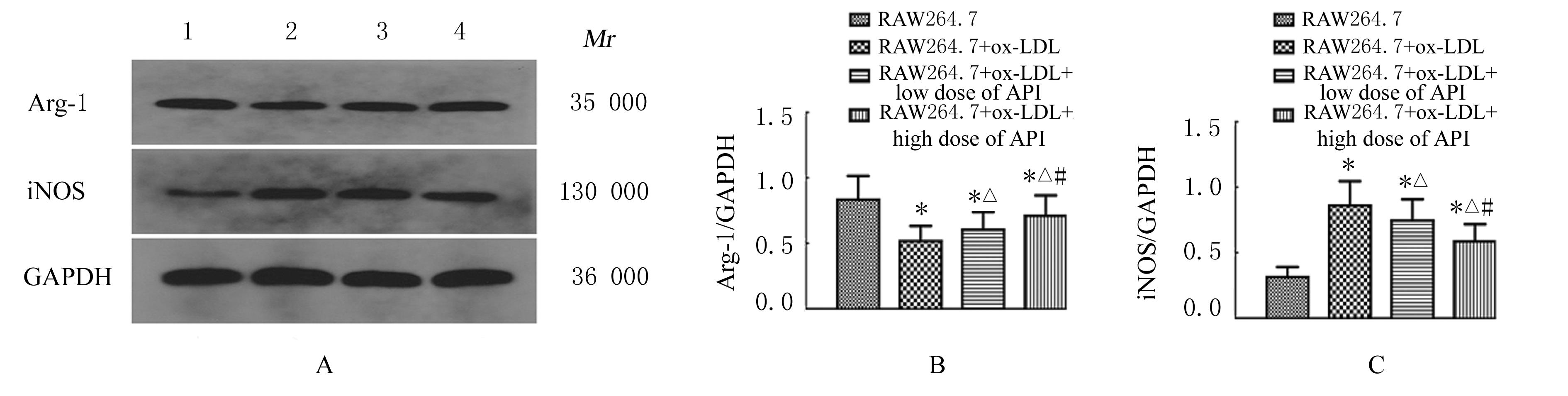
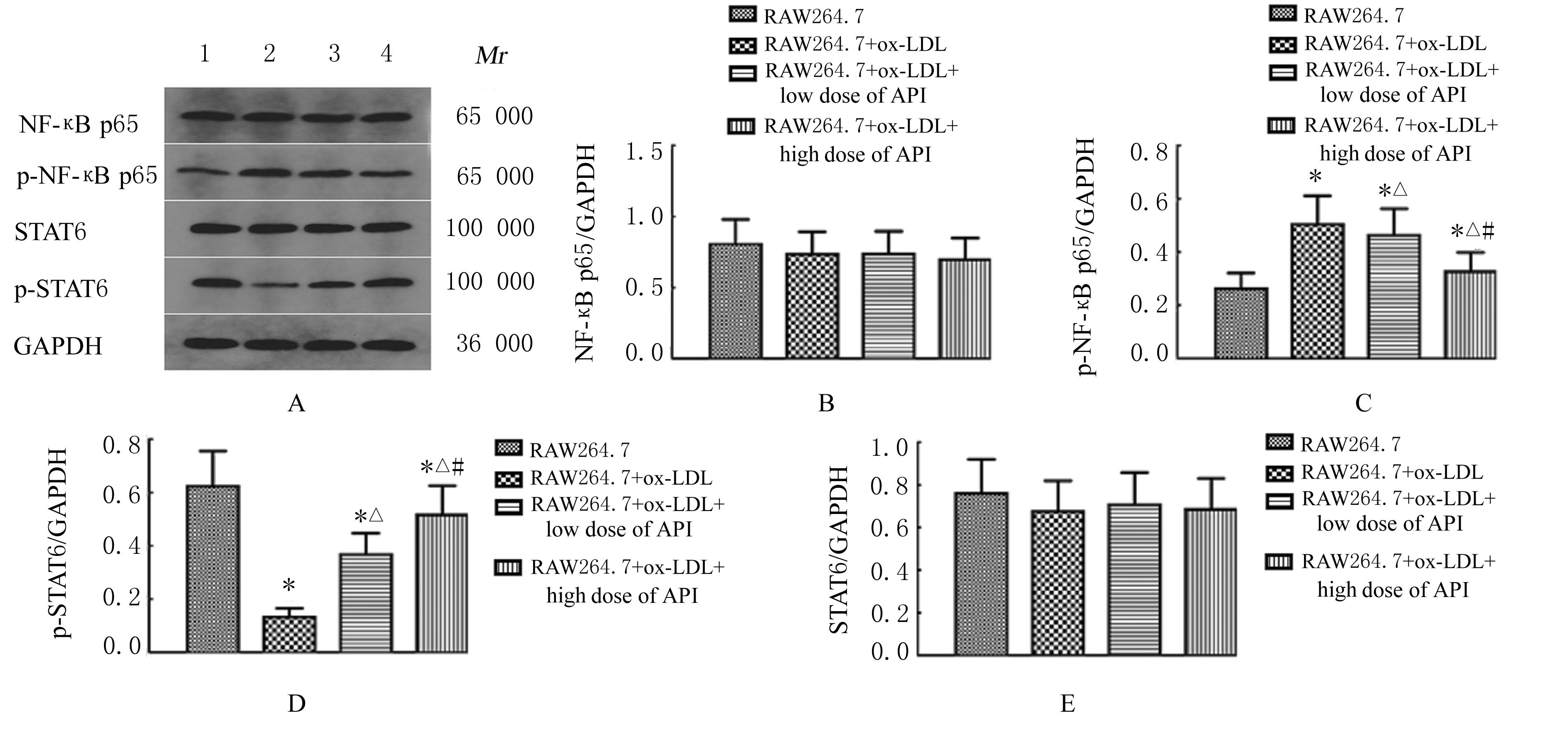
目的 探讨不同浓度芹菜素(API)对氧化低密度脂蛋白(ox-LDL)诱导的小鼠单核巨噬细胞RAW264.7炎症反应和极化的作用,并阐明其可能机制。 方法 将RAW264.7细胞分为RAW264.7组(不做任何处理)和RAW264.7+API组(2、4、8、16 和32 μmol·L-1 API)。药物处理细胞24 h后,CCK-8法检测各组细胞增殖率,选取API实验浓度。将RAW264.7细胞分为RAW264.7组(正常RAW264.7细胞)、RAW264.7+ox-LDL组(0.08 g·L-1 ox-LDL诱导24 h)、RAW264.7+ox-LDL+低剂量API组(2 μmol·L-1 API和0.08 g·L-1 ox-LDL诱导24 h)和RAW264.7+ox-LDL+高剂量API组(8 μmol·L-1 API和0.08 g·L-1 ox-LDL诱导24 h),药物处理细胞24 h,采用油红O染色观察各组RAW264.7细胞中泡沫细胞形态表现,酶联免疫吸附测定(ELISA)法检测各组细胞培养上清液中肿瘤坏死因子α(TNF-α)、白细胞介素1β(IL-1β)、白细胞介素4(IL-4)和白细胞介素10(IL-10)水平,Western blotting法检测各组细胞中核转录因子κB(NF-κB)p65、磷酸化NF-κB(p-NF-κB)p65、诱导型一氧化氮合成酶(iNOS)、信号转导与转录激活因子6(STAT6)、磷酸化STAT6(p-STAT6)和精氨酸酶1(Arg-1)蛋白表达水平。 结果 CCK-8法检测,随着API的浓度增加,RAW264.7细胞增殖率降低(P<0.05),选择无毒的低浓度(2 μmol·L-1)和高浓度(16 μmol·L-1)API作为后续实验API浓度。油红O染色,RAW264.7组极少数RAW264.7细胞被油红O染色;RAW264.7+ox-LDL组较多细胞被染成暗红色,胞内脂质明显增加,表明成功建立了RAW264.7源性泡沫细胞;RAW264.7+ox-LDL+低剂量API组和RAW264.7+ox-LDL+高剂量API组少量细胞被油红O染色。ELISA法检测,与RAW264.7组比较,RAW264.7+ox-LDL组、RAW264.7+ox-LDL+低剂量API组和RAW264.7+ox-LDL+高剂量API组细胞培养上清液中TNF-α和IL-1β水平升高(P<0.05),IL-4和IL-10水平降低(P<0.05);与RAW264.7+ox-LDL组比较,RAW264.7+ox-LDL+低剂量API组和RAW264.7+ox-LDL+高剂量API组RAW264.7细胞培养上清液中TNF-α和IL-1β水平降低(P<0.05),IL-4和IL-10水平升高(P<0.05)。Western blotting法检测,与RAW264.7组比较,RAW264.7+ox-LDL组、RAW264.7+ox-LDL+低剂量API组和RAW264.7+ox-LDL+高剂量API组RAW264.7细胞中iNOS和p-NF-κB p65蛋白表达水平升高(P<0.05),Arg-1和p-STAT6蛋白表达水平降低(P<0.05);与RAW264.7+ox-LDL组比较,RAW264.7+ox-LDL+低剂量API组和RAW264.7+ox-LDL+高剂量API组RAW264.7细胞中iNOS和p-NF-κB p65蛋白表达水平降低(P<0.05),Arg-1和p-STAT6蛋白表达水平升高(P<0.05);与RAW264.7+ox-LDL+低剂量API组比较,RAW264.7+ox-LDL+高剂量API组RAW264.7细胞中iNOS和p-NF-κB p65蛋白表达水平降低(P<0.05),Arg-1和p-STAT6蛋白表达水平升高(P<0.05)。 结论 API能抑制ox-LDL诱导的RAW264.7细胞中泡沫细胞形成和调控巨噬细胞向M2极化,改善炎症反应,其机制可能与NF-κB和STAT6通路有关。

关键词: 芹菜素, 动脉粥样硬化, 核因子κB, 信号转导与转录激活因子6, 巨噬细胞极化

Abstract:

Objective To discuss the effects of different concentrations of apigenin (API) on the inflammatory and polarization response of the mouse mononuclear macrophage cells RAW264.7 induced by oxidized low-density lipoprotein (ox-LDL),and to clarify their possible mechanisms. Methods The RAW264.7 cells were divided into RAW264.7 group (without any treatment)and RAW264.7+API group (2, 4, 8, 16 and 32 μmol·L-1 API).After the cells were treated for 24 h.CCK-8 assay was used to detect the proliferation rates of the cells in various groups.The experimental concentration of API was selected.The RAW264.7 cells were divided into RAW264.7 group(normal RAW264.7 cells), RAW264.7+ox-LDL group(induced with 0.08 g·L-1 ox-LDL for 24 h), RAW264.7+ox-LDL+low dose of API group(induced with 2 μmol·L-1 API and 0.08 g·L-1 ox-LDL for 24 h) and RAW264.7+ox-LDL+high dose of API group. Oil red O staining was used to observe the morphology of the foam cells in various groups. The levels of tumor necrosis factor-α(TNF-α),interleukin-1β(IL-1β), interleukin-4(IL-4), and interleukin-10(IL-10) in the cell culture supernant were detected by enzyme-linked immunosorbent assay(ELISA) method. The expression levels of neclear factor-κB(NF-κB) p65, phosphorylated NF-κB(p-NF-κB) p65,inducible nitric oxide synthase(iNOS), signal transducer and activator of transcription 6(STAT6), phosphorylated STAT-6(p-STAT6), and arginase-1(Arg-1) in the cells in various groups were detected by Western blotting method. Results The results of CCK-8 assay showed that the proliferation rates of the cells were significantly decreased with the increasing of the concentrations of API (P<0.05).The non-toxic low concentration (2 μmol·L-1) and high concentration (8 μmol·L-1) of API were selected to use in the follow-up experiment.The Oil red O staining results showed that few RAW264.7 cells were stained with Oil red O; a lot of RAW264.7 cells in RAW264.7+ox-LDL group were stained as dark red,and the lipids in the cells were increased,indicating the foam cell model was successfully established; a little of RAW264.7 cells in RAW264.7+ox-LDL+low dose of API group and RAW264.7+ox-LDL+high dose of API group were stained with Oil red O.The ELISA results showed that compared with RAW264.7 group, the levels of TNF-α and IL-1β in the cell culture supernatant in RAW264.7+ox-LDL group were significantly increased (P<0.05), while the levels of IL-4 and IL-10 were significantly decreased (P<0.05); compared with RAW264.7+ox-LDL group, the levels of TNF-α and IL-1β in the cell culture supernatant in RAW264.7+ox-LDL+low dose of API group,the levels of IL-4 and IL-10 were significantly increased (P<0.05).The Western blotting results showed that compared with RAW264.7 group, the expression levels of iNOS and p-NF-κB p65 proteins in the RAW264.7 cells in RAW264.7+ox-LDL group were significantly increased (P<0.05), and the expression levels of Arg-1 and p-STAT6 proteins were significantly decreased (P<0.05); compared with RAW264.7+ox-LDL group, the expression levels of iNOS and p-NF-κB p65 proteins in the RAW264.7 cells in RAW264.7+ox-LDL+low dose of API group and RAW264.7+ox-LDL+high dose of API group were decreased(P<0.05),and the expression levels of Arg-1 and p-STAT6 proteins were increased(P<0.05);compared with RAW264.7+ox-LDL+low dose of API group,the expression levels of iNOS and p-NF-κB p65 proteins in the RAW264.7 cells in RAW264.7+ox-LDL+high dose of API group were decreased(P<0.05),and the expression levels of Arg-1 and p-STAT6 proteins were increased(P<0.05). Conclusion API can inhibit the foam cell formation of RAW264.7 cells induced by ox-LDL and improve the inflammatory response by regulating the polarization from macrophages into M2, and their mechanisms may be related to NF-κB and STAT6 pathways.

Key words: Apigenin, Atherosclerosis, Nuclear factor-κB, Signal transducer and activator of transcription 6, Macrophage polarization

中图分类号: 

  • R543.5