吉林大学学报(医学版) ›› 2025, Vol. 51 ›› Issue (6): 1487-1497.doi: 10.13481/j.1671-587X.20250605

• 基础研究 • 上一篇    

血管生成素1和酪氨酸激酶受体2抑制剂对内皮细胞葡萄糖转运的作用及其机制

白冰1,张倩2,蒲涛1,倪宇1,胡婷婷1,胡琳弘1,杨亦彬1()   

  1. 1.遵义医科大学附属医院肾脏内科,贵州 遵义 563000
    2.贵州省遵义市第一人民医院综合医疗科,贵州 遵义 563000
  • 收稿日期:2025-01-11 接受日期:2025-02-22 出版日期:2025-11-28 发布日期:2025-12-15
  • 通讯作者: 杨亦彬 E-mail:yyb1011@sina.com
  • 作者简介:白 冰(1988-),女,贵州省遵义市人,主治医师,医学硕士,主要从事肾脏疾病基础和临床方面的研究。
  • 基金资助:
    国家自然科学基金项目(81860139);贵州省卫健委科学技术基金项目(2024GZWJKJXM1070);贵州省遵义市科技与大数据项目(遵市科合HZ字(2022)358号)

Effect of angiopoietin 1 and tyrosine kinase receptor 2 inhibitor on glucose transportation in endothelial cells and its mechanism

Bing BAI1,Qian ZHANG2,Tao PU1,Yu NI1,Tingting HU1,Linhong HU1,Yibin YANG1()   

  1. 1.Department of Nephrology,Affiliated Hospital,Zunyi Medical University,Zunyi 563000,China
    2.Department of Comprehensive Medical Sciences,First People’s Hospital,Zunyi City,Guizhou Province,Zunyi 563000,China
  • Received:2025-01-11 Accepted:2025-02-22 Online:2025-11-28 Published:2025-12-15
  • Contact: Yibin YANG E-mail:yyb1011@sina.com

摘要:

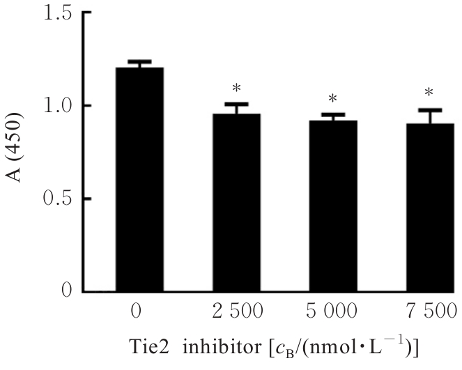
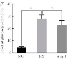
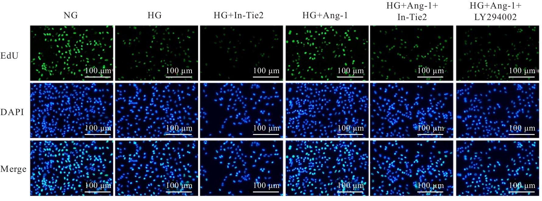
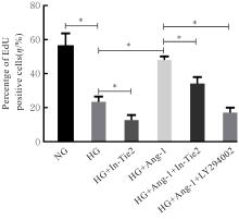
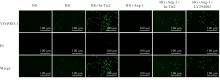
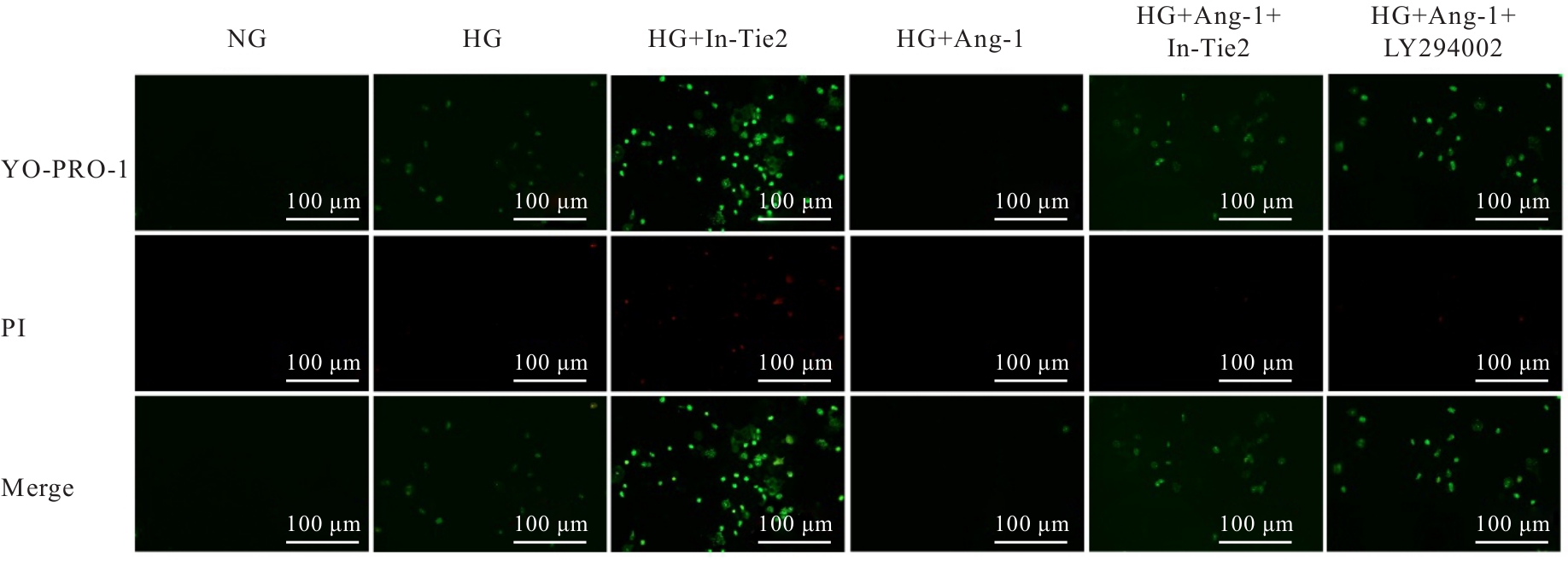
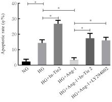
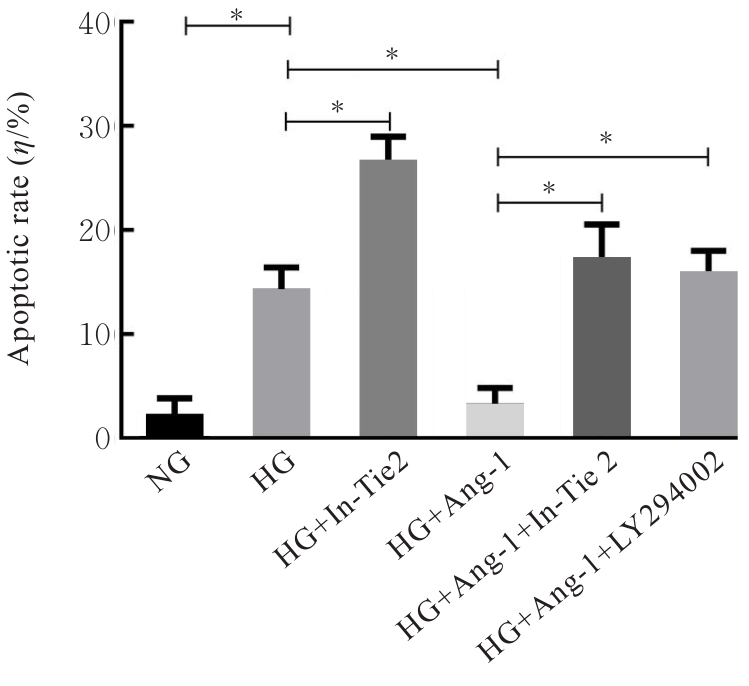
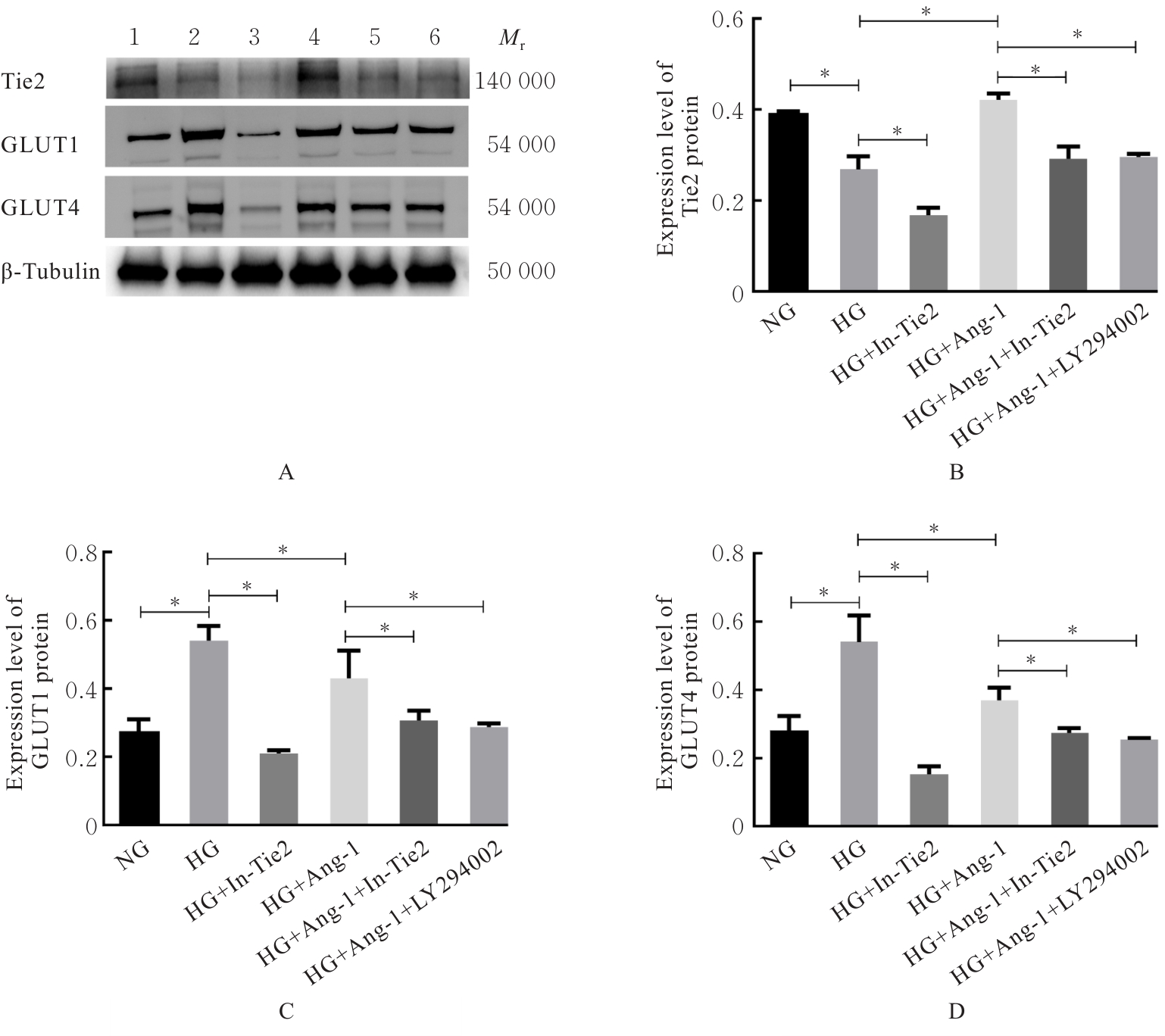
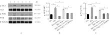
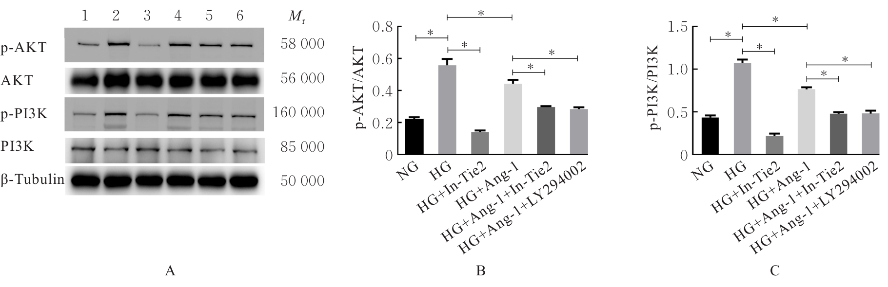
目的 研究血管生成素1(Ang-1)和酪氨酸激酶受体2(Tie2)抑制剂对高糖培养人脐静脉内皮细胞(HUVECs)葡萄糖转运的作用,并阐明其作用机制。 方法 体外高糖(30 mmol·L-1)培养HUVECs,采用0、200、500、1 000和2 000 μg·L-1 Ang-1及0、2 500、5 000和7 500 nmol·L-1 Tie2抑制剂处理细胞,细胞计数试剂盒8(CCK-8)法检测细胞活性,筛选Ang-1和Tie2抑制剂最佳作用浓度。葡萄糖试剂盒检测Ang-1干预细胞后HUVECs上清液中葡萄糖水平。HUVECs细胞随机分为空白对照组(NG组)、高糖作用组(HG组)、HG+Tie2抑制剂组(HG+In-Tie2组)、HG+Ang-1组、HG+Ang-1+Tie2抑制剂组(HG+Ang-1+In-Tie2组)和HG+Ang-1+磷脂酰肌醇3-激酶(PI3K)抑制剂组(HG+Ang-1+LY294002组)。5-乙炔基-2'脱氧尿嘧啶核苷(EdU)法检测各组细胞增殖活性,恶唑黄/碘化丙啶(YO-PRO-1/PI)法检测各组细胞凋亡率,实时荧光定量PCR(RT-qPCR)法检测各组细胞中Ang-1Tie2 mRNA表达水平,Western blotting法检测各组细胞中Tie2、葡萄糖转运蛋白1(GLUT1)和葡萄糖转运蛋白4(GLUT4)蛋白表达水平及磷酸化PI3K(p-PI3K)/PI3K和磷酸化蛋白激酶(p-AKT)/AKT比值。 结果 CCK-8法检测,与0 μg·L-1 Ang-1比较,200 μg·L-1 Ang-1作用HUVECs 48 h后HUVECs细胞活性明显升高(P<0.01);与0 nmol·L-1 Tie2抑制剂比较,2 500、5 000和7 500 nmol·L-1 Tie2抑制剂作用下HUVECs细胞活性明显降低(P<0.01);Ang-1和Tie2抑制剂的最佳浓度分别为200 μg·L-1及2 500 nmol·L-1。与NG组比较,HG组HUVECs上清液中葡萄糖水平明显升高(P<0.01);与HG组比较,Ang-1组HUVECs上清液中葡萄糖水平明显降低(P<0.01)。EdU法检测,与NG组比较,HG组HUVECs增殖活性明显降低(P<0.01);与HG组比较,HG+In-Tie2组HUVECs增殖活性明显降低(P<0.01),HG+Ang-1组HUVECs增殖活性明显升高(P<0.01);与HG+Ang-1组比较,HG+Ang-1+In-Tie2组和HG+Ang-1+LY294002组HUVECs增殖活性均明显降低(P<0.01)。YO-PRO-1/PI法检测,与NG组比较,HG组HUVECs凋亡率明显升高(P<0.01);与HG组比较,HG+In-Tie2组HUVECs凋亡率明显升高(P<0.01),HG+Ang-1组HUVECs凋亡率明显降低(P<0.01);与HG+Ang-1组比较,HG+Ang-1+In-Tie2组和HG+Ang-1+LY294002组HUVECs凋亡率均明显升高(P<0.01)。RT-qPCR法检测,与NG组比较,HG组和HG+In-Tie2组HUVECs中Ang-1Tie2 mRNA表达水平均明显降低(P<0.01);与HG组比较,HG+In-Tie2组HUVECs中Ang-1Tie2 mRNA表达水平均明显降低(P<0.01),HG+Ang-1组HUVECs中Ang-1Tie2 mRNA表达水平均明显升高(P<0.05);与HG+Ang-1组比较, HG+Ang-1+In-Tie2组和HG+Ang-1+LY294002组HUVECs中Ang-1Tie2 mRNA表达水平均明显降低(P<0.05或P<0.01)。Western blotting法检测,与NG组比较,HG组HUVECs中Tie2蛋白表达水平明显降低(P<0.01),GLUT1和GLUT4蛋白表达水平均明显升高(P<0.01);与HG组比较,HG+In-Tie2组HUVECs中Tie2、GLUT1和GLUT4蛋白表达水平均明显降低(P<0.01),HG+Ang-1组HUVECs中Tie2蛋白表达水平明显升高(P<0.01),GLUT1和GLUT4蛋白表达水平均明显降低(P<0.01);与HG+Ang-1组比较,HG+Ang-1+In-Tie2组和HG+Ang-1+LY294002组HUVECs中Tie2、GLUT1和GLUT4蛋白表达水平均明显降低(P<0.01)。与NG组比较,HG组HUVECs中p-PI3K/PI3K和p-AKT/AKT比值均明显升高(P<0.01);与HG组比较,HG+In-Tie2组HUVECs中p-PI3K/PI3K和p-AKT/AKT比值均明显降低(P<0.01),HG+Ang-1组HUVECs中p-PI3K/PI3K和p-AKT/AKT比值均明显降低(P<0.01);与HG+Ang-1组比较,HG+Ang-1+In-Tie2组和HG+Ang-1+LY294002组HUVECs中p-PI3K/PI3K和p-AKT/AKT比值均明显降低(P<0.01)。 结论 Ang-1可下调高糖培养HUVECs中GLUT1和GLUT4的表达,Ang-1与Tie2结合可能过PI3K/AKT信号通路下调GLUT1和GLUT4参与高糖培养HUVECs的葡萄糖转运。

关键词: 糖尿病肾病, 血管生成素1, 葡萄糖转运蛋白, 酪氨酸激酶受体2, 磷脂酰肌醇3-激酶, 蛋白激酶B

Abstract:

Objective To study the effect of angiopoietin-1 (Ang-1) and tyrosine kinase receptor 2 (Tie2) inhibitor on glucose transportation in the human umbilical vein endothelial cells (HUVECs) cultured under high glucose conditions, and to clarify its mechanism. Methods The HUVECs were cultured in high glucose (30 mmol·L?1) in vitro and treated with 0, 200, 500, 1 000, and 2 000 μg·L?1 Ang-1 and 0, 2 500, 5 000, and 7 500 nmol·L?1 Tie2 inhibitor; cell counting kit-8 (CCK-8) method was used to detect the cell activity to screen the optimal concentrations of Ang-1 and Tie2 inhibitor. Glucose kit was used to detect the glucose level in the supernatant of the HUVECs after Ang-1 intervention. The HUVECs were randomly divided into blank control group (NG group), high glucose group (HG group), HG+Tie2 inhibitor group (HG+In-Tie2 group), HG+Ang-1 group, HG+Ang-1+Tie2 inhibitor group (HG+Ang-1+In-Tie2 group), and HG+Ang-1+phosphatidylinositol 3-kinase(PI3K) inhibitor group (HG+Ang-1+LY294002 group). 5-Ethynyl-2'-deoxyuridine (EdU) method was used to detect the proliferation activities of the cells in various groups; YO-PRO-1/PI method was used to detect the apoptotic rates of the cells in various groups; real-time fluorescence quantitative PCR (RT-qPCR) method was used to detect the expression levels of Ang-1 mRNA and Tie2 mRNA in the cells in various groups; Western blotting method was used to detect the expression levels of Tie2, glucose transporter 1 (GLUT1), and glucose transporter 4 (GLUT4) proteins and the ratios of phosphorylated PI3K (p-PI3K)/PI3K and phosphorylated protein kinase B (p-AKT)/AKT in the cells in various groups. Results The CCK-8 assay results showed that compared with 0 μg·L?1 Ang-1 group, the activity of the HUVECs was significantly increased after treated with 200 μg·L?1 Ang-1 for 48 h (P<0.01); compared with 0 nmol·L?1 Tie2 inhibitor group, the activity of the HUVECs was significantly decreased after treated with 2 500、 5 000 and 7 500 nmol·L?1 Tie2 inhibitor (P<0.01); the optimal concentrations of Ang-1 and Tie2 inhibitor were 200 μg·L?1 and 2 500 nmol·L?1, respectively. Compared with NG group, the glucose level in the supernatant of the HUVECs in HG group was significantly increased (P<0.01); compared with HG group, the glucose level in the supernatant of the HUVECs in Ang-1 group was significantly decreased (P<0.01). The EdU assay results showed that compared with NG group, the proliferation activity of the HUVECs in HG group was significantly decreased (P<0.01); compared with HG group, the proliferation activity of the HUVECs in HG+In-Tie2 group was significantly decreased (P<0.01), and the proliferation activity of the HUVECs in HG+Ang-1 group was significantly increased (P<0.01); compared with HG+Ang-1 group, the proliferation activities of the HUVECs in HG+Ang-1+In-Tie2 group and HG+Ang-1+LY294002 group were significantly decreased (P<0.01). The YO-PRO-1/PI assay results showed that compared with NG group, the apoptotic rate of the HUVECs in HG group was significantly increased (P<0.01); compared with HG group, the apoptotic rate of the HUVECs in HG+In-Tie2 group was significantly increased (P<0.01), and the apoptotic rate of the HUVECs in HG+Ang-1 group was significantly decreased (P<0.01); compared with HG+Ang-1 group, the apoptotic rates of the HUVECs in HG+Ang-1+In-Tie2 group and HG+Ang-1+LY294002 group were significantly increased (P<0.01). The RT-qPCR results showed that compared with NG group, the expression levels of Ang-1 mRNA and Tie2 mRNA in the HUVECs in HG group and HG+In-Tie2 group were significantly decreased (P<0.01); compared with HG group, the expression levels of Ang-1 mRNA and Tie2 mRNA in HG+ In-Tie2 group were significantly decreased (P<0.01), and the expression levels of Ang-1 mRNA and Tie2 mRNA in the HUVECs in HG+Ang-1 group were significantly increased (P<0.05); compared with HG+Ang-1 group, the expression levels of Ang-1 mRNA and Tie2 mRNA in the HUVECs in HG+Ang-1+In-Tie2 group and HG+Ang-1+LY294002 group were significantly decreased (P<0.05 or P<0.01). The Western blotting results showed that compared with NG group, the expression level of Tie2 protein in the HUVECs in HG group was significantly decreased (P<0.01), and the expression levels of GLUT1 and GLUT4 proteins were significantly increased (P<0.01); compared with HG group, the expression levels of Tie2, GLUT1, and GLUT4 proteins in the HUVECs in HG+In-Tie2 group were significantly decreased (P<0.01), the expression level of Tie2 protein in the HUVECs in HG+Ang-1 group was significantly increased (P<0.01), and the expression levels of GLUT1 and GLUT4 proteins were significantly decreased (P<0.01); compared with HG+Ang-1 group, the expression levels of Tie2, GLUT1, and GLUT4 proteins in the HUVECs in HG+Ang-1+In-Tie2 group and HG+Ang-1+LY294002 group were significantly decreased (P<0.01). Compared with NG group, the p-PI3K/PI3K and p-AKT/AKT ratios in the HUVECs in HG group were significantly increased (P<0.01); compared with HG group, the p-PI3K/PI3K and p-AKT/AKT ratios in the HUVECs in HG+In-Tie2 group were significantly decreased (P<0.01), and the p-PI3K/PI3K and p-AKT/AKT ratios in the HUVECs in HG+Ang-1 group were significantly decreased (P<0.01); compared with HG+Ang-1 group, the p-PI3K/PI3K and p-AKT/AKT ratios in the HUVECs in HG+Ang-1+In-Tie2 group and HG+Ang-1+LY294002 group were significantly decreased (P<0.01). Conclusion Ang-1 down-regulates the expressions of GLUT1 and GLUT4 in the HUVECs cultured under high glucose conditions; the binding of Ang-1 to Tie2 may down-regulate GLUT1 and GLUT4 via the PI3K/AKT signaling pathway to participate in the glucose transportation in the HUVECs cultured under high glucose conditions.

Key words: Diabetic nephropathy, Angiopoietin-1, Glucose transporter protein, Tyrosine kinase receptor 2, Phosphatidylinositol 3-kinase, Protein kinase B

中图分类号: 

  • R587.2