吉林大学学报(医学版) ›› 2020, Vol. 46 ›› Issue (6): 1117-1123.doi: 10.13481/j.1671-587x.20200602

• 基础研究 • 上一篇    下一篇

芦丁对肝细胞氧化应激损伤的保护作用及其机制

金芳多,张天,张钊,尹学哲,全吉淑()   

  1. 延边大学基础医学院生物化学与分子生物学教研室,吉林 延吉 133002
  • 收稿日期:2020-05-12 出版日期:2020-11-28 发布日期:2022-08-24
  • 通讯作者: 全吉淑 E-mail:quanjs1@sina.com
  • 作者简介:金芳多(1995-),女,吉林省吉林市人,在读医学硕士,主要从事天然成分活性方面的研究。
  • 基金资助:
    国家自然科学基金资助课题(81760659)

Protective effect of rutin on oxidative stress injury of HepG2 cells and its mechanism

Fangduo JIN,Tian ZHANG,Zhao ZHANG,Xuezhe YIN,Jishu QUAN()   

  1. Department of Biochemistry and Molecular Biology,School of Basic Medical Sciences,Yanbian University,Yanji 133002,China
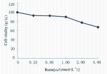
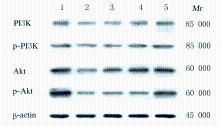
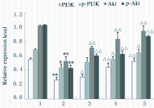
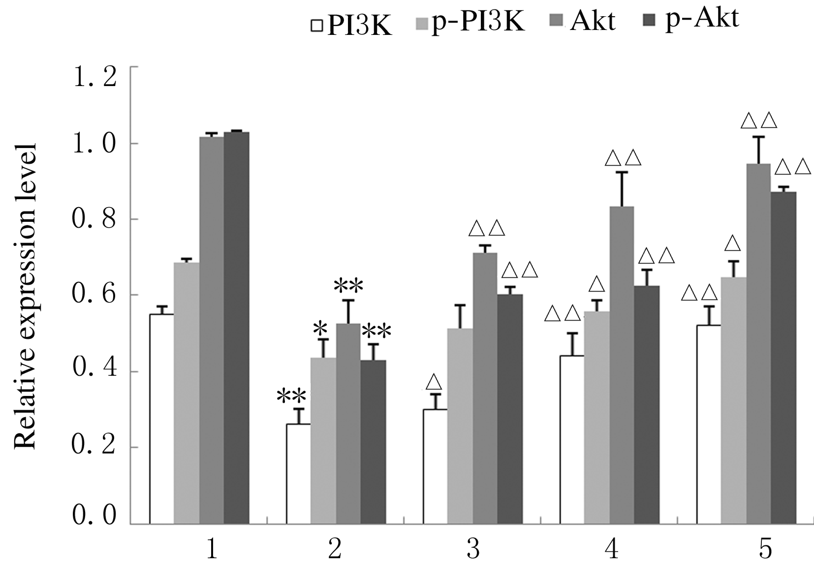
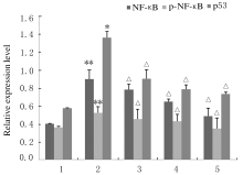
  • Received:2020-05-12 Online:2020-11-28 Published:2022-08-24
  • Contact: Jishu QUAN E-mail:quanjs1@sina.com

摘要: 目的

探讨芦丁对肝癌HepG2细胞氧化应激损伤的保护作用,并阐明其作用机制。

方法

培养人肝癌HepG2细胞,取处于对数生长期的HepG2细胞分为对照组、叔丁基过氧化氢(TBHP)组(给予400 μmol·L-1TBHP)和0.25、0.50、1.00 μmolL-1芦丁组(给予0.25、0.50和1.00 μmol·L-1芦丁+400 μmol·L-1 TBHP)。采用四甲基偶氮唑盐(MTT)法检测各组HepG2细胞活性和各组肝癌HepG2细胞存活率,比色法检测各组肝癌HepG2细胞中丙二醛(MDA)和还原型谷胱甘肽(GSH)水平及超氧化物歧化酶(SOD)活性,采用二氯荧光素乙酰乙酸盐(DCFH-DA)荧光染色法检测各组肝癌HepG2细胞中活性氧(ROS)水平,Western blotting法检测各组肝癌HepG2细胞中磷脂酰肌醇3激酶(PI3K)、磷酸化磷脂酰肌醇3激酶(p-PI3K)、蛋白激酶B(Akt)、磷酸化AktB(p-Akt)、核转录因子κB(NF-κB)、磷酸化NF-κB(p-NF-κB)和p53蛋白表达水平。

结果

MTT检测,芦丁摩尔浓度≤1 μmol·L-1时,肝癌HepG2细胞的存活率均>90%,不显示细胞毒性;当芦丁摩尔浓度>1 μmol·L-1时,肝癌HepG2细胞存活率逐渐降低,均<90%,显示细胞毒性。与对照组比较,TBHP组HepG2细胞存活率明显降低(P<0.01);与TBHP组比较,不同浓度芦丁组HepG2细胞存活率明显升高(P<0.01)。比色法检测,与对照组比较,TBHP组肝癌HepG2细胞中MDA水平明显升高(P<0.01),GSH水平和SOD活性明显降低(P<0.01);与TBHP组比较,不同浓度芦丁组肝癌HepG2细胞中MDA水平明显降低(P<0.01),GSH水平和SOD活性明显升高(P<0.01)。DCFH-DA染色,与对照组比较,TBHP组肝癌HepG2细胞中ROS水平明显升高;与TBHP组比较,不同浓度芦丁组肝癌HepG2细胞中ROS水平降低。Western blotting法检测,与对照组比较,TBHP组肝癌HepG2细胞中PI3K、p-PI3K、Akt和p-Akt蛋白表达水平降低(P<0.05),NF-κB、p-NF-κB和p53蛋白表达水平升高(P<0.05);与TBHP组比较,不同浓度芦丁组肝癌HepG2细胞中PI3K、p-PI3K、Akt和p-Akt蛋白表达水平升高(P<0.05),NF-κB、p-NF-κB和p53蛋白表达水平降低(P<0.05)。

结论

芦丁对肝癌HepG2细胞损伤具有保护作用,其机制可能与调控PI3K/Akt和NF-κB两条信号通路有关联。

关键词: 芦丁, 叔丁基过氧化氢, 氧化应激, 肝细胞损伤, 肝细胞保护

Abstract: Objective

To explore the protective effect of rutin on the oxidative stress injury of liver cancer HepG2 cells, and to clarify its mechanism.

Methods

The liver cancer HepG2 cells in the logarithmic growth phase were divided into control group, tert-butyl hydroperoxide (TBHP) group (400 μmol·L-1 TBHP), 0.25, 0.50,and 1.00 μmol·L-1 rutin groups (0.25, 0.50, 1.00 μmol·L-1 rutin+400 μmol·L-1 TBHP). The vitalities of liver cancer HepG2 cells in various groups and the survival rates of liver cancer HepG2 cells in various groups were detected by MTT assay;the levels of malondialdehyde (MDA) and glutathione (GSH) and activities of superoxide dismutase (SOD) in the liver cancer HepG2 cells in various groups were assessed by colorimetric method; the levels of reactive oxidative species (ROS) in the liver cancer HepG2 cells in various groups were quantified with DCFH-DA immnofluorescence staining. The expression levels of PI3K, p-PI3K, Akt, p-Akt, NF-κB, p-NF-κB,and p53 proteins in the liver cancer HepG2 cells in various groups were mesured by Western blotting method.

Results

The MTT results showed that when the concentrations of rutin were less than or equal to 1.00 μmol·L-1, the vitality of liver cancer HepG2 cells was higher than 90%, and the rutin had no toxic effect; when the concentration of rutin was higher than 1.00 μmol·L-1, the vitality of the liver cancer HepG2 cells was lower than 90%,and the rutin had toxic effect. Compared with control group, the survival rate of the HepG2 cells in TBHP group was significantly decreased (P<0.01); compared with TBHP group, the survival rates of the HepG2 cells in different concertrations of rutin groups were significantly increased (P<0.01). Compared with control group, the level of MDA in the HepG2 cells in TBHP group was significantly decreased (P<0.01), the level of GSH and the activity of SOD were significantly increased (P<0.01); compared with TBHP group, the levels of MDA in the HepG2 cells in different concentrations of rutin groups were significantly increased(P<0.01), the levels of GSH and the activities of SOD in the HepG2 cells in different concertrations of rutin groups were significantly decreased (P<0.01). The results of DCFH-DA immunofluorescence staining showed that the ROS level in the HepG2 cells in TBHP group was higher than that in control group; compared with TBHP group, the ROS levels in the HepG2 cells in different concentrations of rutin groups were gradually decreased(P<0.05). The Western blotting results showed that compared with control group, the expression levels of PI3K, p-PI3K, Akt, and p-Akt proteins in the HepG2 cells in TBHP group were decreased (P<0.05), and the expressions levels of NF-κB, p-NF-κB,and p53 proteins were increased (P<0.05); compared with TBHP group, the expressions levels of PI3K, p-PI3K, Akt, and p-Akt proteins in the HepG2 cells in different concentrations of rutin groups were increased (P<0.05), and the expressions levels of NF-κB, p-NF-κB, and p53 proteins were decreased (P<0.05).

Conclusion

Rutin has the protective effect on TBHP-induced HepG2 cell damage, and its mechanism may be related to the regulation of PI3K/Akt and NF-κB pathways.

Key words: rutin, tert-butyl hydroperoxide, oxidative stress, hepatocyte injury, hepatocyte protection

中图分类号: 

  • R282.7