吉林大学学报(医学版) ›› 2020, Vol. 46 ›› Issue (6): 1150-1154.doi: 10.13481/j.1671-587x.20200607

• 基础研究 • 上一篇    下一篇

GLI1真核表达载体的构建、鉴定及其对肺癌PC9细胞增殖的影响

陈微微,安佳佳,武艳,代娟娟,杜静()   

  1. 滨州医学院附属医院肿瘤研究实验室,山东 滨州 256600
  • 收稿日期:2020-03-10 出版日期:2020-11-28 发布日期:2022-08-24
  • 通讯作者: 杜静 E-mail:djedith@126.com
  • 作者简介:陈微微(1984-),女,吉林省长春市人,技师,医学硕士,主要从事抗肿瘤分子机制的相关研究。
  • 基金资助:
    国家自然科学基金资助课题(31900441);山东省科技厅科学基金资助课题(ZR2019MC026);山东省卫生厅医药卫生科技发展计划项目资助课题(2017WS155);滨州医学院科研计划与科研启动基金资助课题(BY2015KYQD16)

Construction and identification of GLI1 eukaryotic expression vector and its effect on proliferation of lung cancer PC9 cells

Weiwei CHEN,Jiajia AN,Yan WU,Juanjuan DAI,Jing DU()   

  1. Tumor Research Laboratory,Affiliated Hospital,Binzhou Medical University,Bin zhou 256600,China
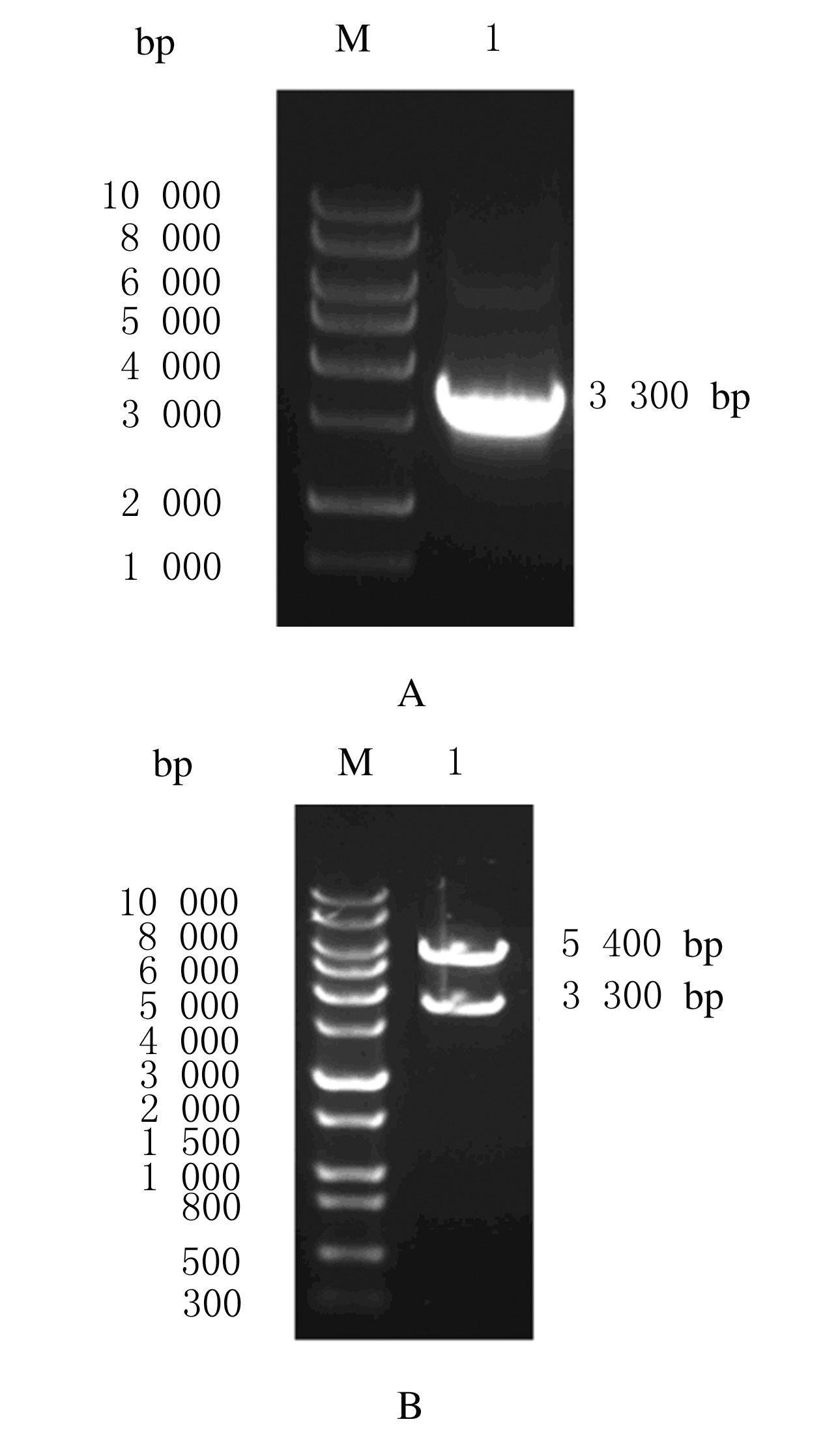
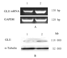
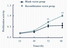
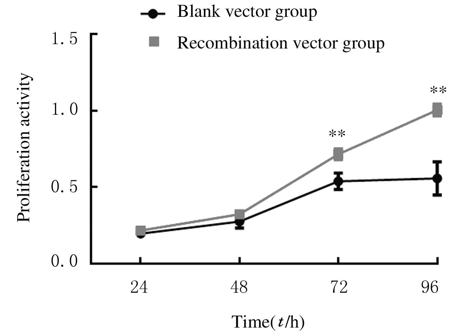
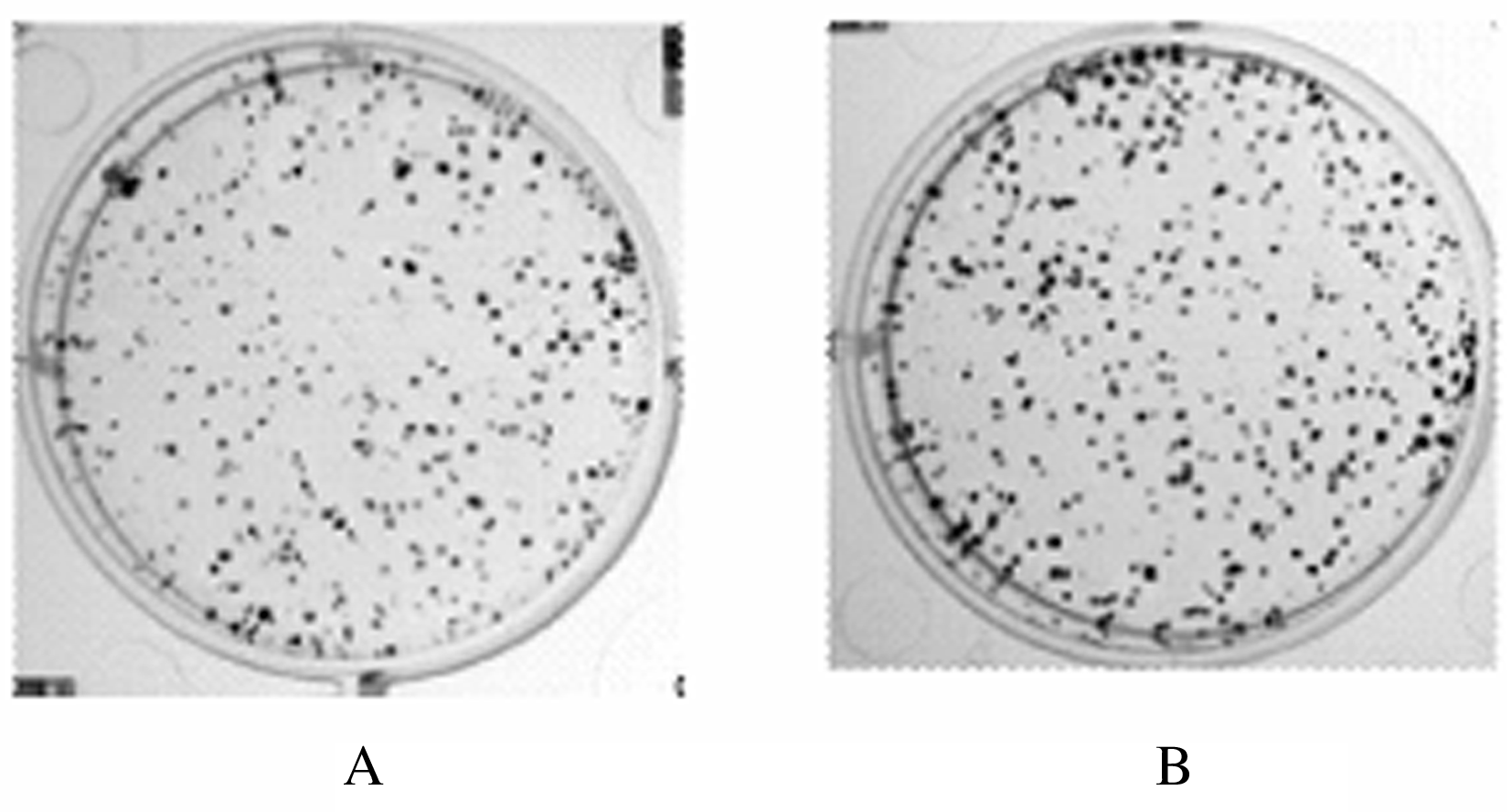
  • Received:2020-03-10 Online:2020-11-28 Published:2022-08-24
  • Contact: Jing DU E-mail:djedith@126.com

摘要: 目的

构建GLI1真核表达载体,探讨过表达GLI1对肺癌PC9细胞增殖的影响。

方法

采用PCR法扩增GLI1基因的全长序列,克隆至pcDNA3.1真核表达载体,将重组质粒pcDNA3.1-GLI1(重组质粒组)和对照质粒pcDNA3.1(空载体组)转染肺癌PC9细胞。采用酶切鉴定法检测GLI1过表达载体的构建情况。通过G418筛选建立稳定过表达GLI1的PC9细胞和空载体对照细胞。RT-PCR法检测2组PC9细胞中GLI1 mRNA表达水平,Western blotting法检测2组PC9细胞中GLI1蛋白表达水平,CCK-8法检测2组PC9细胞增殖活性,细胞克隆形成实验检测2组PC9细胞中克隆形成数。

结果

成功扩增获得GLI1的特异基因片段;构建的GLI1真核表达载体经双酶切鉴定后分别获得目的基因和载体片段。RT-PCR法检测,重组质粒组PC9细胞中GLI1 mRNA表达水平明显高于空载体组(P<0.01)。Western blotting法检测,重组质粒组PC9细胞中GLI1蛋白表达水平明显高于空载体组(P<0.05)。CCK-8法检测,培养72和96 h后,重组质粒组细胞增殖活性明显高于空载体组(P<0.01)。细胞克隆形成实验,培养10 d时,重组质粒组细胞克隆形成数明显多于空载体组(P<0.01)。

结论

成功构建GLI1基因的真核过表达载体和过表达GLI1稳定转染的PC9细胞系, GLI1过表达能够促进肺癌PC9细胞的增殖能力。

关键词: 肺肿瘤, GLI1, 细胞增殖, PC9细胞, 真核表达载体

Abstract: Objective

To construct the GLI1 eukaryotic expression vector,and to discuss the effect of over-expression of GLI1 on the proliferation of lung cancer PC9 cells.

Methods

The full length sequence of GLI1 gene was amplified by PCR method and it was cloned into the pcDNA3.1 eukaryotic expression vector. The PC9 cells were transfected with the recombinant vector pcDNA3.1-GLI1 (recombination vector group) and the control vector pcDNA3.1 (empty vector group). The construction of GLI1 over-expression vector was detected by enzymatic digestion identification method. The cells stably expressed with GLI1 and empty vector were established by G418 screening. The expression levels of GLI1 mRNA in the PC9 cells in two groups were detected by RT-PCR method; the expression levels of GLI1 protein in the PC9 cells in two groups were detected by Western blotting method;the proliferation activities of the PC9 cells in two groups were detected by CCK-8 method; the clone formation number of the PC9 cells in two groups was detected by cell clonal formation assay.

Results

The GLI1 gene fragment was specifically amplified successfully; the target genes and vector fragments were obtained by double enzyme digestion of the GLI1 eukaryotic expression vectors. The RT-PCR results showed that the expression level of GLI1 mRNA in the PC9 cells in recombination vector group was significantly higher than that in empty vector group (P<0.01). The Western blotting results showed that the expression level of GLI1 protein in the PC9 cells in recombination vector group was significantly higher than that in empty vector group (P<0.05). The CCK-8 results showed that the proliferation activities of the PC9 cells in recombination group were significantly higher than those in empty vector group at 72 and 96 h after culture (P<0.01). The clonal formation assay results showed that the clone formation number of the PC9 cells in recombination group was significantly more than that in empty vector group at 10 d of culture (P<0.01).

Conclusion

The GLI1 eukaryotic over-expression vector and the PC9 cells stably over-expressed with GLI1 are established successfully. Over-expression of GLI1 can promote the proliferation ability of lung cancer PC9 cells.

Key words: lung neoplasms, GLI1, cell proliferation, PC9 cells, eukaryotic expression vector

中图分类号: 

  • R734.2