吉林大学学报(医学版) ›› 2021, Vol. 47 ›› Issue (3): 660-668.doi: 10.13481/j.1671-587X.20210316

• 基础研究 • 上一篇    下一篇

骨髓间充质干细胞来源性外泌体携带miR-196b-5p对结肠癌细胞生物学特性的调控作用

李晓辉1,瞿紫微1(),卢昕1,孟庆彬1,陈华涛1,任骏1,谭成沛2   

  1. 1.湖北省武汉第一医院胃肠外科,湖北 武汉 430022
    2.湖北省神农架林区中医医院普外科,湖北 神农架 442000
  • 收稿日期:2020-08-21 出版日期:2021-05-28 发布日期:2021-05-28
  • 通讯作者: 瞿紫微 E-mail:quziwei007@126.com
  • 作者简介:李晓辉(1978-),男,河南省郑州市人,主治医师,医学硕士,主要从事结直肠肿瘤和外科营养方面的研究。
  • 基金资助:
    湖北省自然科学基金知识创新专项(2018CFC814)

Regulatory effect of exosomes carrying miR-196b-5p derived from bone marrow mesenchymal stem cells on biological characteristics of colon cancer cells

Xiaohui LI1,Ziwei QU1(),Xin LU1,Qingbin MENG1,Huatao CHEN1,Jun REN1,Chengpei TAN2   

  1. 1.Department of Gastrointestinal Surgery,Wuhan First Hospital,Hubei Province,Wuhan 430022,China
    2.Department of General Surgery,Shennongjia Forestry Hospital of Traditional Chinese Medicine,Hubei Province,Shennongjia 442000,China
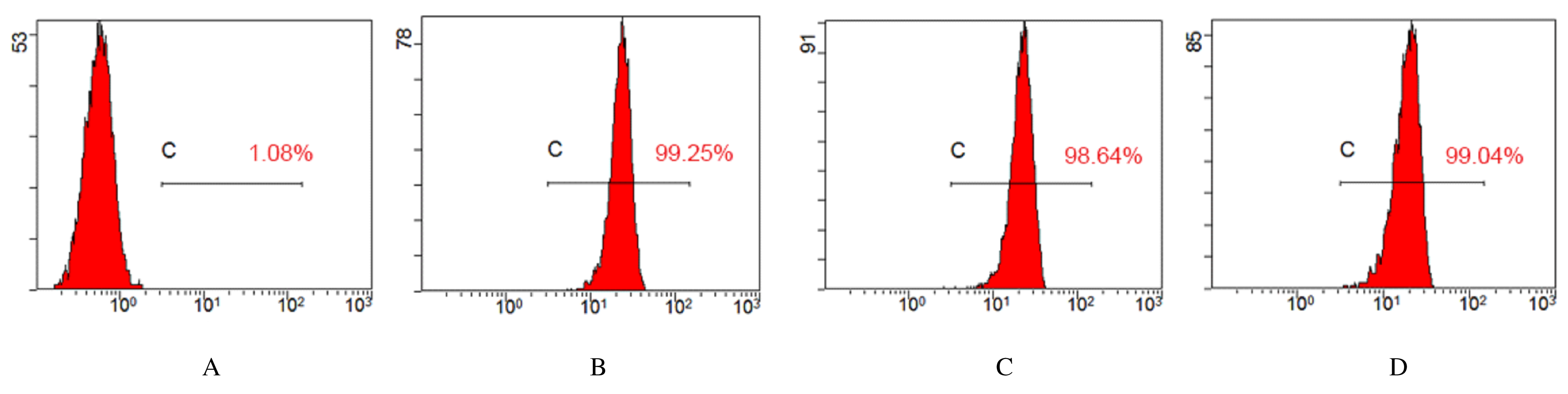
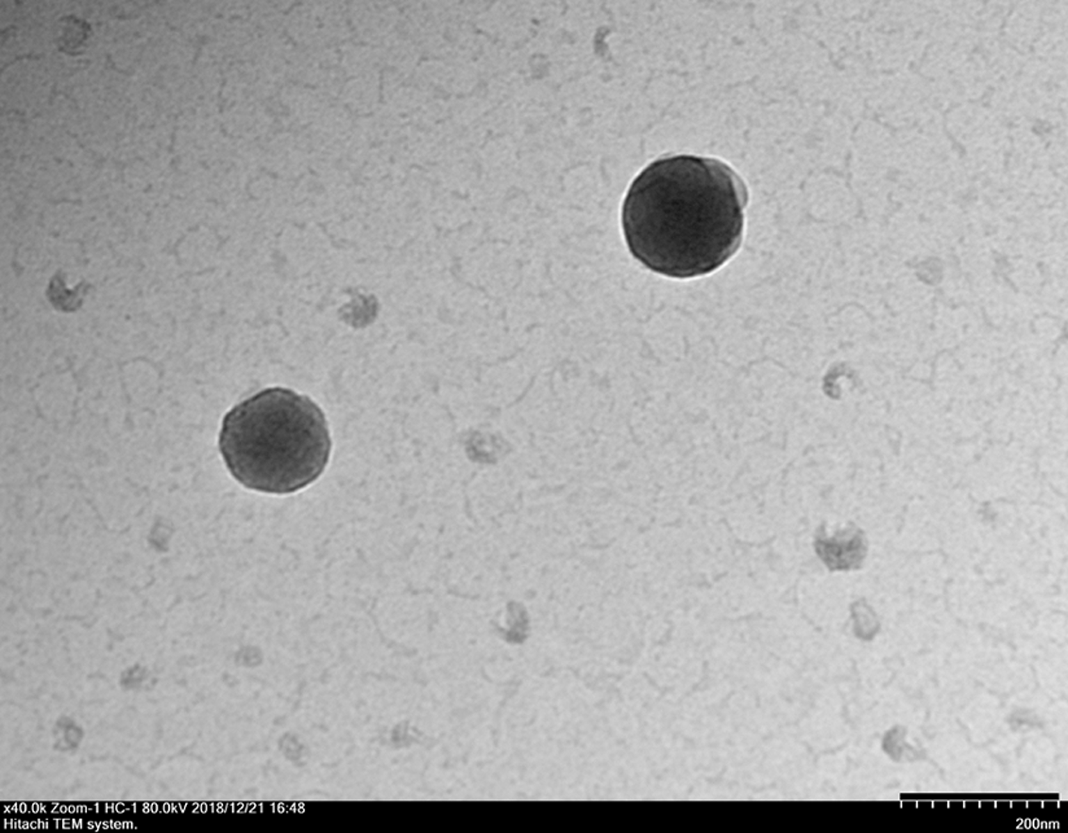
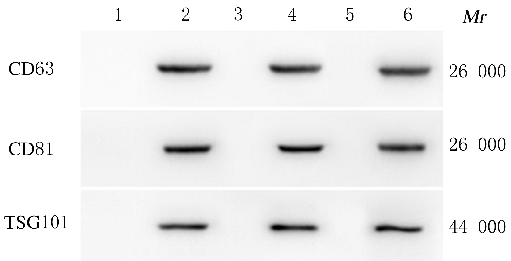
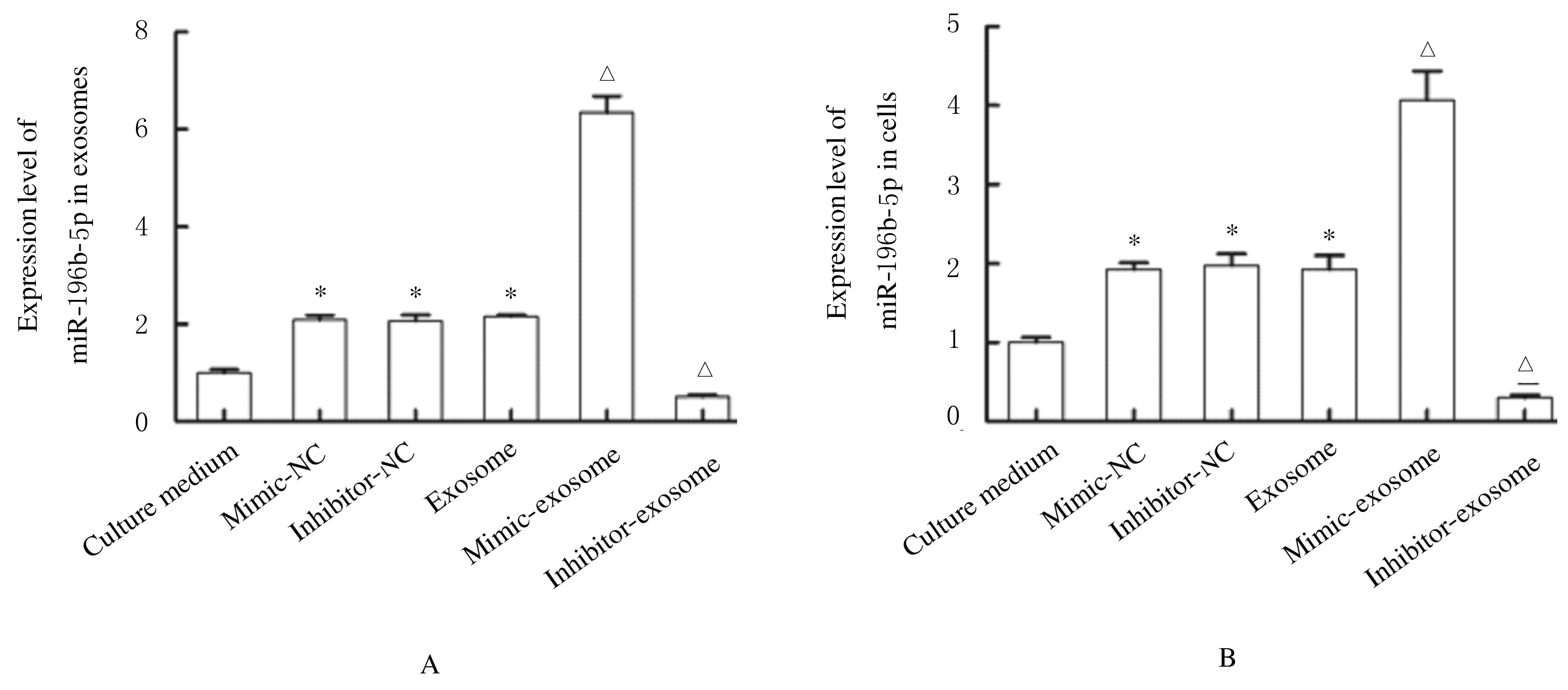
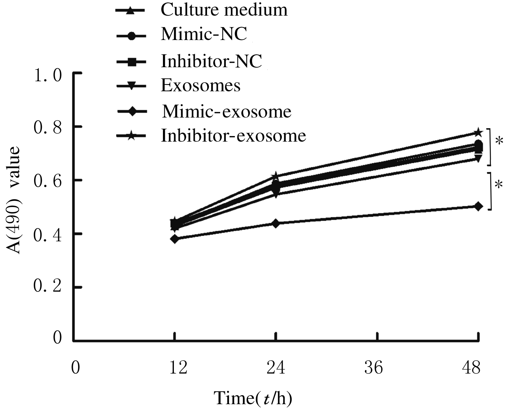
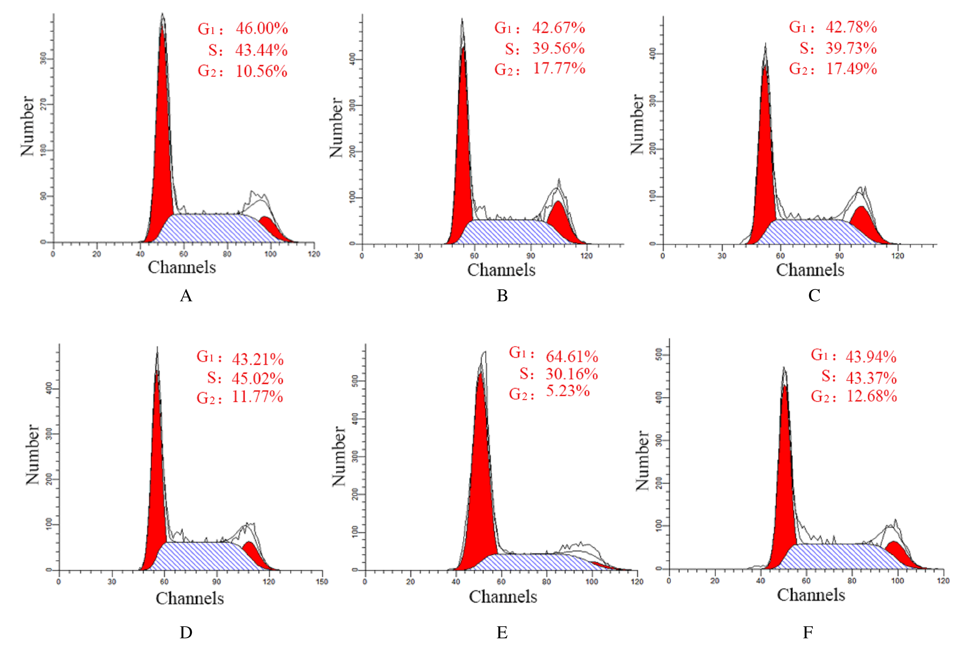
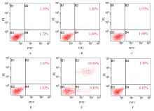
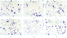
  • Received:2020-08-21 Online:2021-05-28 Published:2021-05-28
  • Contact: Ziwei QU E-mail:quziwei007@126.com

摘要: 目的

探讨骨髓间充质干细胞(MSCs)分泌的携带miR-196b-5p的外泌体对结肠癌细胞生物学特性的影响。

方法

分离培养小鼠骨髓MSCs,采用mimic-196b-5p模拟物(miR-196b-5p miR)和196b-5p抑制物(miR-196b-5p inhibitor)转染MSCs后,收集培养基,分离外泌体。透射电镜下观察外泌体形态表现,采用特定标志物表达鉴定外泌体后采用外泌体干预小鼠结肠癌CT26.WT细胞。将复苏后处于对数生长期的小鼠结肠癌CT26.WT细胞分为培养基组、mimic-NC组、inhibitor-NC组、未转染的外泌体组、mimic-外泌体组和inhibitor-外泌体组。采用MTT法检测各组结肠癌CT26.WT细胞增殖活性,流式细胞术检测各组不同细胞周期结肠癌CT26.WT细胞百分率和细胞凋亡率,Transwell小室实验检测各组结肠癌CT26.WT细胞的迁移和侵袭情况。

结果

与未转染的外泌体组比较,mimic-外泌体组结肠癌细胞增殖活性、迁移细胞数和侵袭细胞数均降低(P<0.05),G1期细胞百分率和细胞凋亡率升高(P<0.05),inhibitor-外泌体组结肠癌细胞增殖活性、迁移细胞数和侵袭细胞数均升高(P<0.05),细胞凋亡率降低(P<0.05)。

结论

MSCs来源携带高水平miR-196b-5p的外泌体可抑制结肠癌细胞增殖、迁移和侵袭,并诱导细胞凋亡。

关键词: 间充质干细胞, 外泌体, 结肠肿瘤, 细胞增殖, 细胞迁移, 细胞凋亡

Abstract: Objective

To investigate the effect of exosomes carrying microRNA-196b-5p secreted by the bone marrow mesenchymal stem cells (MSCs) on the biological characteristics of the colon cancer cells.

Methods

The MSCs were isolated and cultured from bone marrow of the mice. The mi-196b-5p mimic and mi-196b-5p inhibitor were transfected into the MSCs. The culture medium was collected and the exosomes were separated. The morphology of exosomes was observed under transmission electron microscope.After identification of exosomes by expression of specific markers, the colon cancer CT26.WT cells of the mice were intervened with the exosomes. The colon cancer CT26.WT cells of the mice at logarithmic growth phase after resuscitation were divided into medium group, mimic-NC group, inhibitor-NC group,exosome group, mimic-exosome group and inhibitor-exosome group.The proliferation activities of the colon cancer CT26.WT cells in various groups were measured by MTT assay;the percentages of the colon cancer CT26.WT cells at different cell cycles in various groups were detected by flow cytometry;the apoptotic rates of the colon cancer CT26.WT cells in various groups were detected by flow cytometry; the number of migration and invasion colon cancer cells in various groups was evaluated by Transwell chamber assay.

Results

Compared with exosome group, the proliferation activity, the number of migration cells and invasion cells of the colon cancer CT26.WT cells in mimic-exosome group were decreased (P<0.05), and the percentage of colon cancer CT26.WT cells at G1 phase and the apoptotic rate were increased(P<0.05);the proliferation activity, the number of migration and invasion cells of colon cancer CT26.WT cells in inhibitor-exosome group were increased (P<0.05),and the apoptotic rate was decreased(P<0.05).

Conclusion

The exosomes derived from MSCs carring a high level of miR-196b-5p can inhibit the proliferation, migration and invasion of the colon cancer CT26.WT cells, and induce the apoptosis.

Key words: mesenchymal stem cells, exosome, colon cancer, cell proliferation, cell migration, apoptosis

中图分类号: 

  • R735.35