吉林大学学报(医学版) ›› 2022, Vol. 48 ›› Issue (1): 122-128.doi: 10.13481/j.1671-587X.20220115

• 基础研究 • 上一篇    下一篇

桦木酸对胃癌MGC-803细胞迁移和侵袭的抑制作用及其机制

许广松1,蒋海兵2(),盘箐3,李国庆2   

  1. 1.南华大学衡阳医学院附属第二医院普外科,湖南 衡阳 421001
    2.南华大学衡阳医学院 附属第二医院消化内科,湖南 衡阳 421001
    3.永州职业技术学院 病原生物与免疫学教研室,湖南 永州 425100
  • 收稿日期:2021-05-26 出版日期:2022-01-28 发布日期:2022-01-17
  • 通讯作者: 蒋海兵 E-mail:sfxg8902@163.com
  • 作者简介:许广松(1984-),男,湖南省衡阳市人,主治医师,医学硕士,主要从事普外科疾病诊治方面的研究。
  • 基金资助:
    湖南省卫计委2017年度第二批科研计划项目(A2017014);湖南省卫健委科研项目(202103032022);湖南省衡阳市科技局指导性项目(139)

Inhibitory effects of betulinic acid on migration and invasion of gastric cancer MGC-803 cells and their mechanisms

Guangsong XU1,Haibing JIANG2(),Jing PAN3,Guoqing LI2   

  1. 1.Department of General surgery,Second Affiliated Hospital,Hengyang Medical School,University of South China,Hengyang 421001,China
    2.Department of Gastrointestinal Medicine,Second Affiliated Hospital,Hengyang Medical School,University of South China,Hengyang 421001,China
    3.Department of Pathogenic Biology and Immunology,Yongzhou Vocational and Technical College,Yongzhou 425100,China
  • Received:2021-05-26 Online:2022-01-28 Published:2022-01-17
  • Contact: Haibing JIANG E-mail:sfxg8902@163.com

摘要: 目的

探讨桦木酸(BEA)对胃癌MGC-803细胞增殖、凋亡、迁移和侵袭的影响,并阐明其作用机制。

方法

MGC-803细胞分为对照组和不同剂量BEA组,分别采用含0、2.5、5.0、10.0、20.0、40.0和80.0 μmol·L-1 BEA 的DMEM高糖培养基常规培养。采用CCK-8法、流式细胞术、划痕实验和Transwell法分别检测MGC-803细胞增殖率、凋亡率、迁移率和侵袭细胞数;Western blotting法检测各组MGC-803细胞中SMAD同源物7(SMAD7)、转化生长因子β受体1(TGF-βR1)、磷酸化SMAD同源物2(p-SMAD2)、磷酸化SMAD同源物3(p-SMAD3)、性别决定区Y框蛋白4(SOX4)、E盒结合锌指蛋白2(ZEB2)、基质金属蛋白酶9(MMP-9)、Snail和Slug蛋白表达水平。

结果

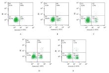
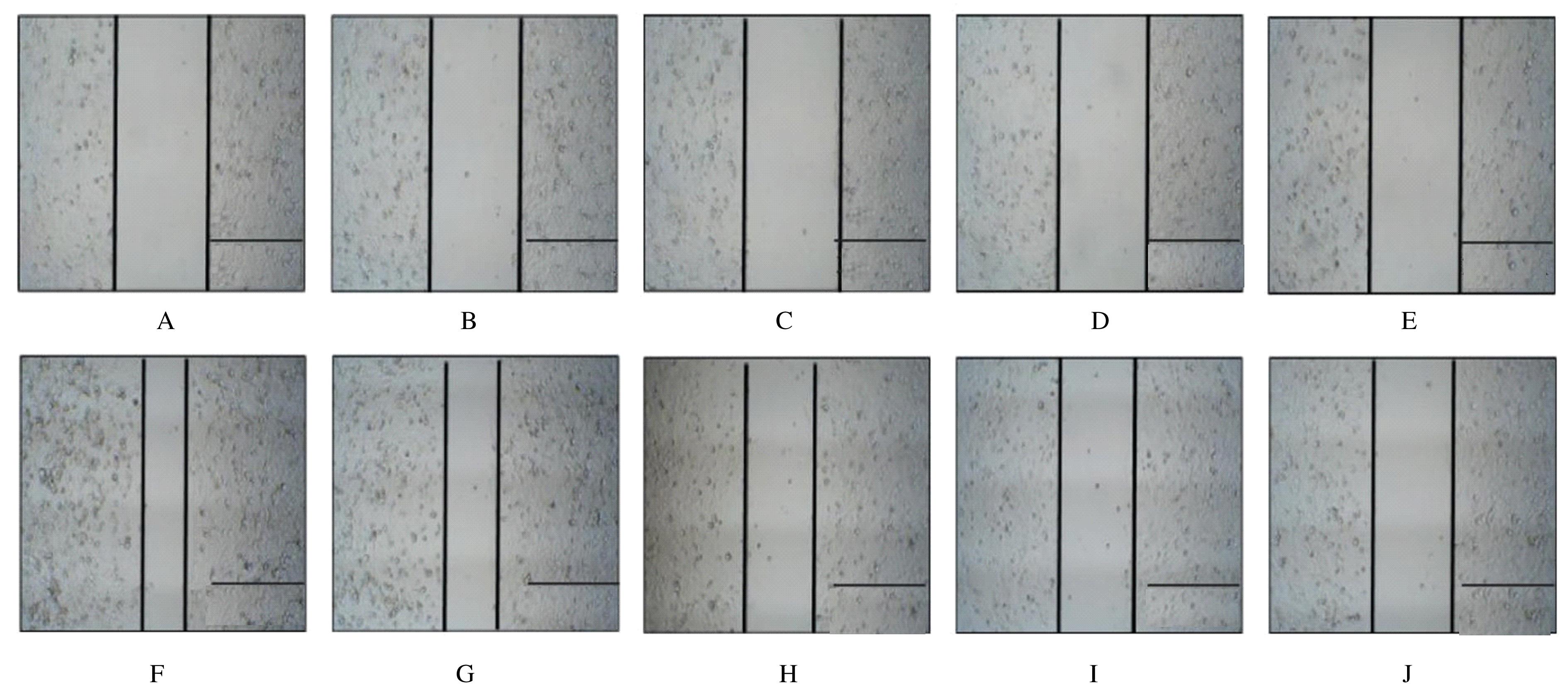
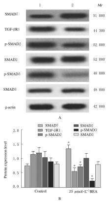
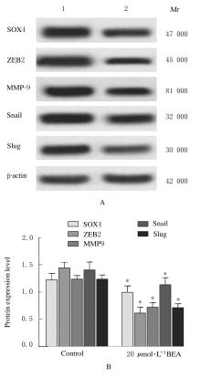
分别培养24、48和72 h后,与对照组比较, 2.5、5.0、10.0、20.0、40.0和80.0 μmol·L-1 BEA组细胞增殖率明显降低(P<0.05);培养72 h后,与对照组比较, 2.5、5.0、10.0和20.0 μmol·L-1 BEA组细胞凋亡率明显升高(P<0.05);培养24 h后,与对照组比较,2.5、5.0、10.0和20.0 μmol·L-1 BEA组细胞迁移率明显降低(P<0.05),侵袭细胞数明显减少(P<0.05)。与对照组比较,培养48 h后,20 μmol·L-1 BEA组细胞中SMAD7蛋白表达水平明显升高(P<0.05),TGF-βR1、p-SMAD2、p-SMAD3、SOX4、ZEB2、MMP-9、Snail和Slug蛋白表达水平明显降低(P<0.05)。

结论

BEA通过上调SMAD7表达以及抑制TGF-β/SMAD信号通路激活,调节胃癌细胞增殖、凋亡、迁移和侵袭。

关键词: 桦木酸, 胃肿瘤, 流式细胞术, 细胞增殖, 细胞凋亡

Abstract: Objective

To investigate the effects of betulinic acid (BEA) on the proliferation, apoptosis, migration and invasion of gastric cancer MGC-803 cells, and to elucidate their mechanisms.

Methods

The MGC-803 cells were divided into control group and different doses of BEA groups,and the cells were cultured with DMEH high sugar medium containing 0,2.5,5.0,10.0,20.0,40.0,and 80.0 μmol·L-1 BEA. The proliferation rates, apoptotic rates, migration rates and the number of invasion MGC-803 cells were detected by CCK-8 method, flow cytometry, scratch test and Transwell method, respectively. The expression levels of SMAD homology 7(SMAD7),transforming growth factor-β receptor 1(TGF-βR1), phosphorylated SMAD2(p-SMAD2),phosphorylated SMAD3(p-SMAD3),SRY-box transcription factor 4(SOX4), zinc finger E-box- binding protein 2(ZEB2), matrix metalloproteinase-9(MMP-9), Snail and Slug proteins in the MGC-803 cells in various groups were detected by Western blotting method.

Results

Compared with control group,the proliferation rates of cells in 2.5, 5.0, 10.0, 20.0, 40.0 and 80.0 μmol·L-1 BEA groups were decreased after 24, 48 and 72 h of incubation(P<0.05).After 72 h of incubation, the apoptotic rates in 2.5, 5.0, 10.0 and 20.0 μmol·L-1 BEA groups were increased compared with control group (P<0.05). After 24 h of incubation, compared with control group, the migration rates in 2.5, 5.0, 10.0 and 20.0 μmol·L-1 BEA groups were decreased (P<0.05),and the number of invasion cells was decreased(P<0.05). The results of Western blotting method revealed that compared with control group,the expression level of SMAD7 protein in 20 μmol·L-1 BEA group was increased(P<0.05),and the expression levels of TGF-βRI, p-SMAD2, p-SMAD3, SOX4, ZEB2, MMP-9, Snail and Slug proteins were decreased after 48 h of incubation.

Conclusion

BEA regulates the proliferation, apoptosis, migration and invasion of gastric cancer cells by up-regulating the expression of SMAD7 and inhibiting the activation of TGF-β/Smad signaling pathway.

Key words: Betulinic acid, Stomach neoplasms, Flow cytometry, Cell proliferation, Apoptosis

中图分类号: 

  • R735.2