吉林大学学报(医学版) ›› 2025, Vol. 51 ›› Issue (6): 1618-1629.doi: 10.13481/j.1671-587X.20250617

• 临床研究 • 上一篇    

基于肝细胞癌中INPP4B基因表达的TCGA数据库生物信息学分析及其实验验证

文丽梅1,2,郭娅丽2,3,马文梅4,薛涛涛2,3,耿若愚2,3,马冲2,3,张新红2,3,杨建华1,2()   

  1. 1.新疆医科大学第一附属医院药学部,新疆 乌鲁木齐 830011
    2.新疆医科大学第一附属医院 新疆药物临床研究重点实验室,新疆 乌鲁木齐 830011
    3.新疆医科大学药学院,新疆 乌鲁木齐 830017
    4.新疆医科大学第一附属医院病理科,新疆 乌鲁木齐 830011
  • 收稿日期:2024-12-26 接受日期:2025-02-17 出版日期:2025-11-28 发布日期:2025-12-15
  • 通讯作者: 杨建华 E-mail:yjh-yfy@163.com
  • 作者简介:文丽梅(1989-),女,新疆维吾尔自治区昌吉市人,副主任药师,医学博士,主要从事新疆特色药用资源的开发和利用方面的研究。
  • 基金资助:
    国家自然科学基金项目(82360795);中国健康促进基金会公立医院高质量发展科研公益项目基金(GL-A005);新疆维吾尔自治区人力资源和社会保障厅2024年度“天山英才”医药卫生高层次人才培养计划项目(TSYC202401B014)

Bioinformatic analysis of TCGA database based on INPP4B gene expression in hepatocellular carcinoma and its experimental validation

Limei WEN1,2,Yali GUO2,3,Wenmei MA4,Taotao XUE2,3,Ruoyu GENG2,3,Chong MA2,3,Xinhong ZHANG2,3,Jianhua YANG1,2()   

  1. 1.Department of Pharmacy,First Affiliated Hospital,Xinjiang Medical University,Urumqi 830011,China
    2.Xinjiang Key Laboratory of Clinical Drug Research,First Affiliated Hospital,Xinjiang Medical University,Urumqi 830011,China
    3.School of Pharmacy,Xinjiang Medical University,Urumqi 830017,China
    4.Department of Pathology,First Affiliated Hospital,Xinjiang Medical University,Urumqi 830011,China
  • Received:2024-12-26 Accepted:2025-02-17 Online:2025-11-28 Published:2025-12-15
  • Contact: Jianhua YANG E-mail:yjh-yfy@163.com

摘要:

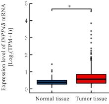
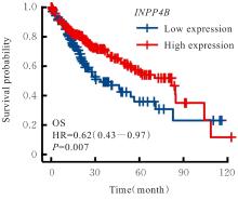
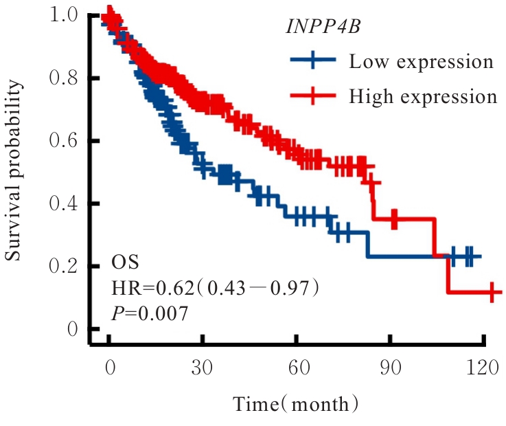
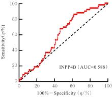
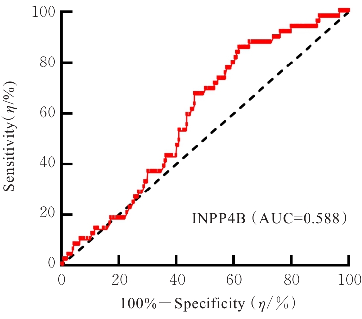
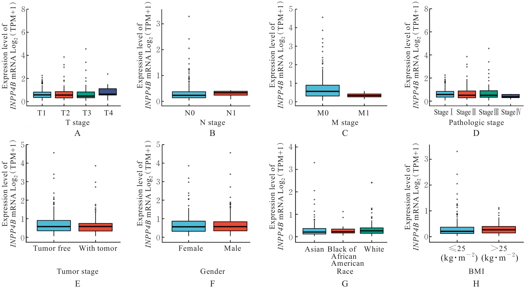
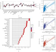
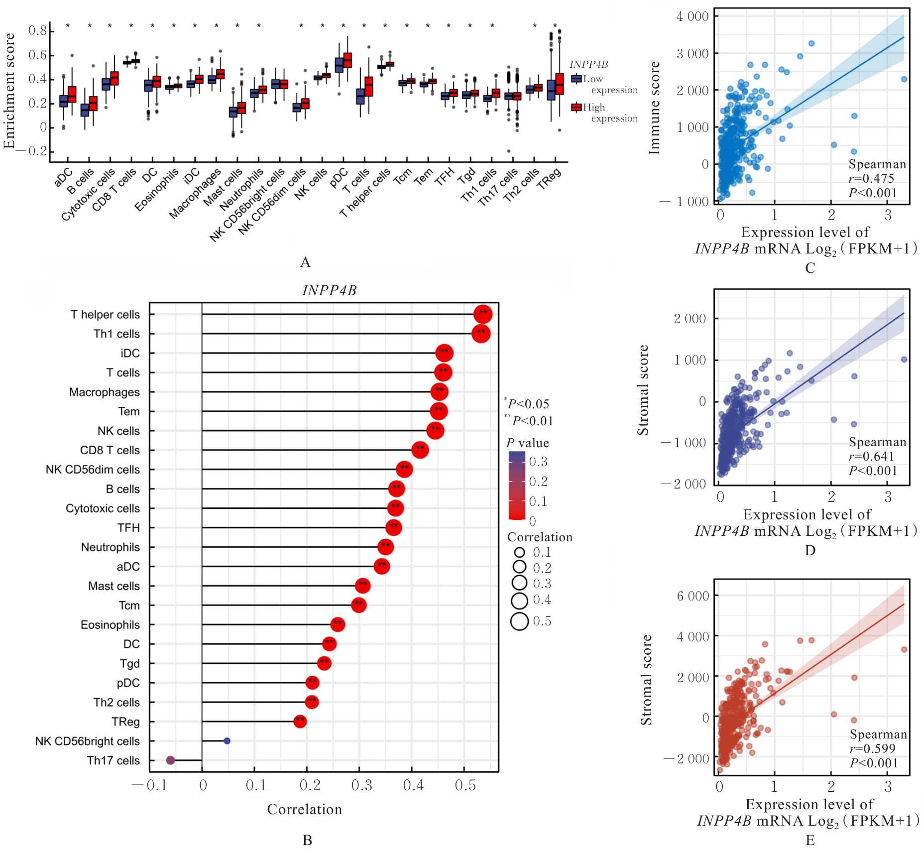
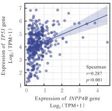
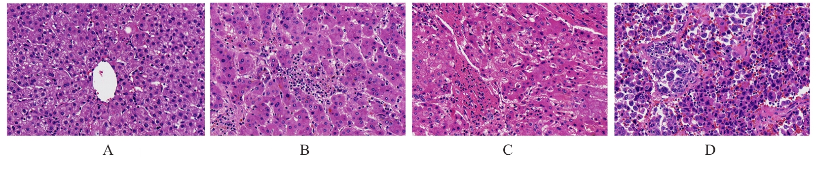
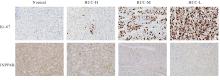
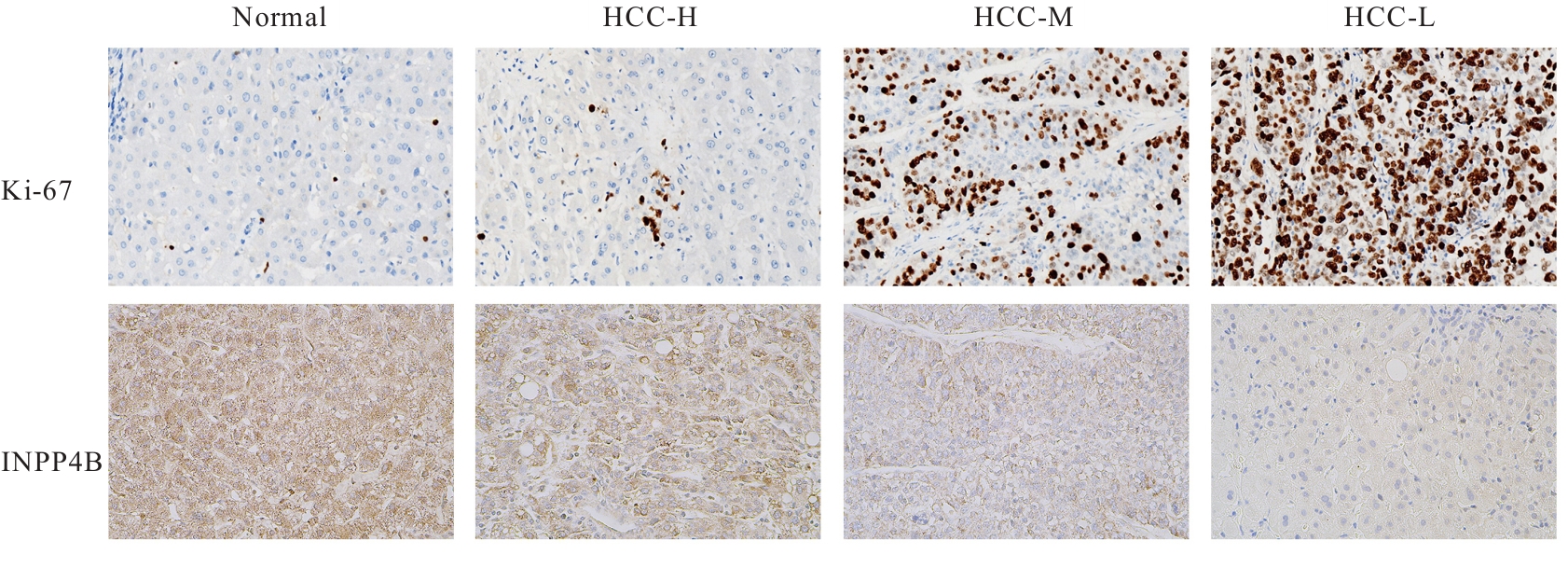
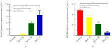
目的 基于癌症基因组图谱(TCGA)数据库和临床样本的实验验证探讨肝细胞癌(HCC)中肌醇多聚磷酸-4-磷酸酶Ⅱ型(INPP4B)基因的表达及临床意义。 方法 基于TCGA数据库中的424例临床样本数据资料(包括HCC组织374例,癌旁组织50例),采用Kaplan-Meier法和Cox回归分析,评估INPP4B基因与HCC患者临床特征及生存预后的关系。分析INPP4B基因与24种免疫细胞数量,肿瘤组织中的基质、肿瘤组织中的免疫细胞浸润和肿瘤纯度,HCC高频突变基因肿瘤蛋白53(TP53)基因表达水平的相关性。收集2022年12月—2023年12月进行手术切除治疗的60例HCC患者临床病理资料及其组织石蜡切片,根据临床诊断分为低分化组(HCC-L组)、中分化组(HCC-M组)和高分化组(HCC-H组),每组20例,选取同时期20例取活检并病理诊断为非肿瘤患者的临床病理资料及其肝组织石蜡切片,作为正常组。HE染色观察各组研究对象HCC组织和正常肝组织病理形态表现,免疫组织化学法检测各组研究对象HCC组织和正常肝组织中Ki-67和INPP4B蛋白表达情况。 结果 与正常组织比较,HCC组织中INPP4B mRNA表达水平明显升高(P<0.01)。与INPP4B低表达组比较,INPP4B高表达组患者的总生存期(OS)均明显延长(P<0.05)。单因素Cox回归分析,肿瘤分期、病理分期、肿瘤状况和残余肿瘤对HCC患者OS存在影响(P<0.05)。单因素回归分析,INPP4B预后风险模型评分危险比(HR)=0.781,95%置信区间(CI):0.552~1.105,P=0.168。INPP4B对HCC患者OS影响的受试者工作特征(ROC)曲线下面积(AUC)值为0.558,表明INPP4B基因预后风险模型在HCC患者生存预后方面具有一定的预测价值。INPP4B mRNA表达水平与TNM分期、Stage分期、患者性别、年龄、种族和体质量指数(BMI)值均无相关关系(P>0.05)。高表达和低表达INPP4B的肿瘤组织中,22种免疫细胞存在统计学差异(P<0.05);INPP4B mRNA表达水平与除辅助性T细胞(Th)17外的23种免疫细胞的数量均呈正相关关系(r>0),其中除自然杀伤细胞(NK)CD56+细胞外,均具有统计学意义(P<0.01);INPP4B与肿瘤组织中的基质(r=0.475)、肿瘤组织中的免疫细胞浸润(r=0.641)和肿瘤纯度(r=0.599)均具有显著的相关性(P<0.01)。INPP4BTP53存在相关性(r=0.287,P<0.01)。HE染色,正常组研究对象肝组织中可见肝小叶结构清晰、完整,细胞排列整齐,轻微炎性细胞浸润;HCC-L组、HCC-M组和HCC-H组患者HCC组织中可见肝小叶被完全破坏,肝细胞脂肪变性明显,炎性细胞大量浸润,部分细胞出现气球样变、小细胞性增生的病变,且HCC分化程度越低,组织破坏程度越严重;免疫组织化学法,与正常组比较,HCC-L组、HCC-M组和HCC-H组患者HCC组织中Ki-67蛋白表达水平均明显升高(P<0.01),且HCC组患者的分化程度越低,Ki-67阳性率越高。在正常组研究对象肝组织中可观察到细胞中棕褐色颗粒平均分布,INPP4B蛋白呈高表达;与正常组比较,HCC-L组、HCC-M组和HCC-H组患者HCC组织中INPP4B蛋白表达水平均明显降低(P<0.01),且HCC组织分化程度越低,INPP4B阳性率越低。 结论 INPP4B是HCC患者预后的保护因素,INPP4B作为一种新的抑癌基因,可能成为治疗HCC新药筛选的潜在靶标。

关键词: 肝细胞癌, 肌醇多聚磷酸-4-磷酸酶Ⅱ型基因, 癌症基因组图谱数据库, 磷脂酰肌醇信号系统, 免疫细胞

Abstract:

Objective To discuss the expression and clinical significance of inositol polyphosphate-4-phosphatase type Ⅱ (INPP4B) gene in hepatocellular carcinoma (HCC) based on The Cancer Genome Atlas (TCGA) database and experimental verification with clinical samples. Methods Based on data from 424 clinical samples in the TCGA database (including 374 HCC tissues and 50 paracarcinoma tissues), Kaplan-Meier method and Cox regression analysis were used to evaluate the relationship between INPP4B gene and the clinical characteristics and survival prognosis of the HCC patients. The correlations between INPP4B gene and the number of 24 types of immune cells, matrix, immune cell infiltration and tumor purity in tumor tissue, and the expression level of the high-frequency mutant gene tumor protein 53(TP53) in HCC were analyzed. The clinicopathological data and paraffin-embedded tissue sections of 60 HCC patients treated with surgical resection from December 2022 to December 2023 were collected. According to clinical diagnosis, they were divided into poorly differentiated group (HCC-L group), moderately differentiated group (HCC-M group) and well-differentiated group (HCC-H group), with 20 cases in each group; 20 patients during the same period who underwent biopsy and were pathologically diagnosed as non-tumor were selected as normal group, and their clinicopathologic data and liver tissue paraffin sections were collected. HE staining was used to observe the pathomorphology of HCC tissue and normal liver tissue of the subjects in various groups; immunohistochemistry method was used to detect the expressions of Ki-67 and INPP4B proteins in the HCC tissue and normal liver tissue of the subjects in various groups. Results The TCGA database analysis results showed that compared with normal tissue, the expression level of INPP4B mRNA in HCC tissue was significantly increased (P<0.01). Compared with INPP4B low expression group, the overall survival(OS) of the patients in INPP4B high expression group was significantly prolonged (P<0.05). The univariate Cox regression analysis results showed that tumor stage, pathological stage, tumor status and residual tumor had impacts on OS of the HCC patients (P<0.05). The univariate regression analysis results showed that the INPP4B prognostic risk model score ratio was HR=0.781, 95% confidence interval(CI): 0.552-1.105, P=0.168. The AUC value for the impact of INPP4B on OS of the HCC patients was 0.558, indicating that the INPP4B gene prognostic risk model had certain predictive value in survival prognosis. The INPP4B mRNA expression level was not correlated with TNM stage, stage, patient gender, age, race or body mass index (BMI) (P>0.05). In tumor tissue with high and low INPP4B expression, 22 types of immune cells showed statistically significant differences (P<0.05); the INPP4B mRNA expression level was positively correlated with the number of 23 types of immune cells except T helper (Th) 17 cells (r>0), among which all Th cells except natural killer (NK) CD56+ cells were statistically significant (P<0.01); INPP4B was significantly correlated with matrix (r=0.475), immune cell infiltration (r=0.641) and tumor purity (r=0.599) in tumor tissue (P<0.01). INPP4B was correlated with TP53r=0.287, P<0.01). The HE staining results showed that clear and complete lobular structure, neatly arranged cells and slight inflammatory cell infiltration were observed in liver tissue of the subjects in normal group; completely destroyed lobular structure, significant hepatocellular steatosis, massive inflammatory cell infiltration, and lesions such as ballooning degeneration and small cell hyperplasia in some cells were observed in HCC tissue of the patients in HCC-L, HCC-M and HCC-H groups, and the lower the HCC differentiation degree, the more severe the tissue destruction; The immunohistochemistry results showed that compared with normal group, the expression levels of Ki-67 protein in HCC tissue of the patients in HCC-L, HCC-M and HCC-H groups were significantly increased (P<0.01), and the lower the differentiation degree of the HCC patients, the higher the Ki-67 positive rate. Brownish-yellow granules evenly distributed in the cells and INPP4B protein was highly expressed in liver tissue of the subjects in normal group; compared with normal group, the expression levels of INPP4B protein in HCC tissue of the patients in HCC-L, HCC-M and HCC-H groups were significantly decreased (P<0.01), and the lower the differentiation degree of the HCC tissue, the lower the INPP4B positive rate. Conclusion INPP4B is a protective factor for the prognosis of HCC patients; as a new tumor suppressor gene, INPP4B may become a potential target for new drug screening in HCC treatment.

Key words: Hepatocellular carcinoma, Inositol polyphosphate-4-phosphatase type Ⅱ gene, The Cancer Genome Atlas database, Phosphatidylinositol signaling system, Immune cells

中图分类号: 

  • R735.7