吉林大学学报(医学版) ›› 2026, Vol. 52 ›› Issue (1): 182-191.doi: 10.13481/j.1671-587X.20260119

• 基础研究 • 上一篇    下一篇

蟛蜞菊内酯对人胰腺癌PANC-1细胞铜死亡的诱导作用

李雨欣1,2,杨露2,李凤金2(),齐玲2()   

  1. 1.大理大学基础医学院病原生物学综合实验室,云南 大理 671000
    2.广州医科大学附属清远医院消化内科,广东 清远 511500
  • 收稿日期:2025-04-15 接受日期:2025-05-31 出版日期:2026-01-28 发布日期:2026-02-24
  • 通讯作者: 李凤金,齐玲 E-mail:wklifengjin@163.com;qiling1718@gzhmu.edu.cn
  • 作者简介:李雨欣(2000-),女,湖南省娄底市人,在读硕士研究生,主要从事胰腺炎和胰腺纤维化发病机制及治疗方面的研究。
  • 基金资助:
    广东省中医药管理局科研项目(20231412);广东省中医药管理局科研项目(20251478);广东省卫健委医学科学技术研究基金项目(A2022163);广东省卫健委医学科学技术研究基金项目(A2024690)

Inductive effect of wedelolactone on cuproptosis in human pancreatic cancer PANC-1 cells

Yuxin LI1,2,Lu YANG2,Fengjin LI2(),Ling QI2()   

  1. 1.Comprehensive Pathogen Biology Laboratory,School of Basic Medical Sciences,Dali University,Dali 671000,China
    2.Department of Gastroenterology,Affiliated Qingyuan Hospital,Guangzhou Medical University,Qingyuan 511500,China
  • Received:2025-04-15 Accepted:2025-05-31 Online:2026-01-28 Published:2026-02-24
  • Contact: Fengjin LI,Ling QI E-mail:wklifengjin@163.com;qiling1718@gzhmu.edu.cn

摘要:

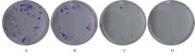
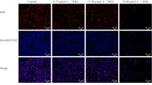
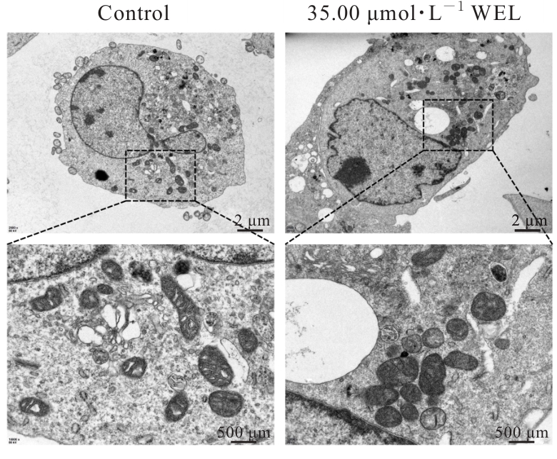
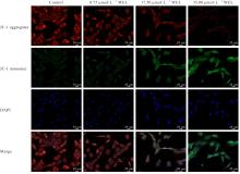
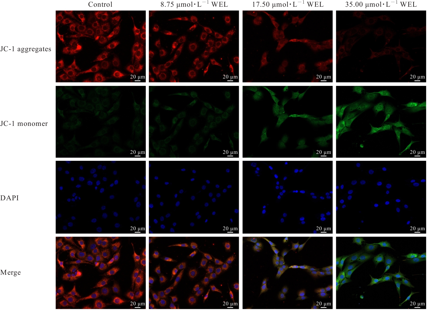
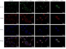
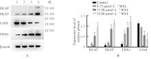
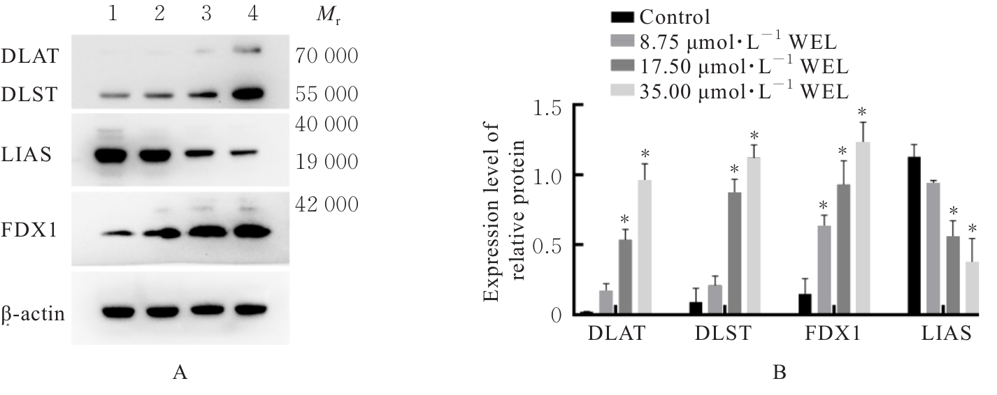
目的 探讨蟛蜞菊内酯(WEL)对人胰腺癌细胞(PANC-1)铜死亡的诱导作用,并阐明其分子机制。 方法 不同浓度(0~300 μmol·L-1)WEL分别处理PANC-1细胞12、24和48 h后,采用细胞计数试剂盒8(CCK-8)法检测不同浓度WEL作用下细胞存活率,确定后续实验用药浓度和作用时间。人胰腺癌PANC-1细胞分为对照组(0 μmol·L-1 WEL)、8.75 μmol·L-1 WEL组、17.50 μmol·L-1 WEL组和35.00 μmol·L-1 WEL组;采用克隆形成实验检测各组PANC-1细胞克隆形成率,5-乙基-2'-脱氧尿嘧啶核苷(EdU)染色法检测各组细胞EdU阳性细胞率,乳酸脱氢酶(LDH)试剂盒检测各组细胞上清液中LDH释放量;利用凋亡抑制剂含半胱氨酸的天冬氨酸蛋白酶(Caspase)抑制剂(Z-VAD-FMK)、铜死亡抑制剂四硫钼酸盐(TTM)、铁死亡抑制剂铁死亡抑素1(Fer-1)和坏死性凋亡抑制剂坏死抑制因子1(Nec-1)与35.00 μmol·L-1 WEL作用PANC-1细胞48 h后,采用CCK-8法检测不同抑制剂作用下的细胞存活率,筛选WEL诱导PANC-1细胞的死亡方式;采用细胞铜(Cu2+)比色法测试盒检测各组细胞内Cu2+水平,透射电镜观察各组PANC-1细胞线粒体超微结构,线粒体膜电位检测试剂盒(JC-1)检测各组细胞线粒体膜电位,免疫荧光染色检测各组细胞中抗二氢硫辛酰胺S-乙酰转移酶(DLAT)表达及线粒体共定位情况,Western blotting法检测各组细胞中铁氧还原蛋白1(FDX1)、硫辛酰合酶(LIAS)、DLAT和二氢硫辛酰胺S-琥珀酰基转移酶(DLST)蛋白表达水平。 结果 CCK-8法,与对照组比较,不同浓度WEL作用PANC-1细胞12、24和48 h后,细胞存活率明显降低(P<0.05),作用48 h时抑制效果最显著,因此选择0、8.75、17.50和35.00 μmol·L-1 WEL作用PANC-1细胞。克隆形成实验,与对照组比较,8.75、17.50和35.00 μmol·L-1 WEL组PANC-1细胞中克隆形成率明显降低(P<0.01)。EdU实验,与对照组比较,8.75、17.50和35.00 μmol·L-1 WEL组PANC-1细胞中EdU阳性细胞率明显降低(P<0.01)。LDH实验,与对照组比较,8.75、17.50和35.00 μmol·L-1 WEL组PANC-1细胞上清液中LDH释放量明显升高(P<0.01)。细胞Cu2+比色法,与对照组比较,35.00 μmol·L-1 WEL组PANC-1细胞Cu2+水平明显升高(P<0.01)。抑制剂干预实验,与对照组比较,35.00 μmol·L-1 WEL组细胞存活率明显升高(P<0.01);与35.00 μmol·L-1 WEL组比较,WEL+Z-VAD-FMK组和WEL+TTM组PANC-1细胞存活率升高(P<0.01)。透射电镜,35.00 μmol·L-1 WEL组PANC-1细胞线粒体膜破裂、嵴数量减少且排列稀疏。JC-1染色,与对照组比较,17.50和35.00 μmol·L-1 WEL组PANC-1细胞中线粒体膜电位明显降低(P<0.01)。免疫荧光染色,与对照组比较,8.75、17.50和35.00 μmol·L-1 WEL组PANC-1细胞中DLAT荧光强度明显增加(P<0.01),并与线粒体存在共定位。Western blotting法,与对照组比较,8.75 μmol·L-1 WEL组PANC-1细胞中FDX1蛋白表达水平明显升高(P<0.01),17.50和35.00 μmol·L-1 WEL组PANC-1细胞中DLAT、DLST及FDX1蛋白表达水平明显升高(P<0.01),而LIAS蛋白表达水平明显降低(P<0.01)。 结论 WEL能够诱导PANC-1细胞发生铜死亡,其作用机制可能与其升高PANC-1细胞中Cu2+水平且上调铜死亡关键蛋白DLAT、DLST和FDX1蛋白表达水平有关。

关键词: 蟛蜞菊内酯, 胰腺肿瘤, 铜死亡, 线粒体, 二氢硫辛酰胺S-乙酰转移酶

Abstract:

Objective To discuss the induction effect of wedelolactone (WEL) on cuproptosis in the human pancreatic cancer cells (PANC-1), and to clarify its molecular mechanism. Methods The PANC-1 cells were treated with different concentrations (0-300 μmol·L-1) of WEL for 12, 24, and 48 h, respectively, cell counting kit-8 (CCK-8) method was used to detect the survival rates of the cells after treated with different concentrations of WEL to determine the drug concentration and action time for subsequent experiments. The human pancreatic cancer PANC-1 cells were divided into control group (0 μmol·L-1 WEL), 8.75 μmol·L-1 WEL group, 17.50 μmol·L-1 WEL group, and 35.00 μmol·L-1 WEL group. Colony formation assay was used to detect the colony formation rates of the PANC-1 cells in various groups; 5-ethynyl-2'-deoxyuridine (EdU) staining was used to detect the EdU positive cell rates of the cells in various groups; lactate dehydrogenase (LDH) kit was used to detect the LDH release in supernatant of the cells in various groups. After treating PANC-1 cells with caspase inhibitor (Z-VAD-FMK), cuproptosis inhibitor tetrathiomolybdate (TTM), ferroptosis inhibitor ferrostatin-1 (Fer-1), and necroptosis inhibitor necrostatin-1 (Nec-1) combined with 35.00 μmol·L-1 WEL for 48 h, CCK-8 method was used to detect the cell survival rates of the PANC-1 cells after treated with different inhibitors, and to screen the death mode induced by WEL; cell copper (Cu2?) colorimetric assay kit was used to detect the intracellular Cu2? levels of the cells in various groups; transmission electron microscope was used to observe the mitochondrial ultrastructure of the PANC-1 cells in various groups; mitochondrial membrane potential assay kit (JC-1) was used to detect the mitochondrial membrane potential of the cells in various groups; immunofluorescence staining was used to detect the expression and mitochondrial co-localization of dihydrolipoamide S-acetyltransferase (DLAT) in the cells in various groups; Western blotting method was used to detect the expression levels of ferredoxin 1 (FDX1), lipoic acid synthetase (LIAS), DLAT, and dihydrolipoamide S-succinyltransferase (DLST) proteins in the cells in various groups. Results The CCK-8 assay results showed that compared with control group, the survival rates of the PANC-1 cells after treated with different concentrations of WEL for 12, 24, and 48 h were significantly decreased (P<0.05), with the most significant inhibitory effect at 48 h; therefore, 0, 8.75, 17.50, and 35.00 μmol·L-1 WEL were selected to treat the PANC-1 cells. The colony formation assay results showed that compared with control group, the colony formation rates of the PANC-1 cells in 8.75, 17.50, and 35.00 μmol·L-1 WEL groups were decreased (P<0.01). The EdU assay results showed that compared with control group, the EdU positive cell rates of the PANC-1 cells in 8.75, 17.50, and 35.00 μmol·L-1 WEL groups were decreased (P<0.01). The LDH assay results showed that compared with control group, the LDH release in supernatant of PANC-1 cells in 8.75, 17.50, and 35.00 μmol·L-1 WEL groups was increased (P<0.01). The cell Cu2? colorimetric assay kit results showed that compared with control group, the Cu2? levels in the PANC-1 cells in 8.75, 17.50, and 35.00 μmol·L?1 WEL groups were increased (P<0.01). The inhibitor intervention assay results showed that compared with control group, the survival rate of the PANC-1 cells in 35.00 μmol·L?1 group was decreased(P<0.01); compared with 35.00 μmol·L?1 WEL group, the survival rates of the PANC-1 cells in WEL+Z-VAD-FMK group and WEL+TTM group were increased (P<0.01). The transmission electron microscope results showed that the mitochondria in the PANC-1 cells in 35.00 μmol·L-1 WEL group exhibited membrane rupture, reduced number of cristae, and sparse arrangement. The JC-1 staining results showed that compared with control group, the mitochondrial membrane potential in the PANC-1 cells in 17.50 and 35.00 μmol·L-1 WEL groups was significantly decreased (P<0.01). The immunofluorescence staining results showed that compared with control group, the DLAT fluorescence intensity in the PANC-1 cells in 17.50 and 35.00 μmol·L-1 WEL groups was significantly increased (P<0.01) and co-localized with mitochondria. The Western blotting method results showed that compared with control group, the expression level of FDX1 protein in the PANC-1 cells in 8.75 μmol·L?1 WEL group was increased (P<0.01), the expression levels of DLAT, DLST, and FDX1 proteins in the PANC-1 cells in 17.50 and 35.00 μmol·L?1 WEL groups were significantly increased (P<0.01), while the expression level of LIAS protein was significantly decreased (P<0.01). Conclusion WEL can induce cuproptosis in the PANC-1 cells, and its mechanism may be related to increasing the Cu2? level and up-regulating the expression levels of key cuproptosis proteins DLAT, DLST, and FDX1 in the PANC-1 cells.

Key words: Wedelolactone, Pancreatic neoplasm, Cuproptosis, Mitochondria, Dihydrolipoamide S-acetyltransferase

中图分类号: 

  • R735.9