吉林大学学报(医学版) ›› 2023, Vol. 49 ›› Issue (2): 315-323.doi: 10.13481/j.1671-587X.20230207

• 基础研究 • 上一篇    下一篇

茯苓酸对人胰腺癌PANC-1细胞迁移、侵袭和上皮间质转化的抑制作用

李锐1,谭晓冬2,胡耀元1()   

  1. 1.中国医科大学附属盛京医院第十二普通外科病房,辽宁 沈阳 110004
    2.中国医科大学附属盛京 医院胰腺甲状腺外科病房,辽宁 沈阳 110004
  • 收稿日期:2022-06-02 出版日期:2023-03-28 发布日期:2023-04-24
  • 通讯作者: 胡耀元 E-mail:yyhu@cmu.edu.cn
  • 作者简介:李 锐(1990-),男,山西省大同市人,医师,医学硕士,主要从事胰腺癌侵袭转移机制及治疗方面的研究。
  • 基金资助:
    辽宁省科技厅重点研发计划项目(2020JH2/10300130)

Inhibitory effect of pachylic acid on migration, invasion, and epithelial-mesenchymal transition of human pancreatic cancer PANC-1 cells

Rui LI1,Xiaodong TAN2,Yaoyuan HU1()   

  1. 1.Department of General Surgery Ward,Affiliated Shengjing Hospital,China Medical University,Shenyang 110004,China
    2.Department of Pancreatic Thyoid Surgery Ward,Affiliated Shengjing Hospital,China Medical University,Shenyang 110004,China
  • Received:2022-06-02 Online:2023-03-28 Published:2023-04-24
  • Contact: Yaoyuan HU E-mail:yyhu@cmu.edu.cn

摘要:

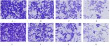
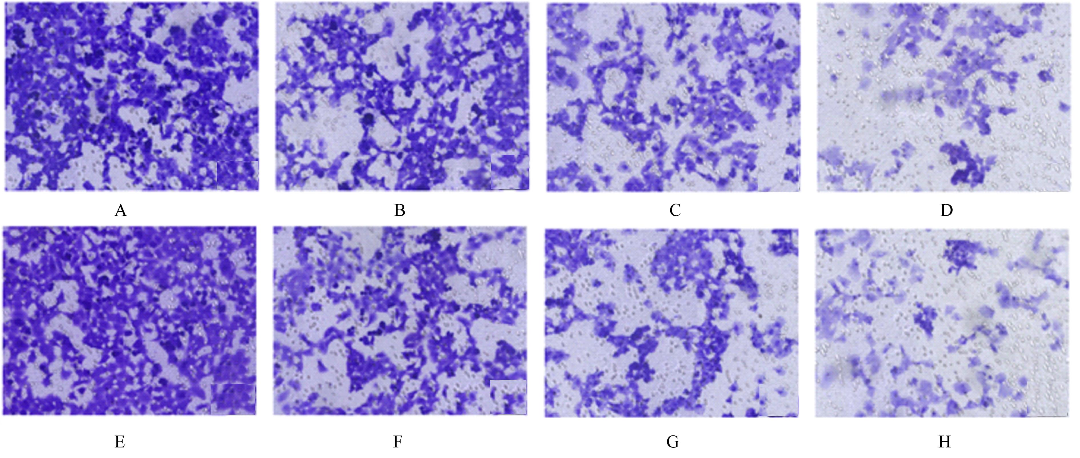
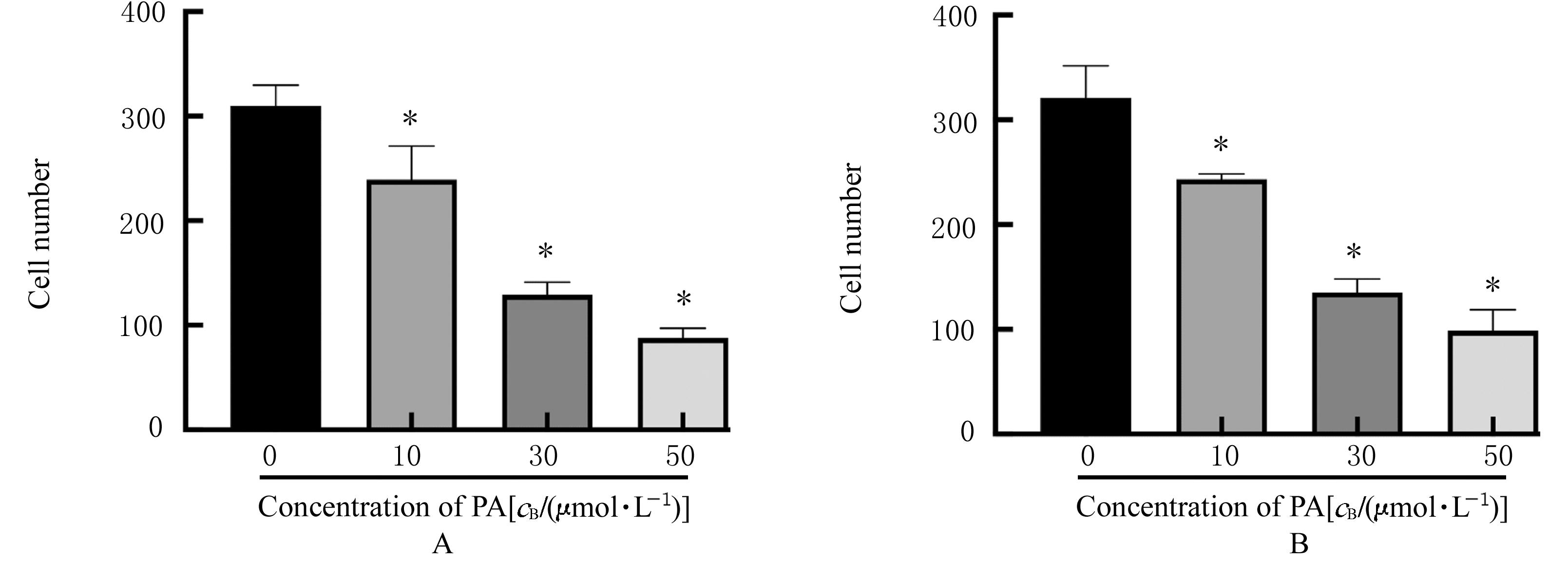
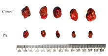
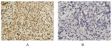
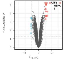
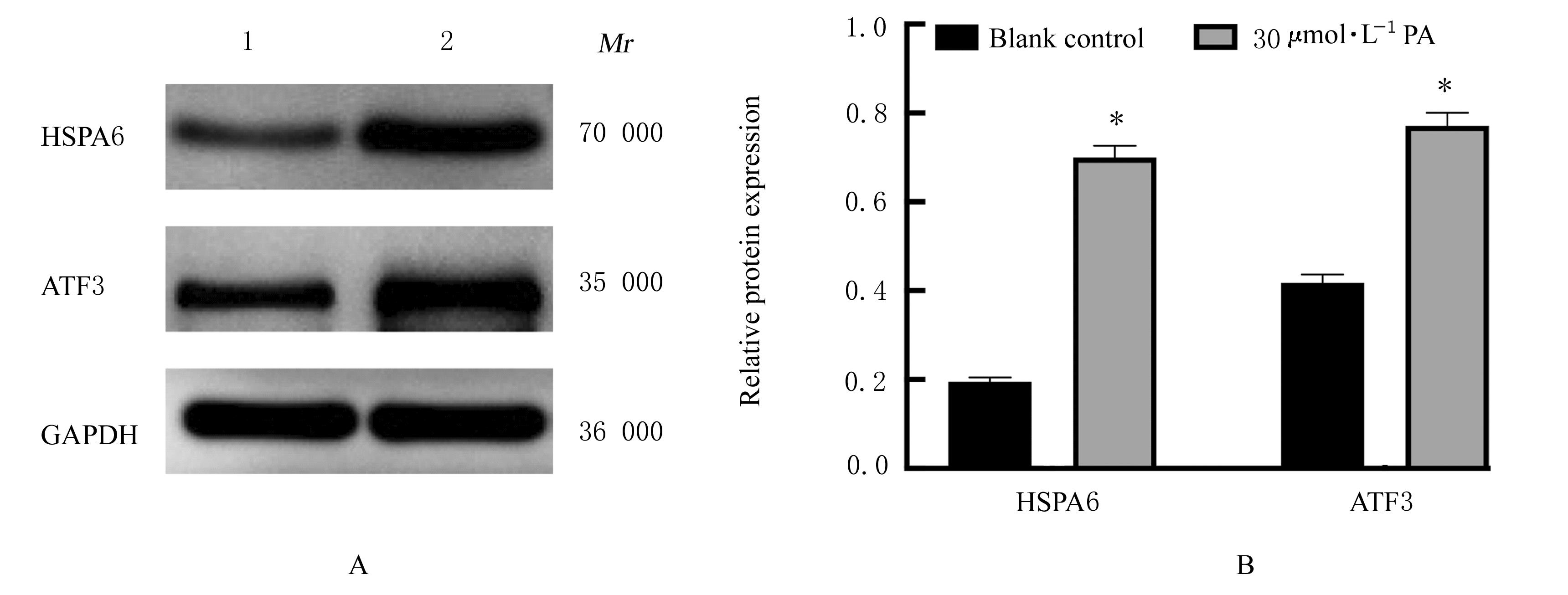
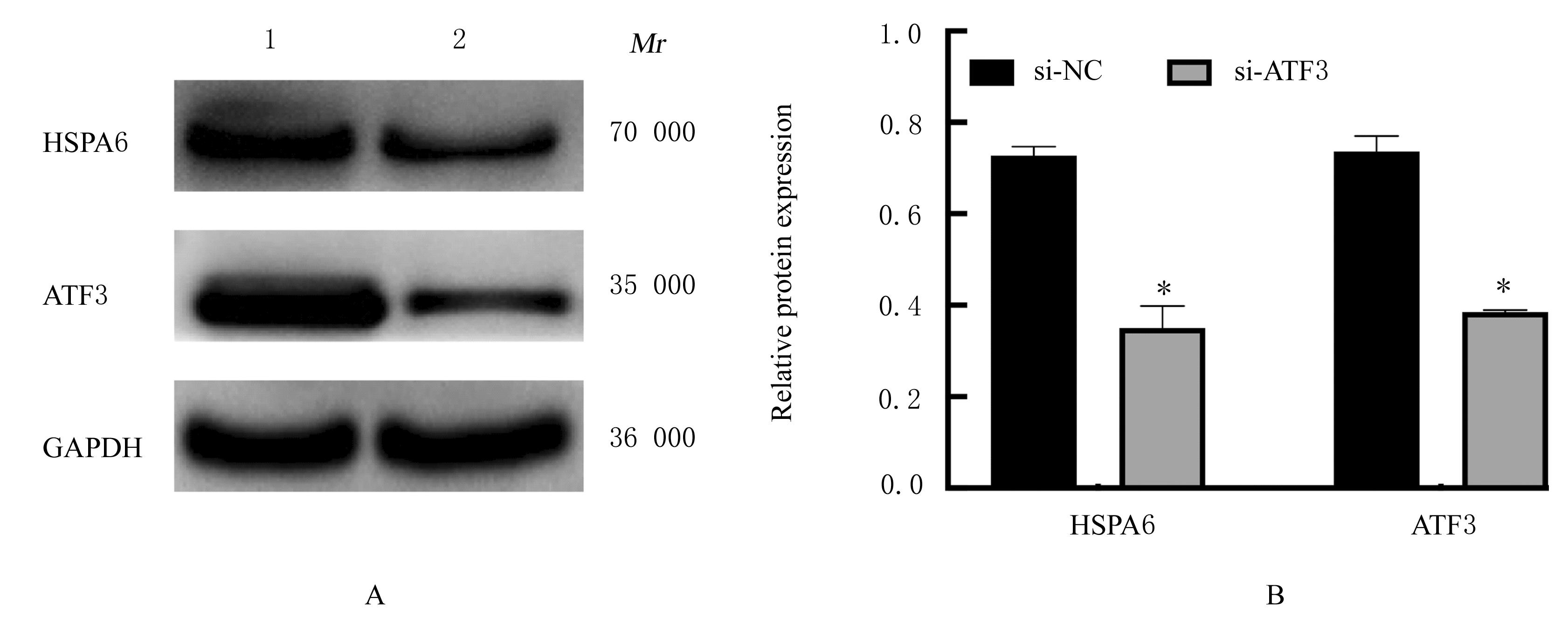
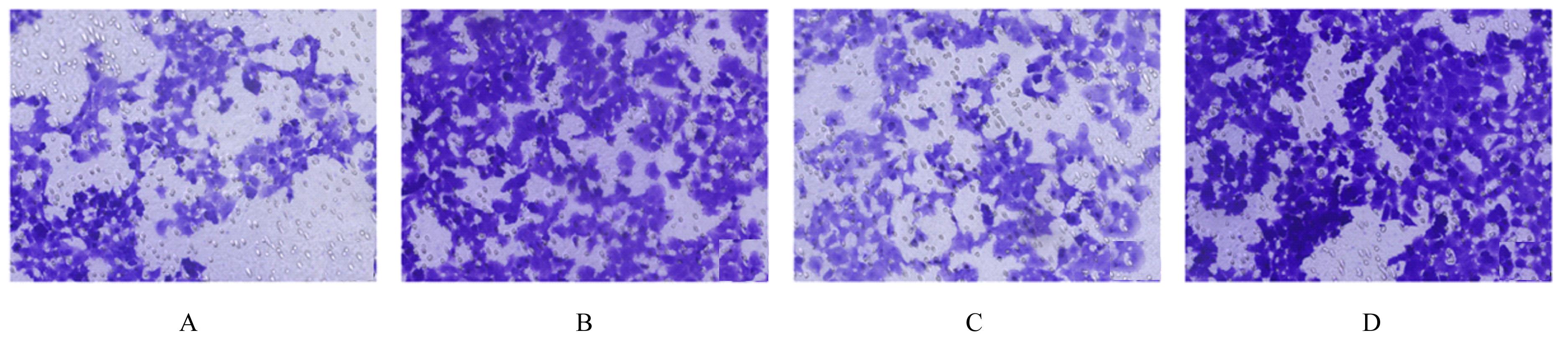
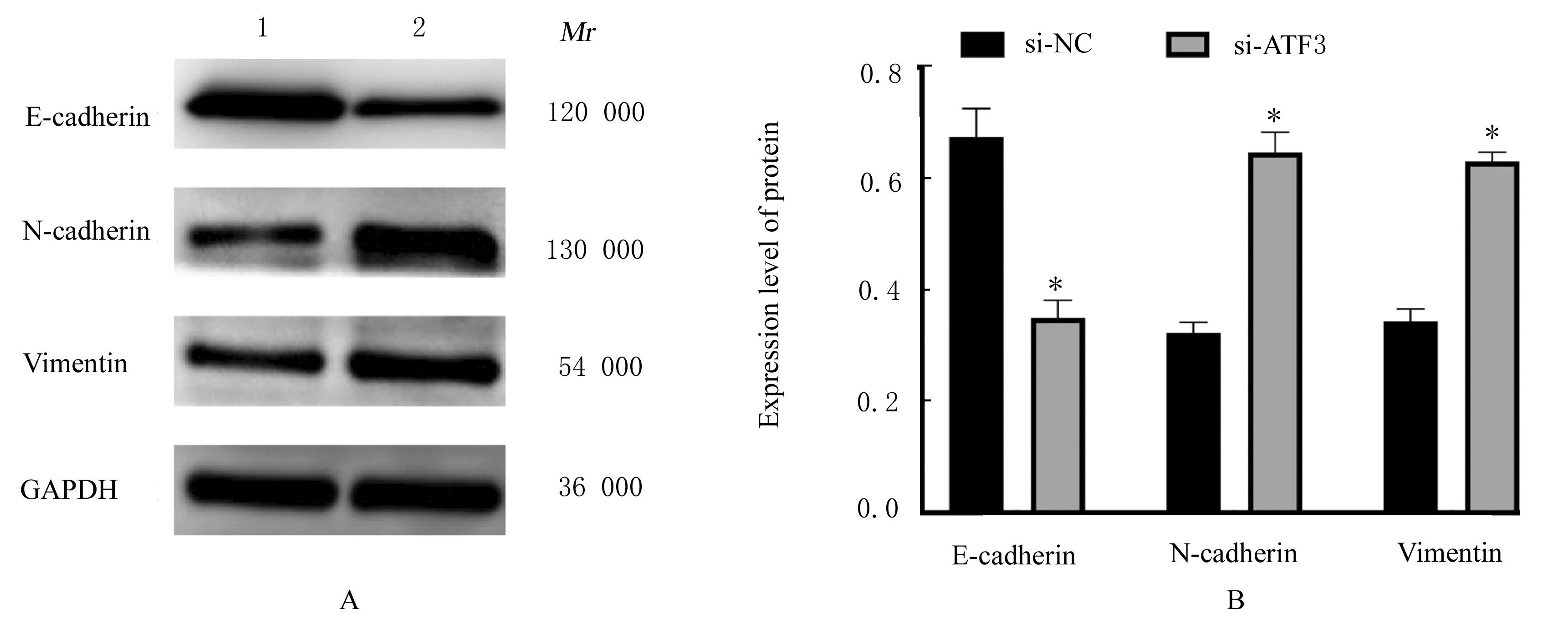
目的 探讨茯苓酸(PA)通过上调活化转录因子3(ATF3)和热休克蛋白家族A成员6(HSPA6)表达对胰腺癌PANC-1细胞迁移、侵袭和上皮间质转化(EMT)的作用,并阐明其可能的作用机制。 方法 胰腺癌PANC-1细胞分为空白对照组和不同浓度(2、5、10、20、30、40和50 μmol·L-1)PA处理组,采用CCK-8法检测各组PANC-1细胞的细胞活性。不同浓度(0、10、30和50 μmol·L-1 )PA作用于PANC-1细胞,采用Transwell小室实验检测PANC-1细胞迁移和侵袭能力,Western blotting法检测PANC-1细胞中EMT相关蛋白表达水平。将10只BALB/c nude裸鼠随机分为对照组和PA组,每组5只,裸鼠皮下注射PANC-1细胞,待肿瘤体积达到60 mm3时,PA组裸鼠腹腔注射25 mg·kg-1 PA,对照组裸鼠注射等量生理盐水,测量肿瘤体积和瘤质量,免疫组织化学法检测各组裸鼠移植瘤组织中Ki-67表达情况。通过GEO2R软件分析GSE64111数据集中PA处理及未处理胰腺癌细胞的差异表达基因。不同浓度(0和30 μmol·L-1 )PA作用于PANC-1细胞,采用Western blotting法检测PANC-1细胞中HSPA6和ATF3蛋白表达水平。将30 μmol·L-1 PA处理的PANC-1细胞分为si-NC组和si-ATF3组,分别转染对照siRNA和ATF3 siRNA,采用Western blotting法检测各组细胞中HSPA6和ATF3蛋白及EMT相关蛋白表达水平,Transwell小室实验检测细胞迁移和侵袭能力。 结果 CCK-8法,与空白对照组比较,不同浓度PA处理组PANC-1细胞的细胞活性呈浓度依赖性降低(P<0.05)。Transwell小室实验,与空白对照组比较,不同浓度PA处理组PANC-1细胞迁移能力和侵袭能力呈浓度依赖性降低(P<0.05)。Western blotting法,与空白对照组比较,不同浓度PA组PANC-1细胞中上皮钙黏素蛋白表达水平升高(P<0.05),神经钙黏素和波形蛋白表达水平降低(P<0.05)。裸鼠成瘤实验,与对照组比较,PA组裸鼠移植瘤体积和瘤质量明显降低(P<0.05);免疫组织化学,与对照组比较,PA组裸鼠移植瘤Ki-67染色较浅。GEO2R软件分析和Western blotting法,与空白对照组比较,PA处理组PANC-1细胞中HSPA6和ATF3蛋白表达水平明显升高(P<0.05)。Western blotting法,与si-NC组比较,si-ATF3组PANC-1细胞中HSPA6、ATF3和上皮钙黏素蛋白表达水平明显降低(P<0.05),神经钙黏素和波形蛋白表达水平明显升高(P<0.05)。Transwell小室实验,与si-NC组比较,si-ATF3组PANC-1细胞迁移和侵袭能力明显增加(P<0.05)。 结论 PA通过上调HSPA6和ATF3表达抑制胰腺癌细胞迁移、侵袭和EMT,从而发挥抗胰腺癌作用。

关键词: 茯苓酸, 胰腺肿瘤, PANC-1细胞, 上皮间质转化, 细胞迁移, 细胞侵袭, 活化转录因子3, 热休克蛋白家族A成员6

Abstract:

Objective To investigate the effect of poiaic acid (PA) on the migration, invasion, and epithelial-mesenchymal transition(EMT) of the pancreatic cancer PANC-1 cells by up regulating the expressions of activating transcription factor 3 (ATF3) and heat shock protein family A member 6(HSPA6),and to clarify its possible mechanism. Methods The pancreatic cancer PANC-1 cells were divided into blank control group and different concentrations (2,5,10,20,30,40, and 50 μmol·L-1)of PA groups,and the activities of the PANC-1 cells in various groups were detected by CCK-8 method. The PANC-1 cells were treated with different concentrations (0, 10, 30, and 50 μmol·L-1) of PA, and the migration and invasion abilities of the PANC-1 cells were detected by Transwell chamber experiment, and the expression level of EMT-related proteins in the PANC-1 cells were detected by Western blotting method. A total of 10 BALB/c nude mice were randomly divided into control group and PA group, and there were 5 mice in each group. The PANC-1 cells were subcutaneously injected into the nude mice. When the tumor volume reached 60 mm3, the nude mice in PA group were intraperitoneally injected with 25 mg·kg-1 PA, and the nude mice in control group were injected with the same volume of normal saline, the tumor volume and tumor weight of the mice in various groups were measured, and the expressions of Ki-67 in xenografted tissue of the mice in various groups were detected by immunohistochemistry. The differentially expressed genes of the PA-treated and PA-untreated pancreatic cancer cells in the GSE64111 Dataset were analyzed by GEO2R software. The PANC-1 cells were treated with different concentrations(0 and 30 μmol·L-1) of PA, and the expression levels of HSPA6 and ATF3 proteins in the PANC-1 cells were detected by Western blotting method. The PANC-1 cells treated with 30 μmol·L-1 PA were randomly divided into si-NC group and si-ATF3 group, and the cells were transfected with control siRNA and ATF3 siRNA, respectively. The expression levels of HSPA6 and ATF3 proteins and EMT-related proteins in the PANC-1 cells in various groups were detected by Western blotting method, and the migration and invasion abilities of the cells were detected by Transwell chamber experiment. Results The CCK-8 experiment results showed that compared with blank control group, the activities of the PANC-1 cells in different concentrations of PA groups were significantly decreased (P<0.05).The results of Transwell chamber experiment showed that compared with blank control group, the migration and invasion abilities of the PANC-1 cells in different concentrations of PA groups were significantly decreased (P<0.05),and showed a concentration depent manner. The Western blotting results showed that compared with blank control group, the expression levels of E-cadherin protein in the PANC-1 cells in different concentrations of PA groups were significantly increased (P<0.05), while the expression levels of N-cadherin and Vimentin proteins were significantly decreased (P<0.05).The results of tumor formation in the nude mice showed that compared with control group, the volume and weight of the xenografted of the mice in PA group were significantly decreased (P<0.05);the immunohistochemistry results showed that the Ki-67 staining in xeografted was shallow; the GEO2R software analysis and Western blotting results showed that compared with blank control group, the expression levels of HSPA6 and ATF3 proteins in the PANC-1 cells in PA group were significantly increased (P<0.05). The Western blotting results showed that compared with si-NC group, the expression levels of ATF3, HSPA6, and E-cadherin proteins in the PANC-1 cells in si-ATF3 group were significantly decreased (P<0.05), while the expression levels of N-cadherin and Vimentin proteins were significantly increased (P<0.05).The Transwell chamber experiment results showed that compared with si-NC group, the migration and invasion abilities of the PANC-1 cells in si-ATF3 group were significantly increased (P<0.05). Conclusion PA inhibits the migration, invasion, and EMT of the pancreatic cancer cells by up-regulating the expressions of HSPA6 and ATF3, thus play an anti-pancreatic cancer effect.

Key words: Pachymic acid, Pancreatic neoplasm, PANC-1 cells, Epithelial-mesenchymal transition, Cell migration, Cell invasion, Activating transcription factor 3, Heat shock protein family A member 6

中图分类号: 

  • R735.9