吉林大学学报(医学版) ›› 2023, Vol. 49 ›› Issue (2): 324-331.doi: 10.13481/j.1671-587X.20230208

• 基础研究 • 上一篇    下一篇

阿托伐他汀对人舌鳞癌CAL-27细胞增殖、凋亡和迁移的影响及其机制

王开,黄汉()   

  1. 锦州医科大学附属第一医院口腔颌面外科,辽宁 锦州 121000
  • 收稿日期:2022-05-12 出版日期:2023-03-28 发布日期:2023-04-24
  • 通讯作者: 黄汉 E-mail:wk201920743@163.com
  • 作者简介:王 开(1997-),女,辽宁省朝阳市人,在读硕士研究生,主要从事口腔颌面部肿瘤基础方面的研究。
  • 基金资助:
    辽宁省科技厅自然科学基金资助项目(20180550549)

Effects of atorvastatin on proliferation, apoptosis, and migration of human tongue squamous cell carcinoma CAL-27 cells and their mechanisms

Kai WANG,Han HUANG()   

  1. Department of Oral Maxillofacial Surgery,First Affiliated Hospital,Jinzhou Medical University,Jinzhou 121000,China
  • Received:2022-05-12 Online:2023-03-28 Published:2023-04-24
  • Contact: Han HUANG E-mail:wk201920743@163.com

摘要:

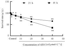
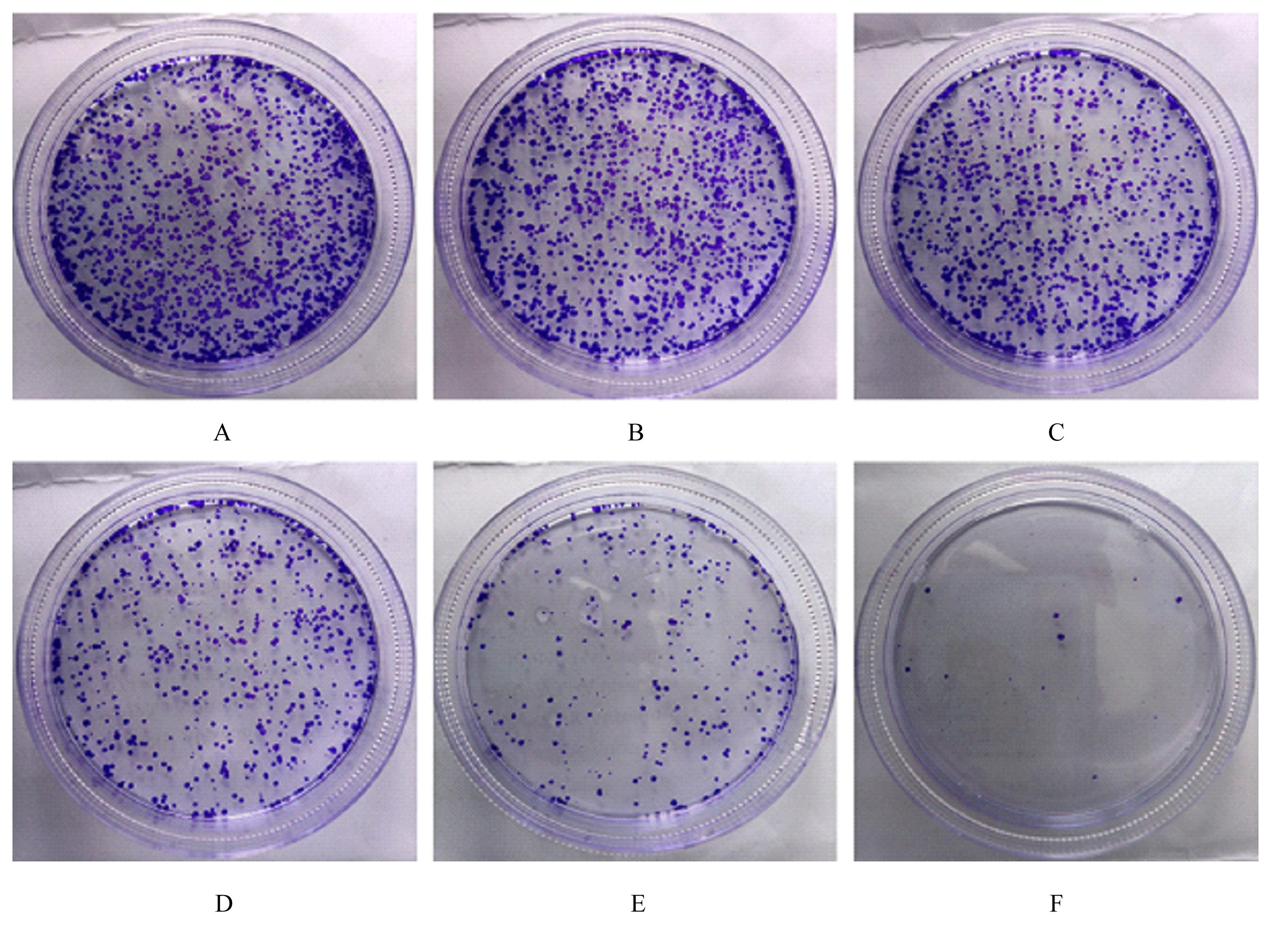
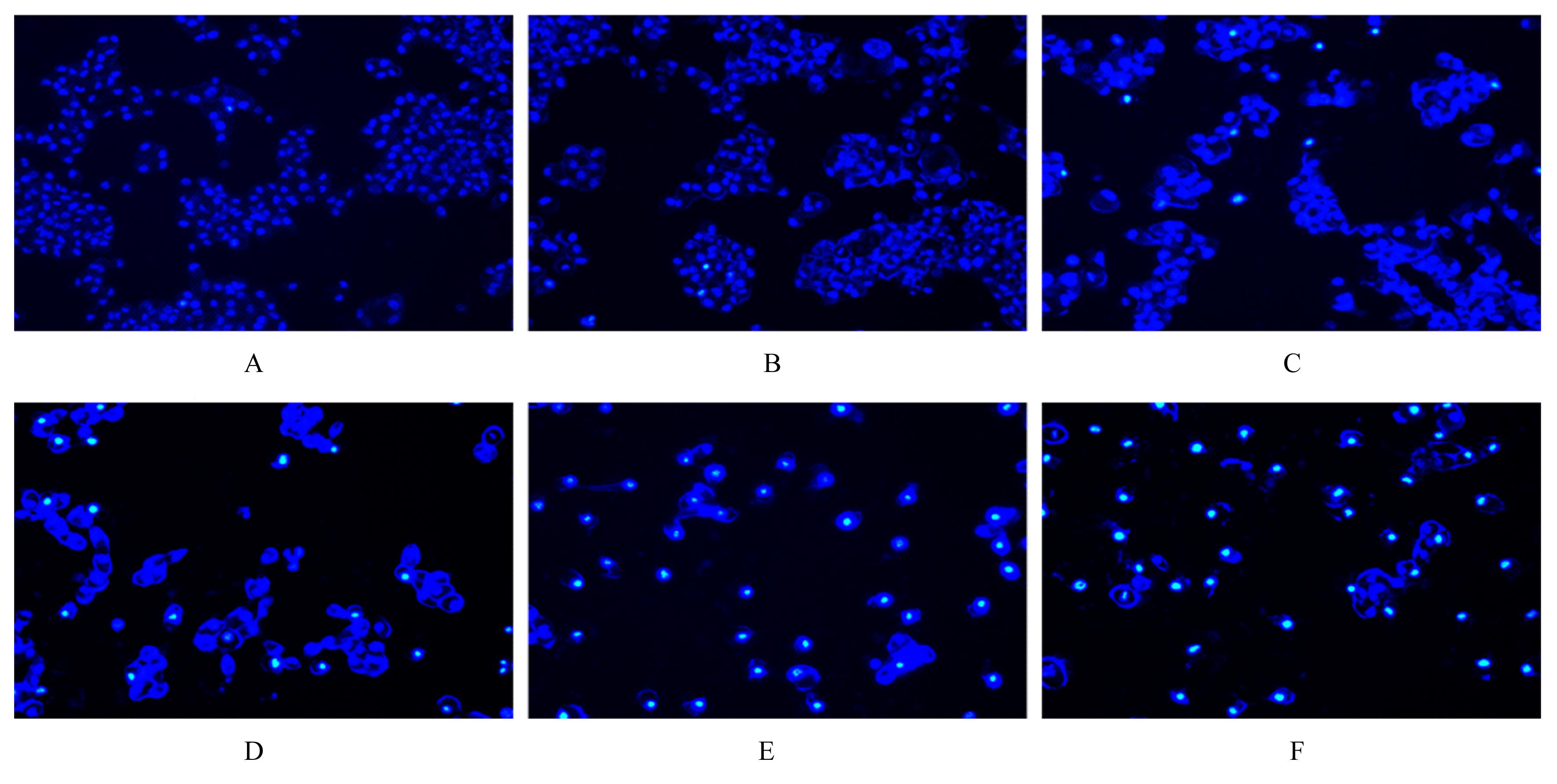
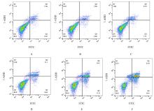
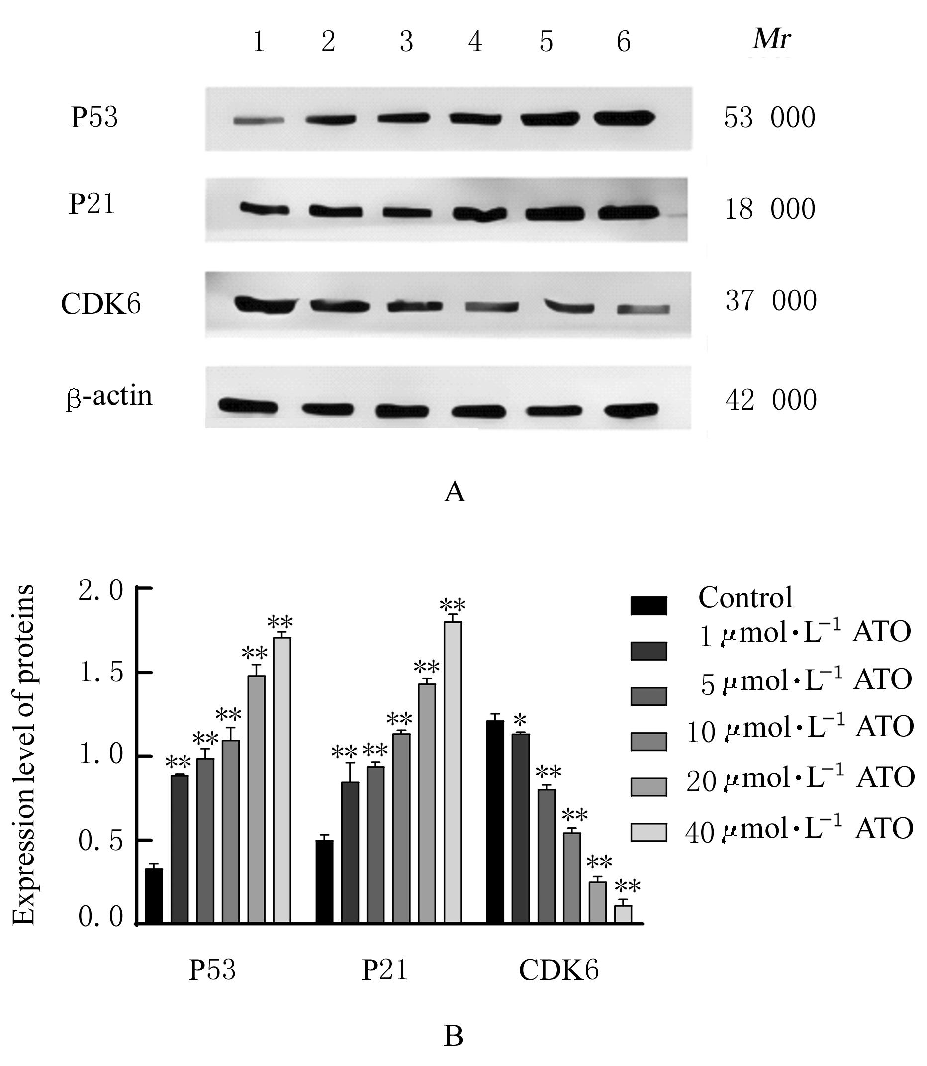
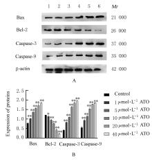
目的 探讨阿托伐他汀(ATO)对人舌鳞癌CAL-27细胞体外增殖、凋亡和迁移的影响,阐明其作用机制。 方法 CAL-27细胞分为对照组和1、5、10、20及40 μmol?L-1 ATO组。ATO作用后,CCK-8法检测各组CAL-27细胞存活率,克隆形成实验检测各组CAL-27细胞克隆形成率,Hoechst33342荧光染色和流式细胞术检测各组CAL-27细胞凋亡率,细胞划痕实验检测各组CAL-27细胞迁移率,Western blotting法检测各组CAL-27细胞中P53、P21、B细胞淋巴瘤2(Bcl-2)、Bcl-2相关X蛋白(Bax)、含半胱氨酸的天冬氨酸蛋白水解酶(Caspase)-3、Caspase-9和周期蛋白依赖性激酶6(CDK6)蛋白表达水平。 结果 培养24和48 h后,与对照组比较不同浓度ATO组细胞存活率明显降低(P<0.05)。培养48 h后,与对照组比较,不同浓度ATO组细胞克隆形成率(P<0.01)和细胞迁移率(P<0.05)明显降低,40 μmol?L-1 ATO组细胞基本丧失克隆和迁移能力;不同浓度ATO组细胞凋亡率明显升高(P<0.05),其中40 μmol?L-1 ATO组细胞几乎全部凋亡。培养48 h后,与对照组比较不同浓度ATO组细胞均出现不同程度细胞核浓染或碎片化,20和40 μmol?L-1 ATO组细胞全部呈现碎片化。与对照组比较,培养48 h后,不同浓度ATO组细胞中P53、P21、Bax、Caspase-3和Caspase-9蛋白表达水平升高(P<0.01),CDK6和Bcl-2蛋白表达水平降低(P<0.05或P<0.01)。 结论 ATO通过P53/P21/CDK6通路抑制CAL-27细胞增殖及迁移并促进凋亡。

关键词: 舌鳞状细胞癌, 阿托伐他汀, 细胞增殖, 细胞凋亡, 细胞迁移

Abstract:

Objective To investigate the effects of atorvastatin (ATO) on the proliferation, apoptosis, and migration of the human tongue squamous cell carcinoma CAL-27 cells in vitro, and to clarify their mechanisms. Methods The CAL-27 cells were divided into control group and 1,5,10,20,and 40 μmol?L-1 ATO groups.After treated with ATO,CCK-8 assay was used to detect the survival rates of the CAL-27 cells in various groups, clone formation assay was used to detect the clone formation rates of the CAL-27 cells in various groups; Hoechst33342 fluorescence staining and flow cytometry were used to detect the apoptotic rates of the CAL-27 cells in various groups; cell scratch test was used to detect the migration rates the CAL-27 cells in various groups; Western blotting method was used to detect the expression levels of P53, P21, B-cell lymphoma-2(Bcl-2),Bcl-2 associated X protein(Bax), cysteinyl aspartate specific proteinase(Caspase)-3, Caspase-9, and cyclin-dependent kinase 6(CDK6) proteins in the CAL-27 cells in various groups. Results Compared with control group, the survival rates of the cells in different concentrations of ATO groups were decreased after cutured for 24 and 48 h(P<0.05).After cultured for 48 h, compared with control group, the clone formation rates (P<0.01) and migration rates (P<0.05) of the cells in different concentrations of ATO groups were decreased,and the cells in 40 μmol?L-1 ATO group basically lost the abilities of cloning and migration; the apoptotic rates in different concentrations of ATO groups were increased (P<0.05),and almost all the cells in 40 μmol?L-1ATO group were apoptotic. After cultured for 48 h, compared with control group, the cells in different concentrations of ATO groups showed nuclear staining or fragmentation,and all the cells in 20 and 40 μmol?L-1 ATO groups were fragmented. Compared with control group, after cultured for 48 h,the expression levels of P53, P21, Bax, Caspase-3, and Caspase-9 proteins in the cells in different concentrations of ATO groups were increased (P<0.01), and the expression levels of CDK6 and Bcl-2 proteins were decreased(P<0.05 or P<0.01). Conclusion ATO inhibits the proliferation and migration of the CAL-27 cells and promotes the apoptosis of the CAL-27 cells through P53 /P21 / CDK6 pathway.

Key words: Tongue squamous cell carcinoma, Atorvastatin, Cell proliferation, Apoptosis, Cell migration

中图分类号: 

  • R739.86