吉林大学学报(医学版) ›› 2023, Vol. 49 ›› Issue (2): 508-513.doi: 10.13481/j.1671-587X.20230228

• 临床医学 • 上一篇    下一篇

腹膜后巨大淋巴管瘤1例报告及文献复习

杨胜男1,王雪1,王雪峰2,赵天宇1,潘颖1(),丁大勇2()   

  1. 1.吉林大学中日联谊医院妇产科,吉林 长春 130033
    2.吉林大学中日联谊医院胃肠外科,吉林 长春 130033
  • 收稿日期:2022-04-17 出版日期:2023-03-28 发布日期:2023-04-24
  • 通讯作者: 潘颖,丁大勇 E-mail:panying424@163.com;dingdy@jlu.edu.cn
  • 作者简介:杨胜男(1998-),女,山东省济宁市人,住院医师,医学硕士,主要从事妇科肿瘤方面的研究。
  • 基金资助:
    吉林省科技厅自然科学基金项目(20200201586JC)

Retroperitoneal giant lymphangioma: A case report and literature review

Shengnan YANG1,Xue WANG1,Xuefeng WANG2,Tianyu ZHAO1,Ying PAN1(),Dayong DING2()   

  1. 1.Department of Obstetrics and Gynecology, China-Japan Union Hospital, Changchun 130033, China
    2.Department of Gastrointestinal Surgery, China-Japan Union Hospital, Changchun 130033, China
  • Received:2022-04-17 Online:2023-03-28 Published:2023-04-24
  • Contact: Ying PAN,Dayong DING E-mail:panying424@163.com;dingdy@jlu.edu.cn

摘要:

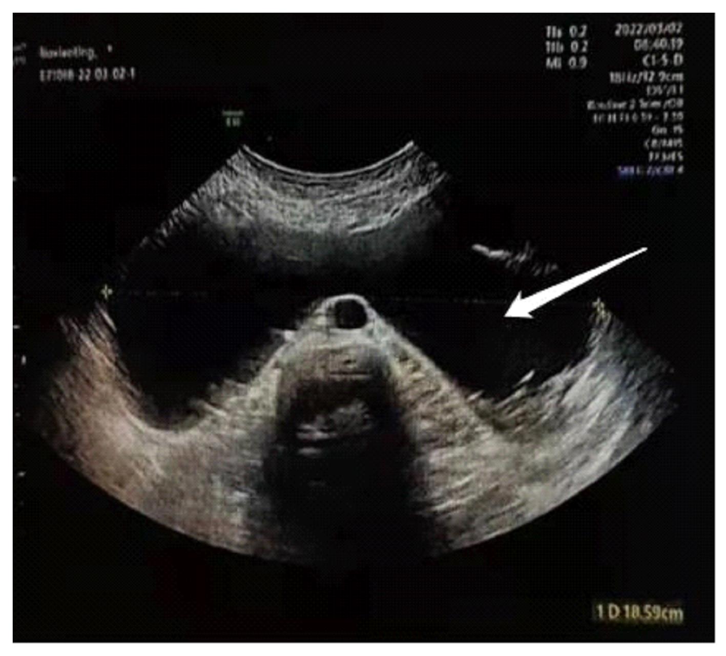
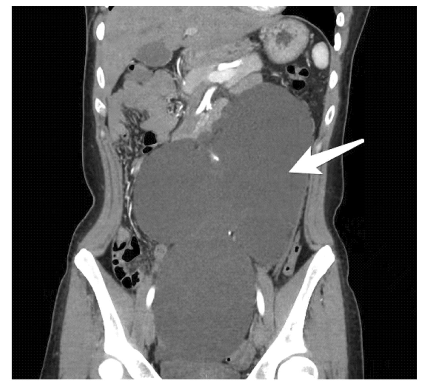
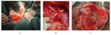
目的 分析腹膜后巨大淋巴管瘤患者的临床表现、影像学特征、治疗措施选择和术后规范化管理,提高临床医师对该病的认识。 方法 收集1例腹膜后巨大淋巴管瘤患者的临床资料,根据患者的专科查体和影像学特征明确临床诊断,并对治疗措施的选择和术后规范化管理进行分析。 结果 患者,女性,33岁,因腹胀2个月入院。专科检查,腹部略膨隆,余未见明显异常;全腹部叩诊以浊音为主,于腋中线处浊音变鼓音,移动性浊音阴性,肠鸣音正常,未闻及过水声。于腹部可触及一包块,大小约17 cm×9 cm×26 cm,质软,边界不清,无活动性,触痛阴性,听诊未闻及血管杂音。入院时妇科彩超,于子宫上方探及巨大无回声,上达剑突下,宽约18.6 cm,形态不规则,内有分隔。腹部计算机断层扫描(CT)示左侧肾盂和输尿管上段扩张,其内可见水样密度影;腹腔内可见团块状囊性占位,大小约17.9 cm×9.0 cm×26.7 cm,病灶局部可见囊壁结节影;增强扫描轻度强化,局部可见点状钙化,余未见异常。考虑腹腔内囊实性占位,不除外压迫左侧输尿管,继发左肾及左侧上段输尿管扩张积水。行腹腔肿物切除术,术中诊断为腹膜后淋巴管瘤,手术过程顺利,术后规范化管理,患者恢复良好出院。 结论 腹膜后巨大淋巴管瘤临床表现特异性较差,目前诊断腹膜后淋巴管瘤最有价值的影像学方式是CT和磁共振成像(MRI),手术治疗是首选的治疗措施。

关键词: 淋巴管瘤, 腹膜后肿瘤, 计算机断层扫描, 磁共振成像, 病例报告

Abstract:

Objective To analyze the clinical manifestation, imaging feature, treatment option and postoperative standardized management of the patient with retroperitoneal giant lymphangioma, and to improve the clinicians’ understanding of this disease. Methods The clinical data of one patient with retroperitoneal giant lymphangioma were collected.The patients was diagnosed according to the patient’s specialist examination and imaging characteristics, and the selection of treatment measures and postoperative standardized management were analyzed. Results The patient, a 33-year-old woman, was admitted to the hospital due to “abdominal distension for 2 months”. The specialist examination results showed the abdomen was slightly swelling, without other obvious abnormalities; the full abdominal percussion was mainly voiced, and the abdominal percussion changed into dulled drums at the midaxillary line,the mobile dullness was negative, the bowel sound was normal, and there were no water sound. There was a lump with the size of 17 cm×9 cm×26 cm which was soft, unclear boundary, inactive, feminine, negative for tenderness, the auscultation did not semell and vascular murmurs. The gynecological color ultrasound results showed that a large anedomic area was detected above the uterus reaching up to the lower part of the process,with a width of 18.6 cm, irregular morphology and with separations inside. The computed tomography(CT) results of the abdomen showed that the left renal pelvis and the upper segment of ureter expended,and the watery density opacity could be seen inside. The massive cystic mass can be seen in the abdominal cavity, about 17.9 cm×9.0 cm×26.7 cm, the cyst nodules shadow could be seen locally, with slight enhancement on the enhanced scan, the punctate calcification could be seen locally in the focus, and the other abnormalities were not found. The intraperitoneal cystic and space occupying in the abdominal cavity was considered, excluding the compression of the left ureter, secondary expansion and hydronephrosis of the left kidney and left upper segment of ureter.The abdminal neoplasm resection was performed, and the patient was diagnosed as retroperitoneal giant lymphangiome the surgical process was still smooth, and the postoperative standardized management was performed and the patient was discharged from the hospital. Conclusion The clinical manifestations of retroperitoneal giant lymphangioma are poor. At present, the most valuable imaging diagonsis method of retroperitoneal giant lymphangioma is CT and magnetic resonance imaging (MRI), and surgical treatment is the preferred treatment measure.

Key words: Lymphangioma, Retroperitoneal neoplasm, Computed tomography, Magnetic resonance imaging, Case report

中图分类号: 

  • R735.4