吉林大学学报(医学版) ›› 2024, Vol. 50 ›› Issue (2): 320-325.doi: 10.13481/j.1671-587X.20240204

• 基础研究 • 上一篇    

苦参总黄酮对醋酸铅诱导小鼠睾丸间质细胞氧化应激的改善作用及对Keap1/Nrf2信号通路的影响

刘玥欣1,来永巍1,王艳春1,徐博2,路倩1,安英2,任旷1,范红艳1()   

  1. 1.吉林医药学院药学院药理学教研室,吉林 吉林 132013
    2.吉林医药学院基础医学院机能实验室,吉林 吉林 132013
  • 收稿日期:2023-03-13 出版日期:2024-03-28 发布日期:2024-04-28
  • 通讯作者: 范红艳 E-mail:583888330@qq.com
  • 作者简介:刘玥欣(1991-),女,吉林省吉林市人,讲师,医学博士,主要从事神经药理学方面的研究。
  • 基金资助:
    吉林省教育厅科学技术研究项目(JJKH20210498KJ)

Improvement effect of total flavonoids of Sophora flavescens on oxidative stress induced by lead acetate in leydig cells in mice and its in effect on Keap1/Nrf2 signaling pathway

Yuexin LIU1,Yongwei LAI1,Yanchun WANG1,Bo XU2,Qian LU1,Ying AN2,Kuang REN1,Hongyan FAN1()   

  1. 1.Department of Pharmacology,College of Pharmacy,Jilin University of Medicine,Jilin 132013,China
    2.Functional Laboratory,College of Basic Medical Sciences,Jilin University of Medicine,Jilin 132013,China
  • Received:2023-03-13 Online:2024-03-28 Published:2024-04-28
  • Contact: Hongyan FAN E-mail:583888330@qq.com

摘要:

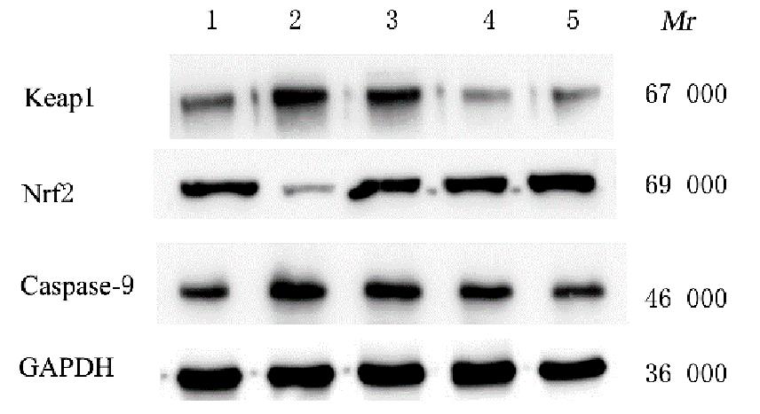
目的 观察苦参总黄酮(SF)对醋酸铅(LA)诱导的小鼠睾丸间质细胞TM3氧化应激的改善作用及其对KELCH状ECH相关蛋白1(Keap1)/细胞中核因子E2相关因子2(Nrf2)信号通路的影响,探讨SF改善由LA诱导的TM3细胞氧化应激的相关作用机制。 方法 利用噻唑蓝(MTT)法检测不同浓度(0、10、20、50和80 μmol·L-1)LA和不同浓度(0、12.5、25.0、50.0和80.0 mg·L-1)SF分别干预24 h后的TM3细胞存活率;将TM3细胞随机分为空白对照组、LA组、LA+低剂量SF组、LA+中剂量SF组和LA+高剂量SF组。除空白对照组外,其他各组均采用50 μmol·L-1 LA诱导TM3细胞24 h建立TM3细胞氧化应激模型,其中LA+低剂量SF组、LA+中剂量SF组和LA+高剂量SF组TM3细胞再分别给予12.5、25.0和50.0 mg·L-1 SF。利用MTT法检测TM3细胞存活率;WST-1法和硫代巴比妥酸(TBA)法检测TM3细胞中超氧化物歧化酶(SOD)活性和丙二醛(MDA)水平;Western blotting法检测Nrf2、Keap1和含半胱氨酸的天冬氨酸蛋白水解酶9(Caspase-9)蛋白表达水平。 结果 MTT法检测,与0 μmol·L-1 LA组比较,10、20、50和80 μmol·L-1 LA组TM3细胞存活率明显降低(P<0.01);与0 mg·L-1 SF组比较,12.5、25.0、50.0和80.0 mg·L-1 SF组TM3细胞存活率明显降低(P<0.05或P<0.01);与LA组比较,LA+低剂量SF组、LA+中剂量SF组和LA+高剂量SF组TM3细胞存活率明显升高(P<0.01); WST-1法和TBA法检测,与LA组比较,LA+低剂量SF组、LA+中剂量SF组和LA+高剂量SF组TM3细胞中SOD活性明显升高(P<0.01),MDA水平明显降低(P<0.01);Western blotting法检测,与LA组比较,LA+低剂量SF组、LA+中剂量SF组和LA+高剂量SF组TM3细胞中Nrf2蛋白表达水平明显升高(P<0.01),Keap1和Caspase-9蛋白表达水平明显降低(P<0.01)。 结论 SF对LA诱导TM3细胞氧化应激具有一定改善作用,并可降低TM3细胞中Keap1蛋白表达水平,升高TM3细胞中Nrf2蛋白表达水平。

关键词: 苦参总黄酮, 醋酸铅, 睾丸间质细胞, 氧化应激, KELCH状ECH相关蛋白1, 核因子E2相关因子2

Abstract:

Objective To observe the improvement effect of total flavonoids of Sophora flavescens (SF) on the oxidative stress in the Leydig’s cells TM3 of the mice induced by lead acetate (LA) and their influences on the Keap1/Nuclear factor E2-related factor 2 (Nrf2) signaling pathway, and to discuss the potential mechanism of the improvement effect of SF on oxidative stress in the LA-induced TM3 cells. Methods The survival rates of the TM3 cells after treated with various concentrations of LA (0, 10, 20, 50, and 80 μmol·L-1) and SF (0, 12.5, 25.0, 50.0, and 80.0 mg·L-1) for 24 h were detected by MTT assay. The TM3 cells were randomly divided into blank control group, LA group, LA + low dose of SF group, LA+middle dose of SF group, and LA+high dose of SF group. Except for blank control group, the TM3 cells in the other groups were induced with 50 μmol·L-1 LA for 24 h to establish the oxidative stress model. The TM3 cells in LA+low dose of SF group, LA+middle dose of SF group, and LA+ high dose of SF group were then treated with 12.5, 25.0, and 50.0 mg·L-1 SF, respectively. The survival rate of the TM3 cells was detected by MTT assay; the activities of superoxide dismutase (SOD) and levels of malondialdehyde (MDA) in the TM3 cells in various groups were detected by WST-1 assay and thiobarbituric acid (TBA) method; the expression levels of Nrf2, Keap1, and Caspase-9 proteins in the TM3 cells in various groups were detected by detected by Western blotting method. Results The MTT assay resuts showed that compared with 0 μmol·L-1 LA group,the survival rates of the TM3 cells in 10, 20, 50, and 80 μmol·L-1 LA groups were decreased (P<0.01); compared with 0 mg·L-1 SF group,the survival rates of the TM3 cells in 12.5, 25.0, 50.0, and 80.0 mg·L-1 SF groups were significantly decreased (P<0.05 or P<0.01); compared with LA group, the survival rates of the TM3 cells in LA+low dose of SF group, LA+middle dose of SF group, and LA+high dose of SF group were significantly increased (P<0.01). The WST-1 assay and TBA method results showed that compared with LA group,the SOD activities and MDA levels in the TM3 cells in LA+low dose of SF group, LA+middle dose of SF group, and LA+high dose of SF group were significantly decreased (P<0.01).The Western blotting results showed that compared with LA group, the expression levels of Nrf2 protein in the TM3 cells in LA+low dose of SF group, LA+middle dose of SF group, and LA+high dose of SF group were increased(P<0.01),and the expression levels of Keap1 and Caspase-9 proteins were decreased (P<0.01). Conclusion SF has certain improvement effect on the oxidative stress in the LA-induced TM3 cells and can reduce the expression level of Keap1 protein and increase the expression level of Nrf2 protein in the TM3 cells.

Key words: Sophoral flavones, Lead acetate, Leydig’s cell, Oxidative stress, Kelch-like ECH-associated protein 1, Nuclear factor erythroid 2-related factor 2

中图分类号: 

  • R96