吉林大学学报(医学版)

• •    

miR-34a-5p对颞叶癫痫大鼠海马神经元凋亡的影响及其机制

李佳芮,杨振林,高帆,郭晶晶,李今子()   

  1. 延边大学附属医院儿科,吉林 延吉 133002
  • 收稿日期:2024-11-14 接受日期:2024-12-27
  • 通讯作者: 李今子 E-mail:yjzli@ybu.edu.cn
  • 作者简介:李佳芮(1987-),女,住院医师,医学硕士,主要从事神经系统疾病基础和临床方面的研究。
  • 基金资助:
    吉林省科技厅自然科学基金项目(JJKH20240693KJ);吉林省卫健委卫生健康科技能力提升项目(2023J022)

Effect of miR-34a-5p on hippocampal neuron apoptosis in rats with temporal lobe epilepsy and its mechanism

Jiarui LI,Zhenlin YANG,Fan GAO,Jingjing GUO,Jinzi LI()   

  1. Department of Pediatrics,Affiliated Hospital,Yanbian University,Yanji 133002,China
  • Received:2024-11-14 Accepted:2024-12-27
  • Contact: Jinzi LI E-mail:yjzli@ybu.edu.cn

摘要:

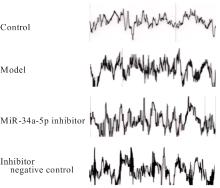
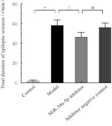
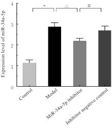
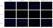
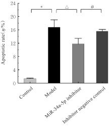
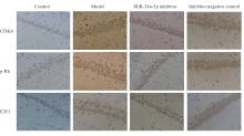
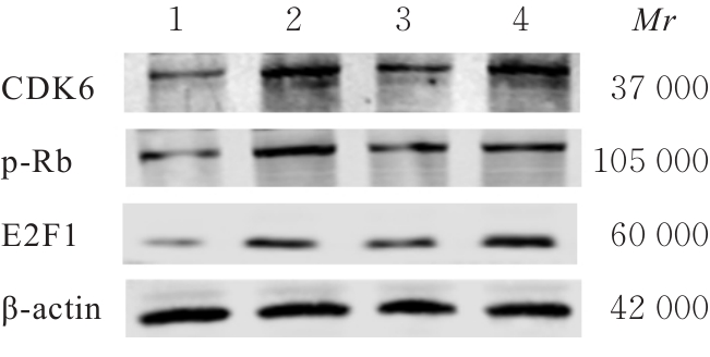
目的 探讨微小RNA-34a-5p (miR-34a-5p)对颞叶癫痫大鼠海马组织中神经元凋亡的影响,并阐明其作用机制。 方法 52只雄性SD大鼠随机分为对照组、模型组、miR-34a-5p抑制剂组和抑制剂阴性对照组,每组13只。采用PONEMAH 6.X实验动物遥测平台记录各组大鼠脑电图,实时荧光定量PCR(RT-qPCR)法检测各组大鼠海马组织中 miR-34a-5p表达水平,HE染色观察各组大鼠海马组织形态表现,TUNEL法检测各组大鼠海马神经元凋亡率,免疫组织化学法检测各组大鼠海马组织中CDK6、p-Rb和E2F1蛋白表达情况,Western blotting法检测各组大鼠海马组织中细胞周期蛋白依赖性激酶6(CDK6)、磷酸化视网膜母细胞瘤蛋白(p-Rb)和E2F转录因子1(E2F1)蛋白表达水平。 结果 对照组大鼠无异常表现;模型组、miR-34a-5p 抑制剂组和抑制剂阴性对照组大鼠出现不同程度流口水、颤抖、血泪,凝视和咀嚼震颤,随后点头眨眼,最后有前肢痉挛、直立及跌倒。与对照组比较,模型组大鼠癫痫发作总时间明显延长(P<0.01);与模型组比较,miR-34a-5p 抑制剂组大鼠癫痫发作总时间缩短(P<0.01);与miR-34a-5p 抑制剂组比较,抑制剂阴性对照组大鼠癫痫发作总时间延长(P<0.05)。RT-qPCR法,与对照组比较,模型组大鼠海马组织中miR-34a-5p表达水平升高(P<0.01);与模型组比较,miR-34a-5p 抑制剂组大鼠海马组织中miR-34a-5p表达水平升高(P<0.01);与miR-34a-5p抑制剂组比较,抑制剂阴性对照组大鼠海马组织中miR-34a-5p表达水平升高(P<0.05)。HE染色,与对照组比较,模型组细胞排列紊乱;与模型组比较,miR-34a-5p抑制剂组细胞排列整齐;与miR-34a-5p 抑制剂组比较,抑制剂阴性对照组细胞形态不规整。TUNEL染色,与对照组比较,模型组大鼠海马CA1区神经元凋亡率升高(P<0.01);与模型组比较,miR-34a-5p抑制剂组大鼠海马CA1区神经元凋亡率降低(P<0.05);与miR-34a-5p抑制剂组比较,抑制剂阴性对照组大鼠海马CA1区神经元凋亡率升高(P<0.05)。免疫组织化学法,与对照组比较,模型组大鼠海马组织中CDK6、p-Rb和E2F1蛋白阳性表达率均升高(P<0.01);与模型组比较,miR-34a-5p抑制剂组大鼠海马组织CA1区中CDK6、p-Rb和E2F1蛋白阳性表达率降低(P<0.05);与miR-34a-5p抑制剂组比较,抑制剂阴性对照组大鼠海马组织中CDK6、p-Rb和E2F1蛋白阳性表达率均升高(P<0.05或P<0.01)。Western blotting法,与对照组比较,模型组大鼠海马组织中CDK6、p-Rb和E2F1蛋白表达水平升高(P<0.01);与模型组比较,miR-34a-5p抑制剂组大鼠海马组织中CDK6、p-Rb和E2F1蛋白表达水平降低(P<0.05或P<0.01);与miR-34a-5p抑制剂组比较,抑制剂阴性对照组大鼠海马组织中CDK6、p-Rb和E2F1蛋白表达水平升高(P<0.05或P<0.01)。 结论 颞叶癫痫大鼠海马组织中miR-34a-5p表达上调,神经元凋亡率升高,抑制miR-34a-5p表达能减少海马神经元凋亡,其机制可能与miR-34a-5p调控海马组织中CDK6、p-Rb和E2F1蛋白表达有关。

关键词: 微小RNA-34a-5p, 癫痫, 细胞凋亡, 细胞周期蛋白依赖性激酶6, 磷酸化视网膜母细胞瘤蛋白, E2F转录因子1

Abstract:

Objective To discuss the effect of microRNA-34a-5p (miR-34a-5p) on the neuron apoptosis in hippocampus tissue of the rats with temporal lobe epilepsy, and to clarify its mechanism. Methods Fifty-two male SD rats were randomly divided into control group, model group, miR-34a-5p inhibitor group, and inhibitor negative control group, and there were 13 rats in each group. The PONEMAH 6.X experimental animal telemetry platform was used to record the electroencephalogram (EEG) of the rats in various groups; real-time fluorescence quantitative PCR (RT-qPCR) was used to detect the expression levels of miR-34a-5p in hippocampus tissue of the rats in various groups; HE staining was used to observe the morphology of hippocampus tissue of the rats in various groups; TUNEL method was used to detect the apoptotic rates of neurons in hippocampus tissue of the rats in various groups; immunohistochemistry method was used to determine the expressions of CDK6, p-Rb, and E2F1 proteins in hippocampus tissue of the rats in various group; Western blotting method was used to detect the expression levels of cyclin-dependent protein kinase 6 (CDK6), phosphorylated retinoblastoma protein (p-Rb), and E2F transcription factor 1 (E2F1) proteins in hippocampus tissue of the rats in various groups. Results No abnormalities were observed in the rats in control group; the rats in model group, miR-34a-5p inhibitor group, and inhibitor negative control group exhibited varying degrees of drooling, trembling, bloody tears, staring, chewing tremors, followed by nodding and blinking, and finally forelimb convulsions, standing upright, and falling. Compared with control group, the total duration of epileptic seizures of the rats in model group was significantly prolonged (P<0.01); compared with model group, the total duration of epileptic seizures of the rats in miR-34a-5p inhibitor group was shortened (P<0.01); compared with miR-34a-5p inhibitor group, the total duration of epileptic seizures of the rats in inhibitor negative control group was prolonged (P<0.05). The RT-qPCR results showed that compared with control group, the expression level of miR-34a-5p in hippocampus tissue of the rats in model group was increased (P<0.01); compared with model group, the expression level of miR-34a-5p in hippocampus tissue of the rats in miR-34a-5p inhibitor group was increased (P<0.01); compared with miR-34a-5p inhibitor group, the expression level of miR-34a-5p in hippocampus tissue of the rats in inhibitor negative control group was increased (P<0.05). The HE staining results showed that compared with control group, the cell arrangement in model group was disordered; compared with model group, the cell arrangement in miR-34a-5p inhibitor group was orderly; compared with miR-34a-5p inhibitor group, the cell morphology in inhibitor negative control group was irregular. The TUNEL staining results showed that compared with control group, the apoptotic rate of neurons in CA1 region of hippocampus tissue of the rats in model group was increased (P<0.01); compared with model group, the apoptotic rate of neurons in CA1 region of hippocampus tissue of the rats in miR-34a-5p inhibitor group was decreased (P<0.05); compared with miR-34a-5p inhibitor group, the apoptotic rate of neurons in CA1 region of hippocampus tissue of the rats in inhibitor negative control group was increased (P<0.05). The immunohistochemistry results showed that compared with control group, the positive expression rates of CDK6, p-Rb and E2F1 proteins in hippocampus tissue of the rats model group were increased (P<0.01); compared with model group, the positive expression rates of CDK6, p-Rb and E2F1 proteins in hippocampus tissue of the rats in miR-34a-5p inhibitor group were decreased (P<0.05); compared with miR-34a-5p inhibitor group, the positive expression rates of CDK6, p-Rb and E2F1 proteins in hippocampus tissue of the rats in inhibitor negative group were increased (P<0.05 or P<0.01).The Western blotting results showed that compared with control group, the expression levels of CDK6, p-Rb, and E2F1 proteins in hippocampus tissue of the rats in model group were increased (P<0.01); compared with model group, the expression levels of CDK6, p-Rb, and E2F1 proteins in hippocampus tissue of the rats in miR-34a-5p inhibitor group were decreased (P<0.05 or P<0.01); compared with miR-34a-5p antagomir group, the expression levels of CDK6, p-Rb, and E2F1 proteins in hippocampus tissue of the rats in inhibitor negative control group were increased (P<0.05 or P<0.01). Conclusion The expression of miR-34a-5p is upregulated in the hippocampal tissue of temporal lobe epilepsy rats, and hippocampal neuron apoptosis is increased. Inhibition of miR-34a-5p expression can reduce the hippocampal neuron apoptotic rate, and its mechanism may be related to the regulation of CDK6, p-Rb, and E2F1 protein expressions in the hippocampus tissue by miR-34a-5p.

Key words: MicroRNA-34a-5p, Epilepsy, Apoptosis, Cyclin-dependent protein kinase 6, Phosphorylated retinoblastoma protein, E2F transcription factor 1

中图分类号: 

  • R742.1