吉林大学学报(医学版) ›› 2025, Vol. 51 ›› Issue (3): 642-652.doi: 10.13481/j.1671-587X.20250309

• 基础研究 • 上一篇    

M2巨噬细胞通过调控NF-κB信号通路对非小细胞肺癌A549细胞上皮-间质转化和顺铂耐药的促进作用

王星翔1,赵颖1,任俏同1,王鹤霏2,蒲刚1,历春1()   

  1. 1.北华大学基础医学院免疫学教研室,吉林 吉林 132013
    2.吉林大学第一医院肿瘤妇科,吉林 长春 130021
  • 收稿日期:2024-06-18 接受日期:2024-08-24 出版日期:2025-05-28 发布日期:2025-07-18
  • 通讯作者: 历春 E-mail:lichunjl@126.com
  • 作者简介:王星翔(2000-),男,山西省忻州市人,在读硕士研究生,主要从事肿瘤免疫方面的研究。
  • 基金资助:
    吉林省卫健委卫生健康科技能力提升项目(2023JC033)

Promotive effect of M2 macrophages on epithelial-mesenchymal transition and cisplatin resistance in non-small cell lung cancer A549 cells by regulating NF-κB signaling pathway

Xingxiang WANG1,Ying ZHAO1,Qiaotong REN1,Hefei WANG2,Gang PU1,Chun LI1()   

  1. 1.Department of Immunology,School of Basic Medical Sciences,Beihua University,Jilin 132013,China
    2.Department of Oncological Gynecology,First Hospital,Jilin University,Changchun 130021,China
  • Received:2024-06-18 Accepted:2024-08-24 Online:2025-05-28 Published:2025-07-18
  • Contact: Chun LI E-mail:lichunjl@126.com

摘要:

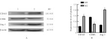
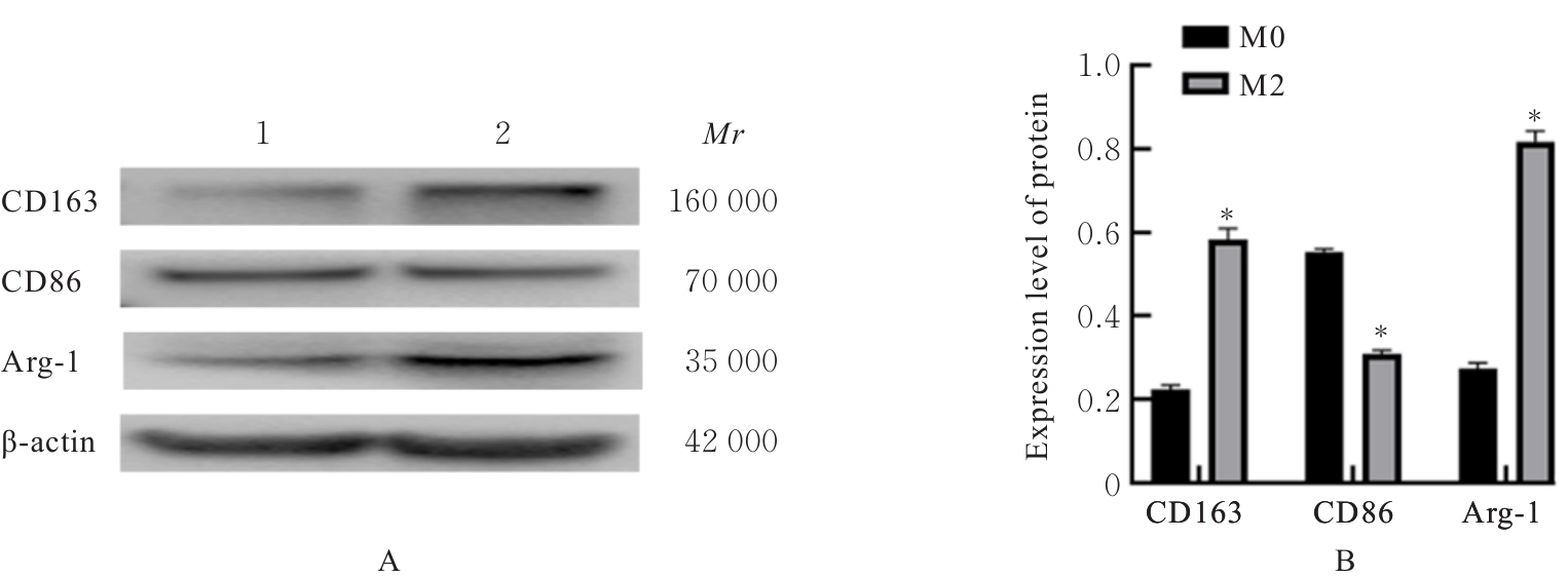
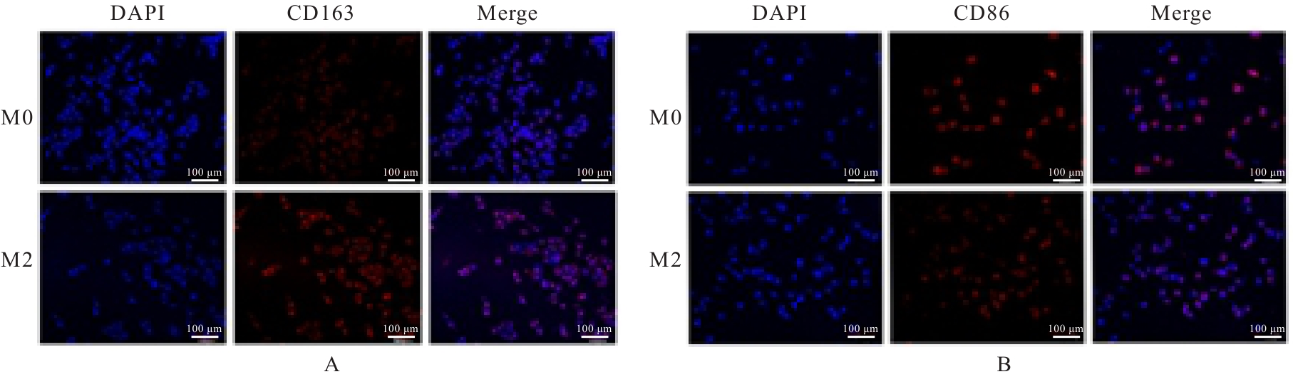
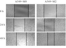
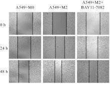
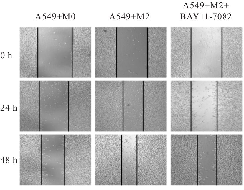
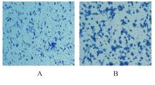
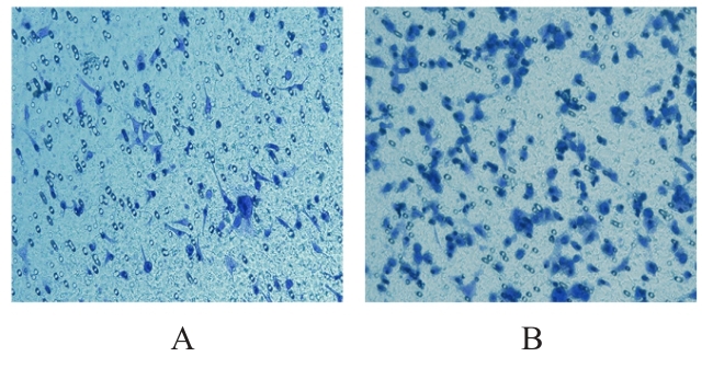
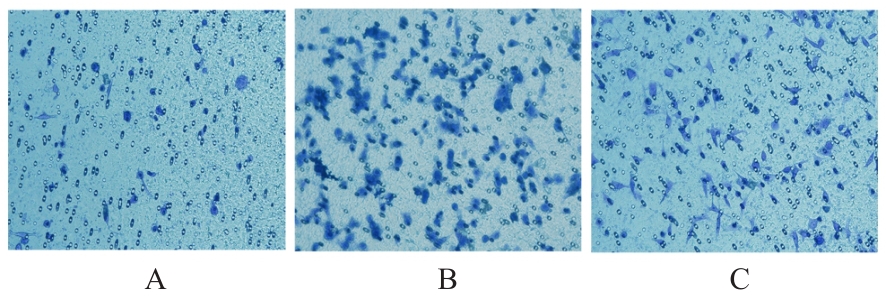
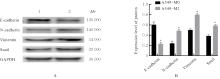
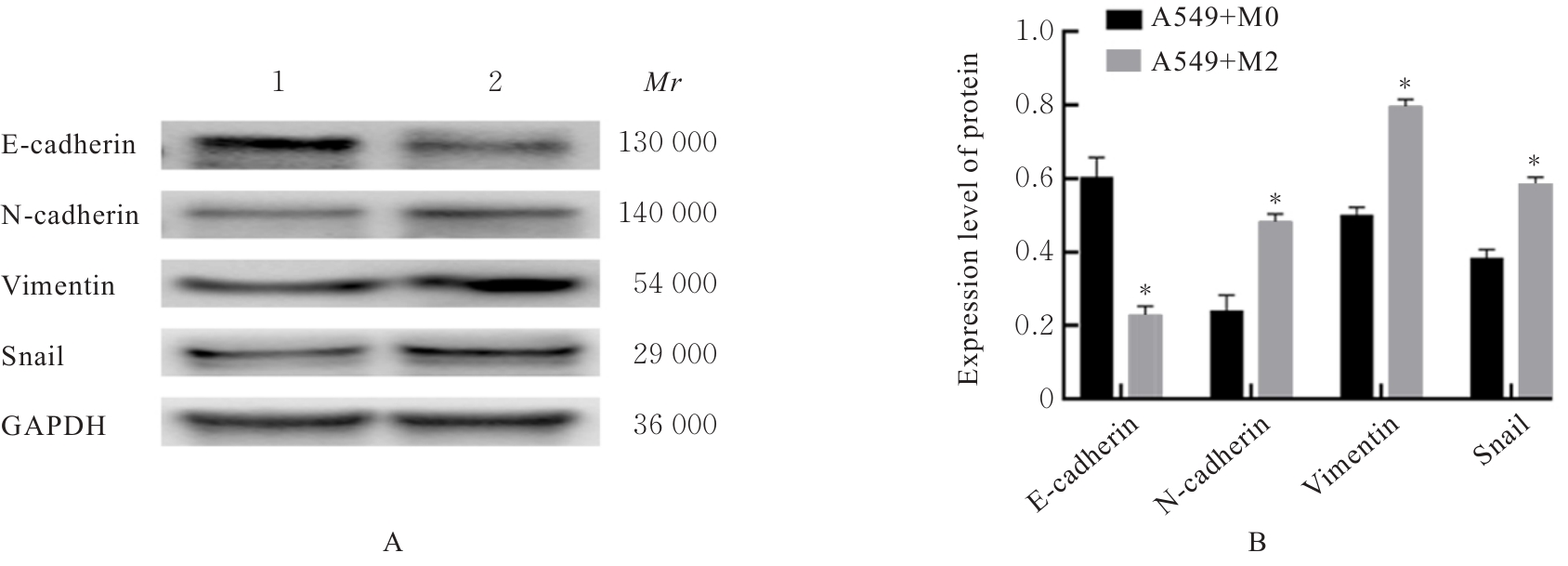
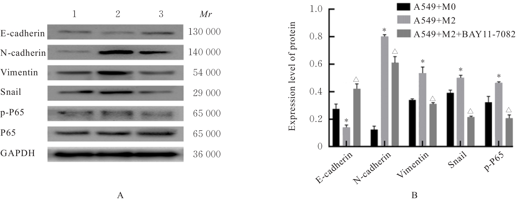
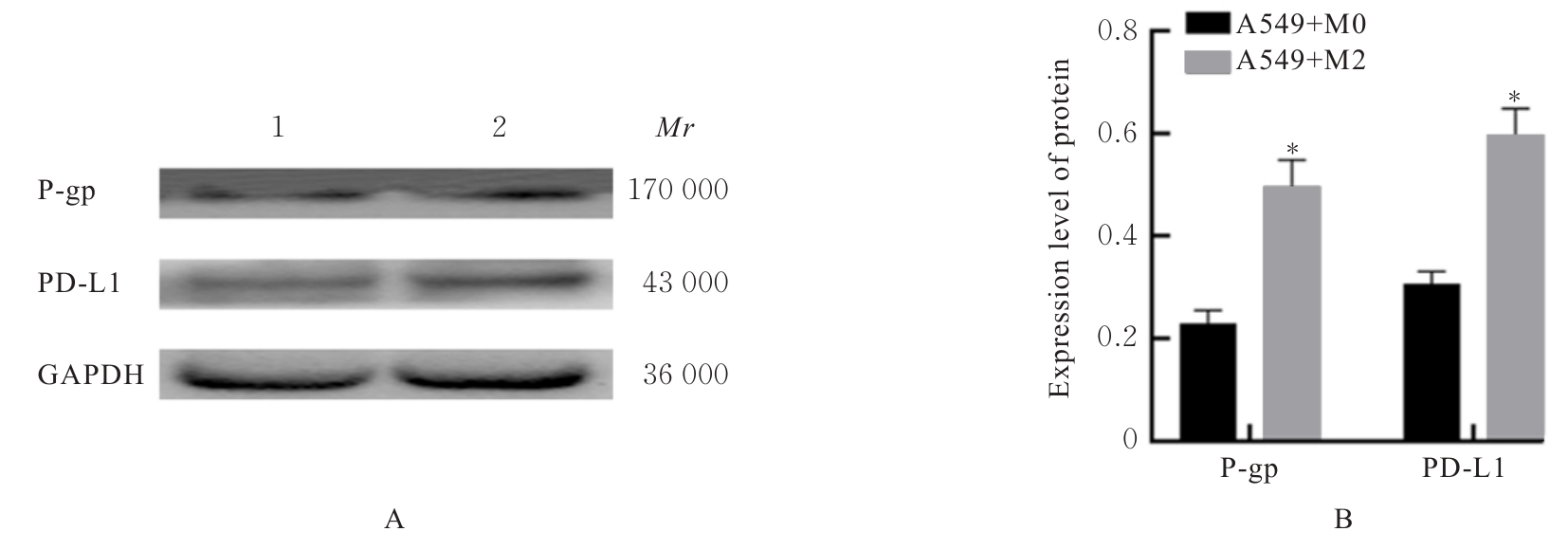
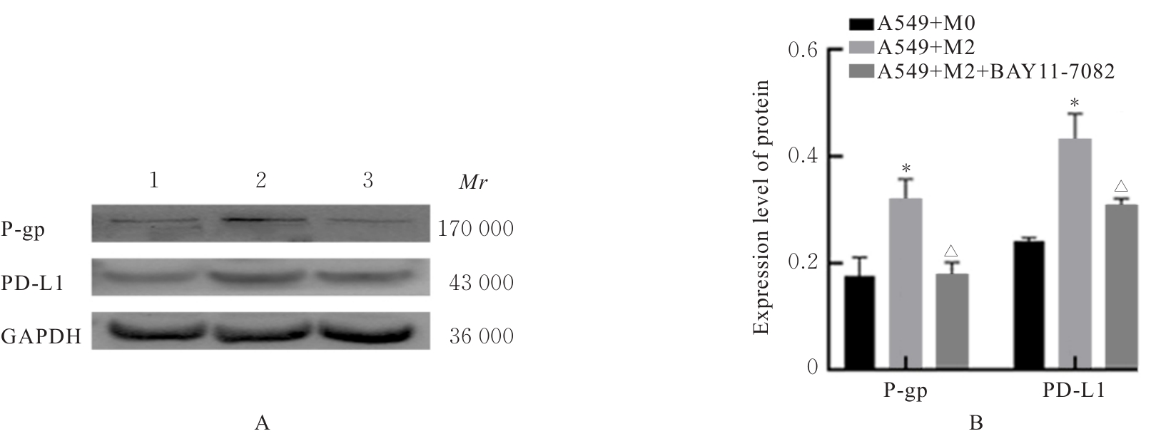
目的 探讨M2巨噬细胞在非小细胞肺癌(NSCLC)上皮-间质转化(EMT)和顺铂(DDP)耐药中的作用,阐明核因子κB(NF-κB)信号通路的调控机制。 方法 选取人单核细胞白血病THP-1细胞,通过佛波酯(PMA)诱导分化为M0巨噬细胞,白细胞介素(IL)-4和IL-13联合诱导M0巨噬细胞分化为M2巨噬细胞。采用Western blotting法和免疫荧光法检测M0和M2巨噬细胞中CD163、CD86及精氨酸酶1(Arg-1)蛋白表达情况。选取人NSCLC细胞A549,采用Transwell小室分别与M0和M2巨噬细胞非接触式共培养,细胞分为A549+M0组(A549细胞与M0巨噬细胞共培养)、A549+M2组(A549细胞与M2巨噬细胞共培养)和A549+M2+BAY11-7082组(A549细胞与M2巨噬细胞共培养后加入10 mmol·L-1 NF-κB抑制剂BAY11-7082)。细胞划痕实验检测各组A549细胞划痕愈合率,Transwell小室实验检测各组A549细胞中侵袭细胞数,细胞计数试剂盒8(CCK-8)法检测共培养体系中DDP处理的各组A549细胞生长抑制率和半数抑制浓度(IC50)值,Western blotting法检测各组A549细胞中波形蛋白(Vimentin)、E钙黏蛋白(E-cadherin)、N钙黏蛋白(N-cadherin)、转录因子Snail、磷酸化P65(p-P65)、P糖蛋白(P-gp)和细胞程序性死亡配体1(PD-L1)蛋白表达水平。 结果 Western blotting法,与M0组比较,M2组巨噬细胞中CD163和Arg-1蛋白表达水平均明显升高(P<0.05),CD86蛋白表达水平明显降低(P<0.05)。免疫荧光法,与M0组比较,M2组巨噬细胞中CD163蛋白表达增强,CD86蛋白表达减弱。细胞划痕实验,培养24和48 h时,与A549+M0组比较,A549+M2组A549细胞划痕愈合率均明显升高(P<0.05);3组共培养体系中,与A549+M0组比较,A549+M2组A549细胞划痕愈合率明显升高(P<0.05);与A549+M2组比较,A549+M2+BAY11-7082组A549细胞中划痕愈合率明显降低(P<0.05)。Transwell小室实验,与A549+M0组比较,A549+M2组A549细胞中侵袭细胞数明显增加(P<0.05);与A549+M2组比较,A549+M2+BAY11-7082组A549细胞中侵袭细胞数明显减少(P<0.05);3组共培养体系中,与A549+M0组比较,A549+M2组A549细胞中侵袭细胞数明显增加(P<0.05)。CCK-8法,2.50、5.00、10.00、20.00和40.00 mg·L-1 DDP处理后,与A549+M0组比较,A549+M2组A549细胞生长抑制率均明显降低(P<0.05或P<0.01),IC50值均明显升高(P<0.01);3组共培养体系中,与A549+M0组比较,A549+M2组A549细胞生长抑制率均明显降低(P<0.05或P<0.01),IC50值均明显升高(P<0.01);与A549+M2组比较,A549+M2+BAY11-7082组A549细胞生长抑制率明显升高(P<0.05),IC50值均明显降低(P<0.05)。Western blotting法,与A549+M0组比较,A549+M2组A549细胞中E-cadherin蛋白表达水平明显降低(P<0.05),N-cadherin、Vimentin和Snail蛋白表达水平均明显升高(P<0.05);3组共培养体系中,与A549+M0组比较,A549+M2组A549细胞中E-cadherin蛋白表达水平明显降低(P<0.05),N-cadherin、Snail、Vimentin和p-P65蛋白表达水平均明显升高(P<0.05);与A549+M2组比较,A549+M2+BAY11-7082组A549细胞中,E-cadherin蛋白表达水平明显升高(P<0.05),Vimentin、N-cadherin和p-P65蛋白表达水平均明显降低(P<0.05)。与A549+M0组比较,A549+M2组A549细胞中P-gp和PD-L1蛋白表达水平均明显升高(P<0.05);3组共培养体系中,与A549+M0组比较,A549+M2组A549细胞中P-gp和PD-L1蛋白表达水平均明显升高(P<0.05);与A549+M2组比较,A549+M2+BAY11-7082组A549细胞中P-gp和PD-L1蛋白表达水平均明显降低(P<0.05)。 结论 M2巨噬细胞可调控NSCLC细胞EMT促进肿瘤侵袭转移,调控P-gp和PD-L1蛋白表达促进DDP耐药,其作用机制可能与NF-κB信号通路有关。

关键词: 肿瘤相关巨噬细胞, 非小细胞肺癌, 核因子κB, 上皮-间质转化, 顺铂

Abstract:

Objective To discuss the role of M2 macrophages in epithelial-mesenchymal transition (EMT) and cisplatin (DDP) resistance in the non-small cell lung cancer (NSCLC), and to clarify the regulatory mechanism of nuclear factor κB (NF-κB) signaling pathway. Methods The human monocytic leukemia THP-1 cells were selected and differentiated into M0 macrophages by phorbol myristate acetate (PMA) induction, followed by M2 macrophage polarization through interleukin (IL)-4 and IL-13 stimulation. Western blotting and immunofluorescence methods were used to detect the protein expression levels of CD163, CD86, and arginase-1 (Arg-1) in M0 and M2 macrophages.The human NSCLC A549 cells were co-cultured non-contactly with M0 or M2 macrophages in Transwell chambers, and the cells were divided into A549+M0 group (A549 cells co-cultured with M0 macrophages), A549+M2 group (A549 cells co-cultured with M2 macrophages), and A549+M2+BAY11-7082 group (A549 cells co-cultured with M2 macrophages and treated with 10 mmol·L-1 NF-κB inhibitor BAY11-7082). Wound healing assay was used to detect the wound healing rate of the A549 cells in various groups; Transwell assay was used to detect the number of invasion A549 cells in various groups; cell counting kit-8 (CCK-8) assay was used to detect the inhibitory rate of proliferation and half maximal inhibitory concentration (IC50) value of the A549 cells after treated with DDP in the co-culture system; Western blotting method was used to detect the expression levels of vimentin, E-cadherin, N-cadherin, transcription factor Snail, phosphorylated P65 (p-P65), P-glycoprotein (P-gp), and programmed death-ligand 1 (PD-L1) proteins in the A549 cells in various groups. Results The Western blotting results showed that compared with M0 group, the expression levels of CD163 and Arg-1 proteins in the macrophages in M2 group were significantly increased (P<0.05), while the expression level of CD86 protein was significantly decreased (P<0.05). The immunofluorescence results showed that compared with M0 group, the expression of CD163 protein in the macrophages in M2 group was enhanced and the expression of CD86 protein was weakened. The wound healing assay results showed that at 24 and 48 h of culture, compared with A549+M0 group, the wound healing rate of the A549 cells in A549+M2 group was significantly increased (P<0.05); in the co-culture system, compared with A549+M0 group, the wound healing rate of the A549 cells in A549+M2 group was significantly increased (P<0.05); compared with A549+M2 group, the wound healing rate of the A549 cells in A549+M2+BAY11-7082 group was significantly decreased (P<0.05). The Transwell assay results showed that compared with A549+M0 group, the number of invasion A549 cells in A549+M2 group was significantly increased (P<0.05); compared with A549+M2 group, the number of invasion A549 cells in A549+M2+BAY11-7082 group was significantly decreased (P<0.05); in the co-culture system, compared with A549+M0 group, the number of invasion A549 cells in A549+M2 group was significantly increased (P<0.05). The CCK-8 assay results showed that after treated with 2.50, 5.00, 10.00, 20.00, and 40.00 mg·L-1 DDP, compared with A549+M0 group, the inhibitory rate of proliferation of the A549 cells in A549+M2 group was significantly decreased (P<0.05 or P<0.01), and the IC50 value was significantly increased (P<0.01); in the co-culture system, compared with A549+M0 group, the inhibitory rate of proliferation of the A549 cells in A549+M2 group was significantly decreased (P<0.05 or P<0.01), and the IC50 value was significantly increased (P<0.01); compared with A549+M2 group, the inhibitory rate of proliferation of the A549 cells in A549+M2+BAY11-7082 group was significantly increased (P<0.05), and the IC50 value was significantly decreased (P<0.05). The Western blotting results showed that compared with A549+M0 group, the expression level of E-cadherin proteins in the A549 cells in A549+M2 group was significantly decreased (P<0.05), while the expression levels of N-cadherin, vimentin, and Snail proteins were significantly increased (P<0.05); in the co-culture system, compared with A549+M0 group, the expression levels of vimentin, Snail, N-cadherin, and p-P65 proteins in the A549 cells in A549+M2 group were significantly increased (P<0.05), while the expression level of E-cadherin proteins was significantly decreased (P<0.05); compared with A549+M2 group, the expression levels of vimentin, N-cadherin, and p-P65 proteins in the A549 cells in A549+M2+BAY11-7082 group were significantly decreased (P<0.05), while the expression level of E-cadherin proteins was significantly increased (P<0.05); compared with A549+M0 group, the expression levels of P-gp and PD-L1 proteins in the A549 cells in A549+M2 group were significantly increased (P<0.05); in the co-culture system, compared with A549+M0 group, the expression levels of P-gp and PD-L1 proteins in the A549 cells in A549+M2 group were significantly increased (P<0.05); compared with A549+M2 group, the expression levels of P-gp and PD-L1 proteins in the A549 cells in A549+M2+BAY11-7082 group were significantly decreased (P<0.05). Conclusion The M2 macrophages can regulate EMT in the NSCLC cells to promote the invasion and metastasis of tumor, and modulate the expressions of P-gp and PD-L1 to enhance DDP resistance, which is associated with the NF-κB signaling pathway.

Key words: M2 macrophage, Non-small cell lung cancer, Nuclear factor-κB, Epithelial-mesenchymal transition, Cisplatin

中图分类号: 

  • R734.2