吉林大学学报(医学版) ›› 2024, Vol. 50 ›› Issue (4): 881-890.doi: 10.13481/j.1671-587X.20240401

• 基础研究 •    下一篇

基于M2型巨噬细胞来源的Siglec15对食管鳞癌细胞恶性生物学行为影响的生物信息学分析及实验验证

任祎琳1,臧翌辰1,薛乐乐1,杨凯歌1,陈素芳1,王魏楠1,罗成华1,梁伟华1,王良海1,李锋1,胡建明1,2()   

  1. 1.石河子大学第一附属医院病理科,新疆 石河子 832002
    2.石河子大学医学院病理学系,新疆 石河子 832002
  • 收稿日期:2023-09-01 出版日期:2024-07-28 发布日期:2024-08-01
  • 通讯作者: 胡建明 E-mail:jianming.120@163.com
  • 作者简介:任祎琳(1996-),女,陕西省延安市人,医学硕士,主要从事肿瘤微环境和免疫方面的研究。
  • 基金资助:
    国家自然科学基金项目(81960435);新疆生产建设兵团指导性科技计划项目(2022DZ003)

Bioinformatics analysis based on effect of M2 macrophage-derived Siglec15 on malignant biological behaviour of esophageal squamous cell carcinoma cells and its experimental validation

Yilin REN1,Yichen ZANG1,Lele XUE1,Kaige YANG1,Sufang CHEN1,Weinan WANG1,Chenghua LUO1,Weihua LIANG1,Lianghai WANG1,Feng LI1,Jianming HU1,2()   

  1. 1.Department of Pathology, First Affiliated Hospital, Shihezi University, Shihezi 832002, China
    2.Department of Patholegy, School of Medical Sciences, Shihezi University, Shihezi 832002, China
  • Received:2023-09-01 Online:2024-07-28 Published:2024-08-01
  • Contact: Jianming HU E-mail:jianming.120@163.com

摘要:

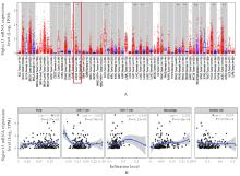
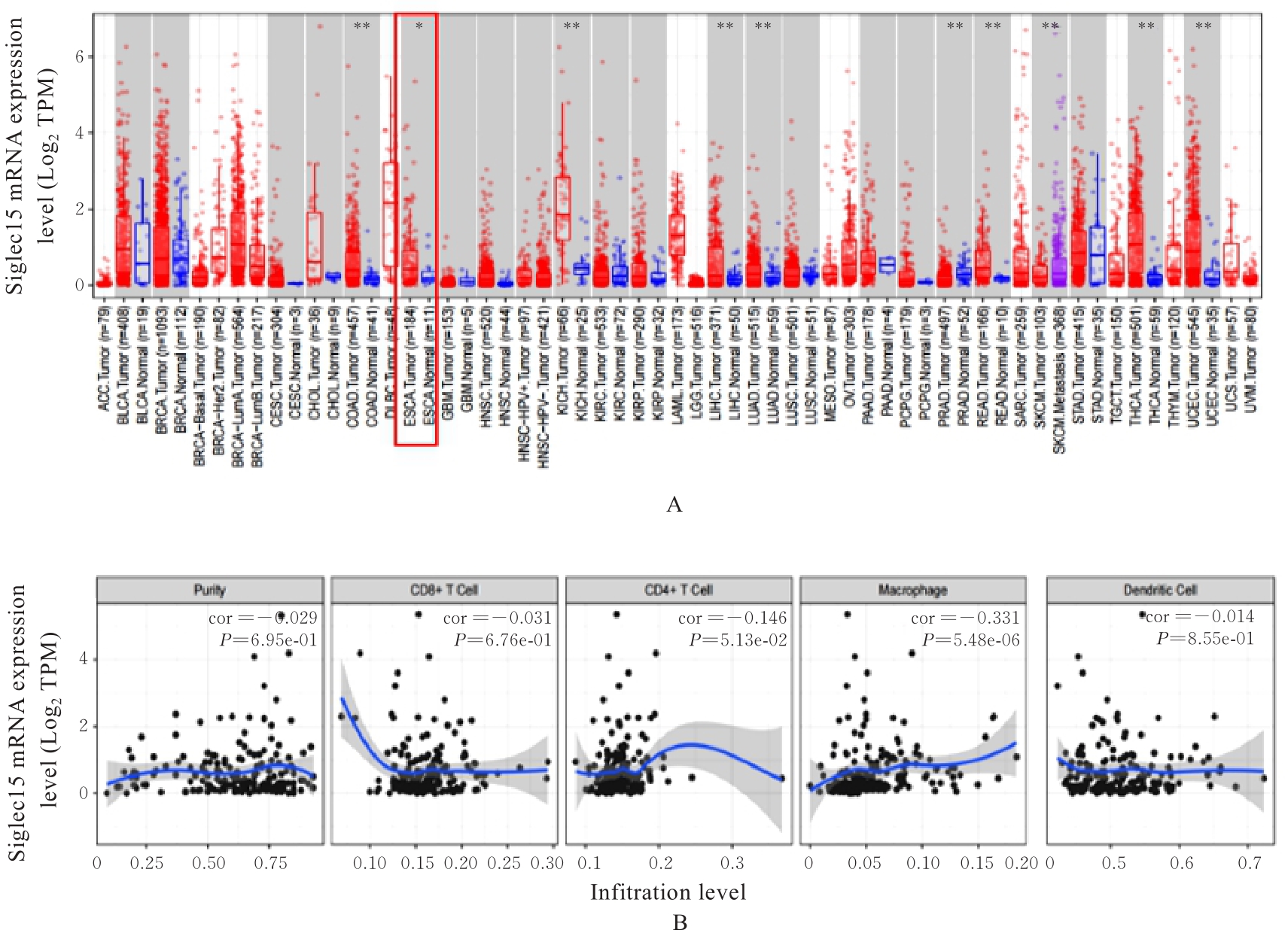
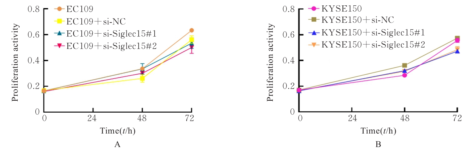
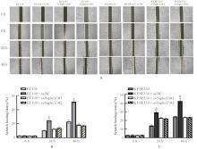
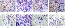
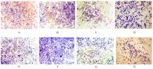
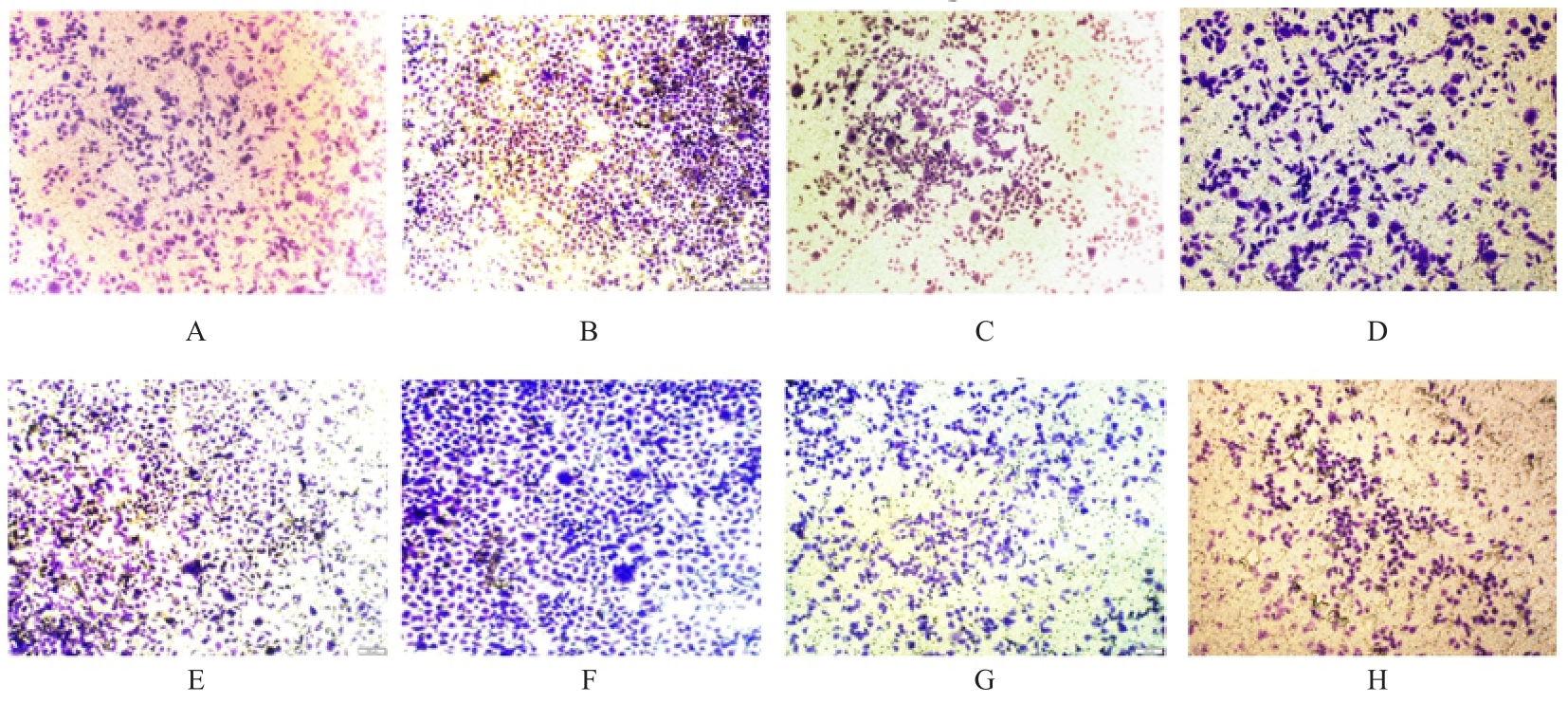
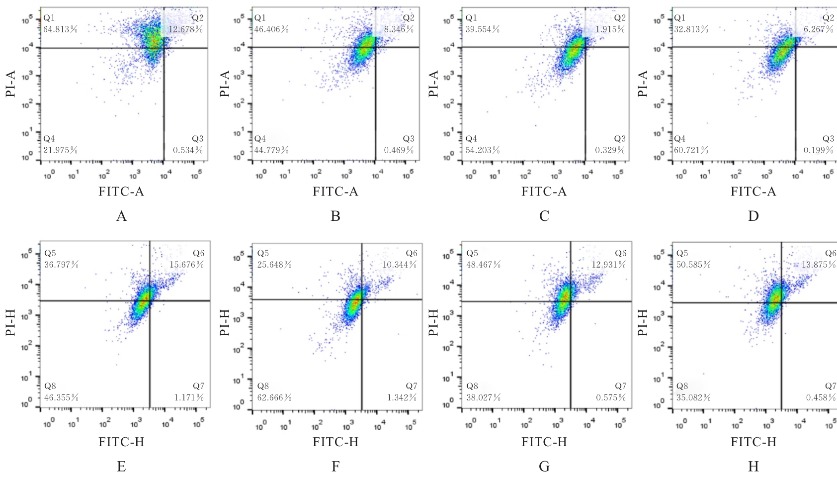
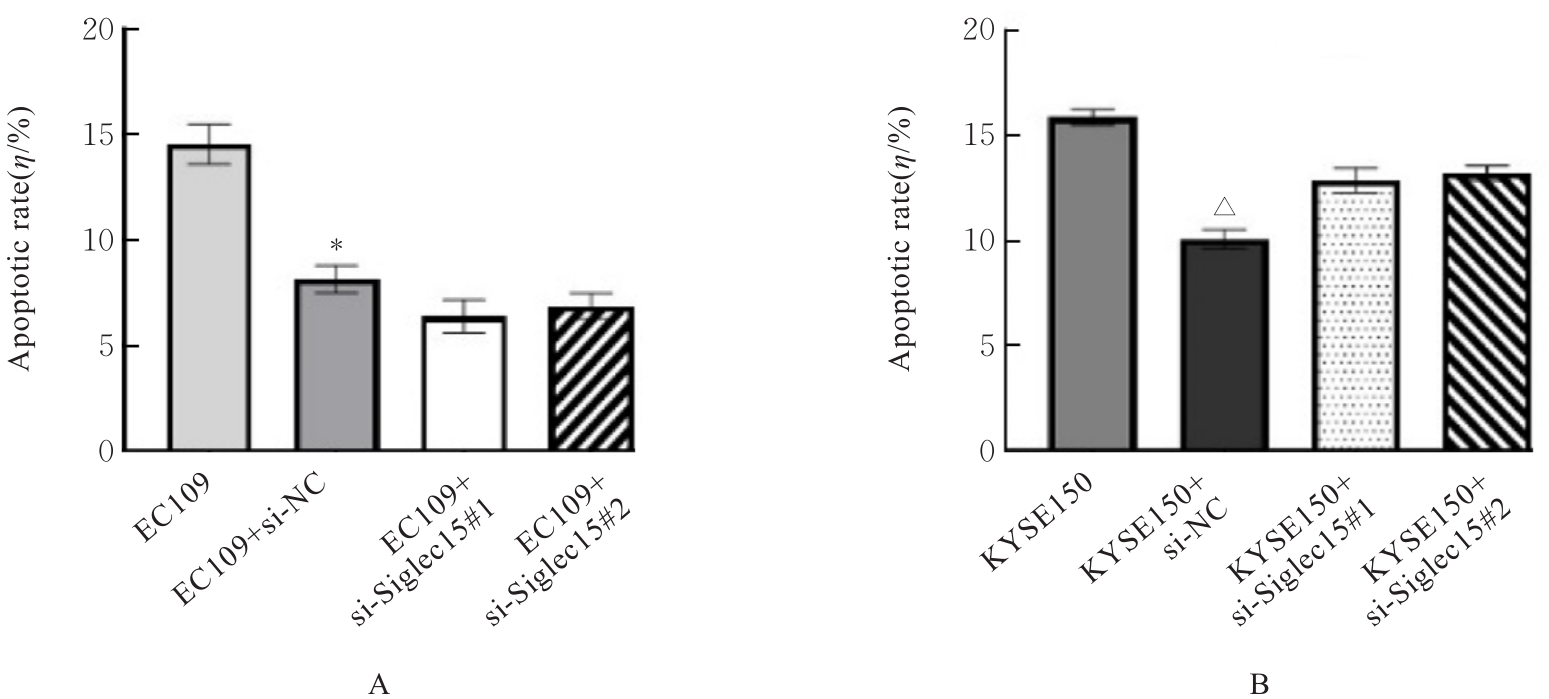
目的 采用生物信息学方法分析M2型肿瘤相关巨噬细胞(M2-TAMs)来源唾液酸结合免疫球蛋白样凝集素15(Siglec15)促进食管鳞状细胞癌(ESCC)恶性生物学行为的作用,并通过细胞实验对其进行验证。 方法 应用肿瘤免疫评价资源(TIMER)数据库分析Siglec15在泛癌和癌旁正常组织中的表达差异及免疫浸润情况,实时荧光定量PCR(RT-qPCR)法检测M2-TAMs和ESCC EC109及KYSE150细胞中Siglec15 mRNA表达水平。在M2-TAMs与ESCC细胞非接触性共培养基础上,分别设置EC109/KYSE150组、EC109/KYSE150+si-NC组(转染si-NC序列)和EC109/KYSE150+si-Siglec15组(分别转染si-Siglec15#1和si-Siglec15#2序列),采用CCK-8法检测各组细胞增殖活性,细胞划痕实验检测各组细胞划痕愈合率,Transwell小室实验检测各组细胞中迁移和侵袭细胞数,流式细胞术检测各组细胞凋亡率。 结果 生物信息学分析,与癌旁正常组织比较,食管癌、结肠癌和头颈部鳞状细胞癌等泛癌组织中Siglec15 mRNA表达水平升高(P<0.05或P<0.01),且食管癌组织中Siglec15 mRNA表达水平与巨噬细胞浸润呈明显正相关关系(P<0.05);与EC109细胞和KYSE150细胞比较,M2-TAMs中Siglec15 mRNA表达水平明显升高(P<0.01)。EC109/KYSE150组、EC109/KYSE150+si-NC组和EC109/KYSE150+si-Siglec15组细胞增殖率比较差异均无统计学意义(P>0.05)。与EC109/KYSE150组比较,24和48 h时EC109/KYSE150+si-NC组细胞划痕愈合率升高(P<0.01),迁移和侵袭细胞数增加(P<0.05),细胞凋亡率降低(P<0.01);与EC109/KYSE150+si-NC组比较,EC109/KYSE150+si-Siglec15#1组和EC109/KYSE150+si-Siglec15#2组细胞划痕愈合率降低(P<0.05),迁移和侵袭细胞数减少(P<0.05),细胞凋亡率差异无统计学意义(P>0.05)。 结论 M2-TAMs来源Siglec15可能是促进ESCC细胞迁移和侵袭的关键因子。

关键词: 食管鳞状细胞癌, 唾液酸结合免疫球蛋白样凝集素15, 肿瘤相关巨噬细胞, 细胞迁移, 细胞侵袭

Abstract:

Objective To discuss the effect of sialic acid-binding immunoglobulin-like lectin-15 (Siglec15) derived from M2 tumor-associated macrophages (M2-TAMs) on promoting the malignant biological behavior of the esophageal squamous cell carcinoma (ESCC) through bioinformatics analysis, and to validate the findings through cell experiment. Methods The Tumor Immune Estimation Resource (TIMER) online Database was used to analyze the expression differences and immune infiltration of Siglec15 in pan-cancer and adjacent normal tissues. Real-time fluorescence quantitative PCR (RT-qPCR) method was used to detect the expression levels of Siglec15 mRNA in M2-TAMs and ESCC EC109 and KYSE150 cells. Based on the non-contact co-culture of M2-TAMs and ESCC cells, the following groups were set up,such as EC109/KYSE150 group, EC109/KYSE150+si-NC group (transfected with si-NC sequence), and EC109/KYSE150+si-Siglec15 group (transfected with si-Siglec15#1 and si-Siglec15#2 sequences). CCK-8 method was used to detect the proliferation activities of the cells in various groups; wound healing assay was used to detect the wound healing rates of the cells in various groups; Transwell chamber assay was used to detect the numbers of migration and invasion cells in various groups; flow cytometry was used to detect the apoptotic rates of the cells in various groups. Results The bioinformatics analysis results showed that compared with adjacent normal tissue, the expression levels of Siglec15 mRNA in pan-cancer tissues such as esophageal cancer, colon cancer, and head and neck squamous cell carcinoma tissues were increased (P<0.05 or P<0.01), and the expression level of Siglec15 mRNA in esophageal cancer tissue was significantly positively correlated with the infiltration of the macrophages (P<0.05). Compared with the EC109 cells and KYSE150 cells, the expression level of Siglec15 mRNA in M2-TAMs was significantly increased (P<0.01). There was no significant difference in the proliferation rate of the cells among EC109/KYSE150 group, EC109/KYSE150+si-NC group, and EC109/KYSE150+si-Siglec15 group (P>0.05). Compared with EC109/KYSE150 group, after treated for 24 and 48 h, the wound healing rate of the cells in EC109/KYSE150+si-NC group was increased (P<0.01), the numbers of migration and invasion cells were increased (P<0.05), and the apoptotic rate was decreased (P<0.01). Compared with EC109/KYSE150+si-NC group, the wound healing rates of the cells in EC109/KYSE150+si-Siglec15#1 group and EC109/KYSE150+si-Siglec15#2 group were decreased (P<0.05), the numbers of migration and invasion cells were decreased (P<0.05), and the apoptotic rates of the cells had no significant difference (P>0.05). Conclusion Siglec15 derived from M2-TAMs may be a key factor in promoting the migration and invasion of the ESCC cells.

Key words: Esophageal squamous cell carcinoma, Sialic acid-binding immunoglobulin-like lectin-15, Tumor-associated macrophage, Cell migration, Cell invasion

中图分类号: 

  • R735.1