Journal of Jilin University(Medicine Edition) ›› 2026, Vol. 52 ›› Issue (3): 680-693.doi: 10.13481/j.1671-587X.20260311
• Research in basic medicine • Previous Articles
Effect of NGFR gene expression differences on promotion of apoptosis of squamous cell carcinoma and adenocarcinoma cells by PD-1 inhibitor-activated T lymphocytes
Yang LI1,Shiyang LI2,Hongmei ZHANG3,Qianqian LIU1,Yubing CHEN1( )
)
- 1.Department of Radiotherapy,Second Hospital,Jilin University,Changchun 130041,China
2.Department of Radiation,Jilin Provincial Tumor Hospital,Changchun 130012,China
3.Scientific Research Center,China-Japan Union Hospital,Jilin University,Changchun 130033,China
-
Received:2025-12-01Accepted:2026-01-12Online:2026-05-28Published:2026-06-08 -
Contact:Yubing CHEN E-mail:yb0707@163.com

CLC Number:
- R734.2
Cite this article
Yang LI,Shiyang LI,Hongmei ZHANG,Qianqian LIU,Yubing CHEN. Effect of NGFR gene expression differences on promotion of apoptosis of squamous cell carcinoma and adenocarcinoma cells by PD-1 inhibitor-activated T lymphocytes[J].Journal of Jilin University(Medicine Edition), 2026, 52(3): 680-693.
share this article
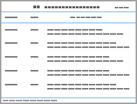
Tab.1
Primer sequences and product lengths"
| Gene | Primer sequence(5'-3') | Product length (bp) |
|---|---|---|
| NGFR | F:ACCTCCAGAACAAGACCTCATAG | 224 |
| R:CAATAGACAGGGATGAGGTTGTC | ||
| GADPH | F:AATCCCATCACCATCTTC | 218 |
| R:AGGCTGTTGTCATACTTC |
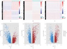
Fig. 1
Heatmaps and volcano plots of DEGs in squamous cell carcinoma and adenocarcinoma tissues"
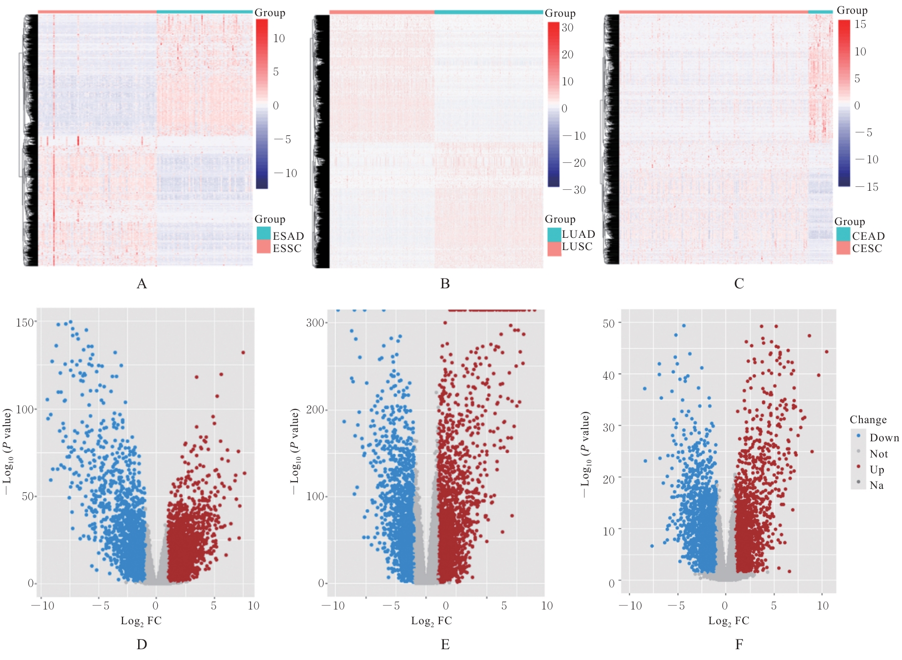
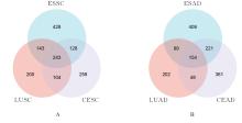
Fig. 2
Venn diagrams of intersection of DEGs in squamous cell carcinoma(A) and adenocarcinoma(B) tissues"
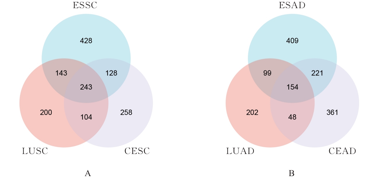
Fig. 3
Chord diagrams of functional enrichment of GO functional enrichment (A) and KEGG signaling pathway enrichment(B) of 397 DEGs"
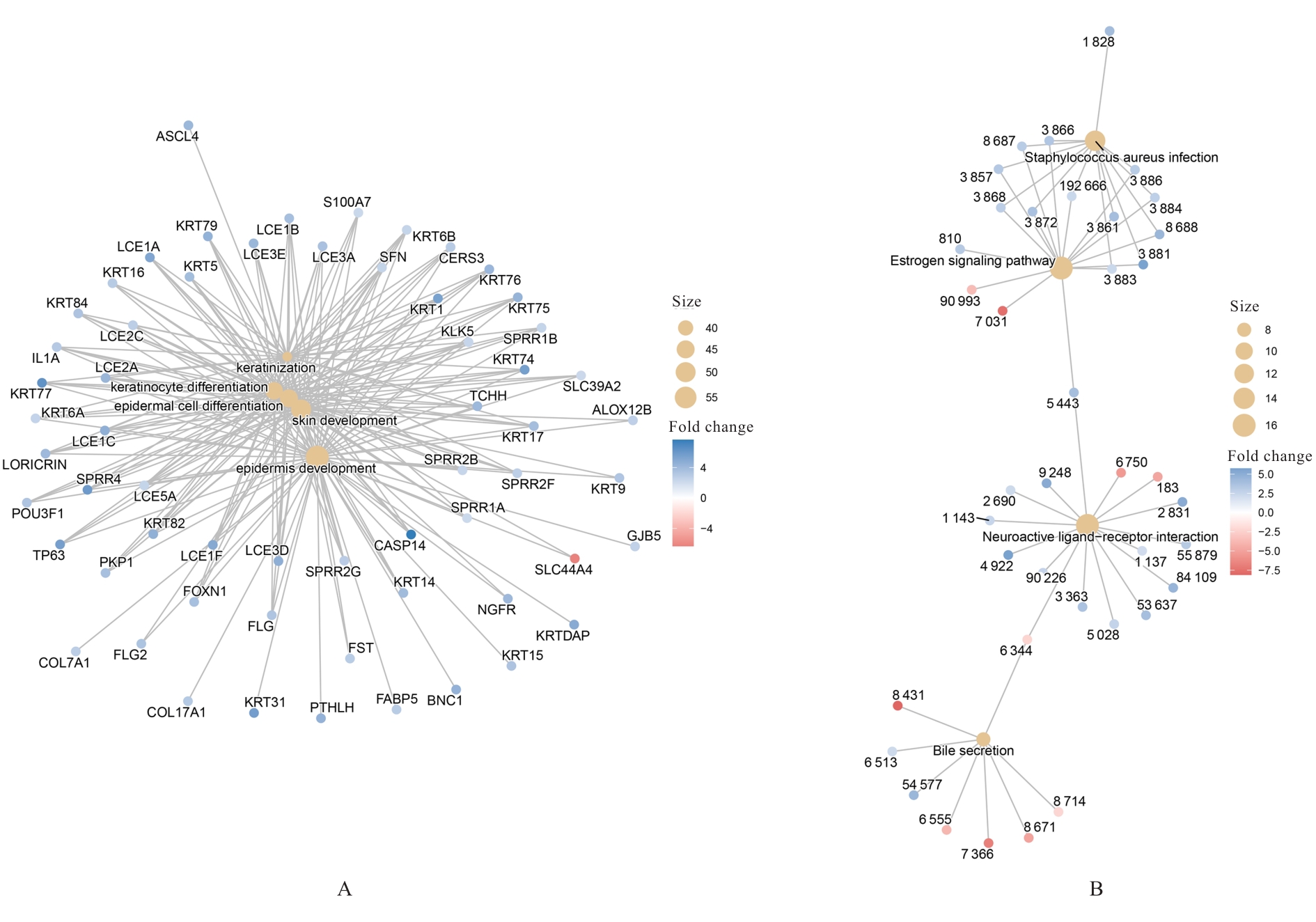
Fig. 4
Kaplan-Meier survival curves of GHR, NGFR, NTRK2, COL4A6, and IL20RB genes"
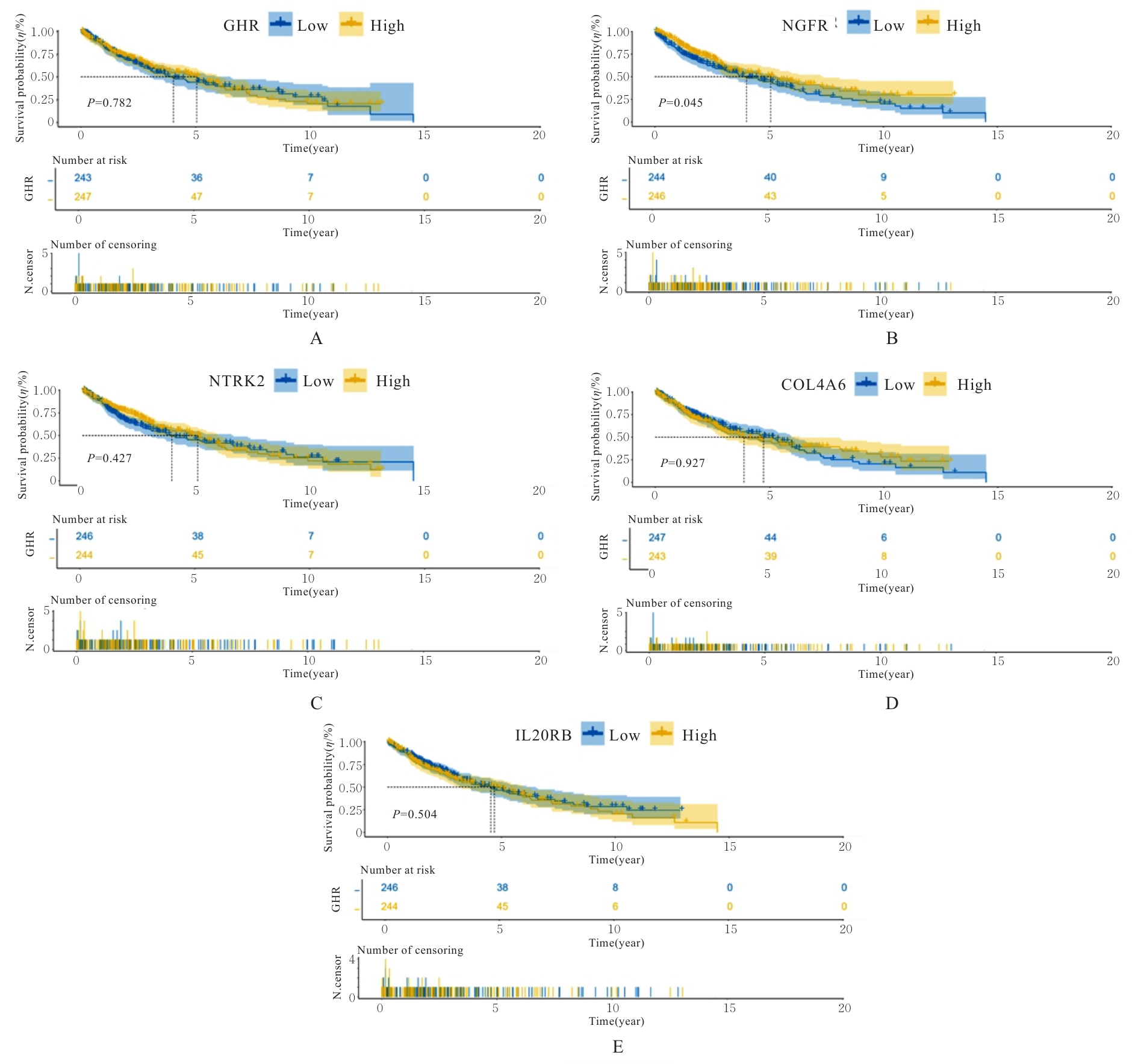
Fig. 5
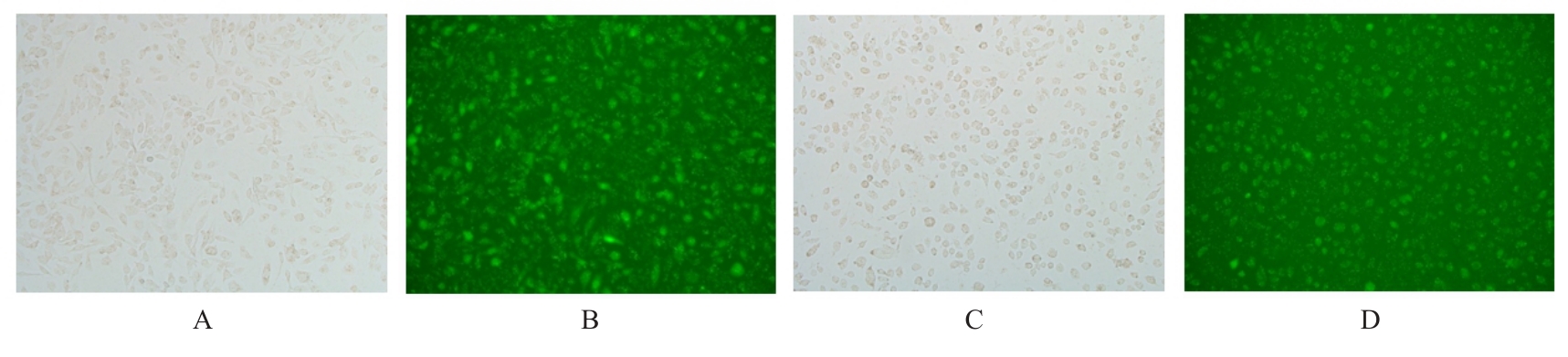
Expressions of protein in tumor cells 6 h after transfected with NGFR siRNA (×100)"
Tab. 2
Expression levels of NGFR mRNA in tumor cells in various groups"
| Group | Expression level of NGFR mRNA | P |
|---|---|---|
| Control | ||
| A549 | 1.000±0.110 | - |
| SK-MES-1 | 1.000±0.110 | - |
| HeLa | 1.000±0.140 | - |
| SiHa | 1.000±0.010 | - |
| OE-NC | ||
| A549 | 0.878±0.215 | 0.381 |
| HeLa | 1.167±0.095 | 0.093 |
| OE-NGFR | ||
| A549 | 2.522±0.543 | 0.008 |
| HeLa | 112.997±23.750 | 0.015 |
| Si-NC | ||
| SK-MES-1 | 0.924±0.110 | 0.358 |
| SiHa | 0.984±0.277 | 0.929 |
| Si-NGFR | ||
| SK-MES-1 | 0.188±0.028 | <0.001 |
| SiHa | 0.130±0.022 | <0.001 |
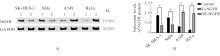
Fig. 6
Electrophoregram(A) and histogram(B) of expression of NGFR protein in tumor cells in various groups"
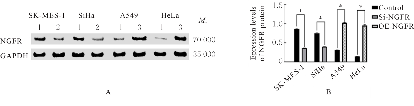
Fig. 7
Apoptotic rates and relative increase in folds apoptotic rates of A549 cells in various groups"
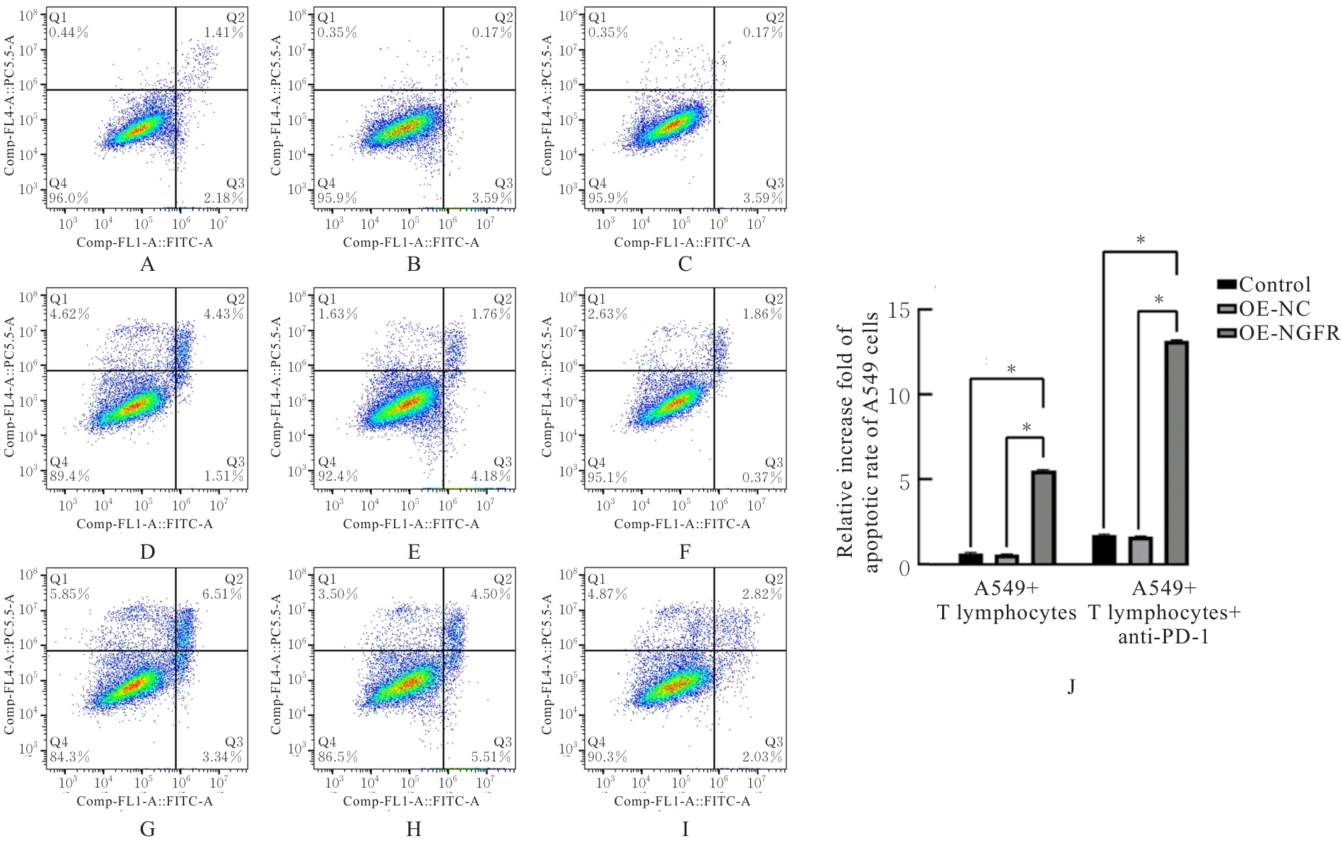
Fig. 8
Apoptotic rates and relative increase folds in apoptotic rates of SK-MES-1 cells in various groups"
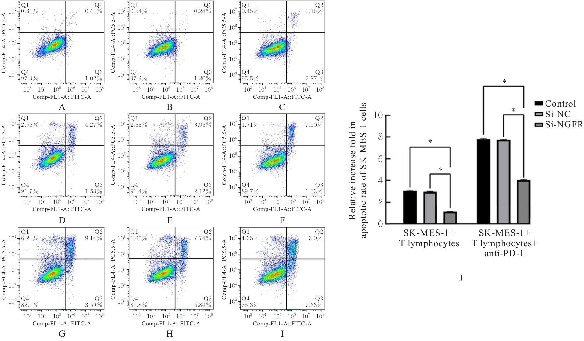
Fig. 9
Apoptotic rates and of relative increase folds in apoptotic rates of HeLa cells in various groups"
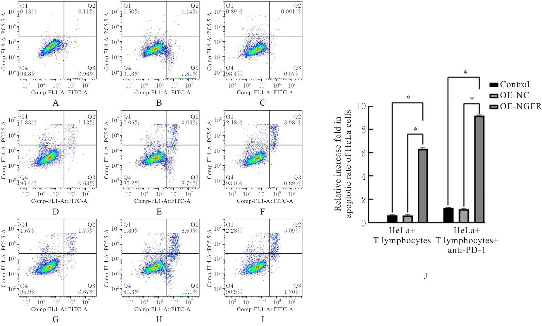
Fig. 10
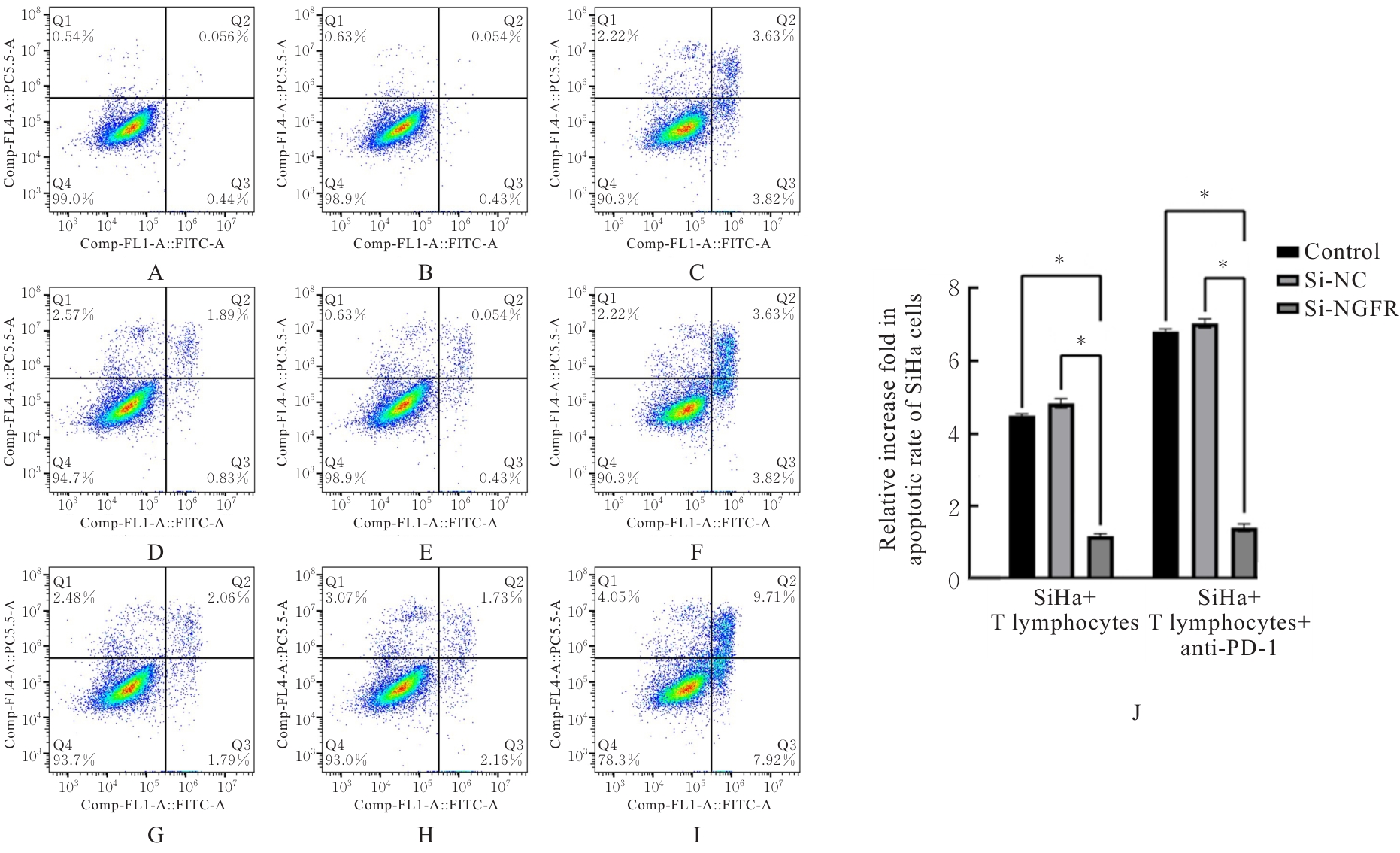
Apoptotic rates and relative increase folds in apoptotic rates of SiHa cells in various groups"
| [1] | ANTONIA S J, BORGHAEI H, RAMALINGAM S S, et al. Four-year survival with nivolumab in patients with previously treated advanced non-small-cell lung cancer: a pooled analysis[J]. Lancet Oncol, 2019, 20(10): 1395-1408. |
| [2] | HAMID O, ROBERT C, DAUD A, et al. Five-year survival outcomes for patients with advanced melanoma treated with pembrolizumab in KEYNOTE-001[J]. Ann Oncol, 2019, 30(4): 582-588. |
| [3] | ROBERT C, SCHACHTER J, LONG G V, et al. Pembrolizumab versus ipilimumab in advanced melanoma[J]. N Engl J Med, 2015, 372(26): 2521-2532. |
| [4] | SEIWERT T Y, BURTNESS B, MEHRA R, et al. Safety and clinical activity of pembrolizumab for treatment of recurrent or metastatic squamous cell carcinoma of the head and neck (KEYNOTE-012): an open-label, multicentre, phase 1b trial[J]. Lancet Oncol, 2016, 17(7): 956-965. |
| [5] | BRAHMER J, RECKAMP K L, BAAS P, et al. Nivolumab versus docetaxel in advanced squamous-cell non-small-cell lung cancer[J]. N Engl J Med, 2015, 373(2): 123-135. |
| [6] | BORGHAEI H, PAZ-ARES L, HORN L, et al. Nivolumab versus docetaxel in advanced nonsquamous non-small-cell lung cancer[J]. N Engl J Med, 2015, 373(17): 1627-1639. |
| [7] | SUN J M, SHEN L, SHAH M A, et al. Pembrolizumab plus chemotherapy versus chemotherapy alone for first-line treatment of advanced oesophageal cancer (KEYNOTE-590): a randomised, placebo-controlled, phase 3 study[J]. Lancet, 2021, 398(10302): 759-771. |
| [8] | KOJIMA T, SHAH M A, MURO K, et al. Randomized phase Ⅲ KEYNOTE-181 study of pembrolizumab versus chemotherapy in advanced esophageal cancer[J]. J Clin Oncol, 2020, 38(35): 4138-4148. |
| [9] | CHUNG H C, ROS W, DELORD J P, et al. Efficacy and safety of pembrolizumab in previously treated advanced cervical cancer: results from the phase Ⅱ KEYNOTE-158 study[J]. J Clin Oncol, 2019, 37(17): 1470-1478. |
| [10] | RECK M, RODRÍGUEZ-ABREU D, ROBINSON A G, et al. Pembrolizumab versus chemotherapy for PD-L1-positive non-small-cell lung cancer[J]. N Engl J Med, 2016, 375(19): 1823-1833. |
| [11] | RIZVI N A, HELLMANN M D, SNYDER A, et al. Cancer immunology. Mutational landscape determines sensitivity to PD-1 blockade in non-small cell lung cancer[J]. Science, 2015, 348(6230): 124-128. |
| [12] | CHAROENTONG P, FINOTELLO F, ANGELOVA M, et al. Pan-cancer immunogenomic analyses reveal genotype-immunophenotype relationships and predictors of response to checkpoint blockade[J]. Cell Rep, 2017, 18(1): 248-262. |
| [13] | SALEM M E, PUCCINI A, XIU J, et al. Comparative molecular analyses of esophageal squamous cell carcinoma, esophageal adenocarcinoma, and gastric adenocarcinoma[J]. Oncologist, 2018, 23(11): 1319-1327. |
| [14] | RONAGHY A, HU S M, TANG Z Y, et al. Myeloid neoplasms associated with t(3;12)(q26.2;p13) are clinically aggressive, show myelodysplasia, and frequently harbor chromosome 7 abnormalities[J]. Mod Pathol, 2021, 34(2): 300-313. |
| [15] | YI M, NIU M K, XU L P, et al. Regulation of PD-L1 expression in the tumor microenvironment[J]. J Hematol Oncol, 2021, 14(1): 10. |
| [16] | JIANG X J, WANG J, DENG X Y, et al. Role of the tumor microenvironment in PD-L1/PD-1-mediated tumor immune escape[J]. Mol Cancer, 2019, 18(1): 10. |
| [17] | BRUNO F, ABONDIO P, MONTESANTO A, et al. The nerve growth factor receptor (NGFR/p75NTR): a major player in Alzheimer’s disease[J]. Int J Mol Sci, 2023, 24(4): 3200. |
| [18] | BOTHWELL M. Recent advances in understanding context-dependent mechanisms controlling neurotrophin signaling and function[J]. F1000Res, 2019, 8: F1000FacultyRev-F1000Facult1658. |
| [19] | GARCÍA-SILVA S, BENITO-MARTÍN A, NOGUÉS L, et al. Melanoma-derived small extracellular vesicles induce lymphangiogenesis and metastasis through an NGFR-dependent mechanism[J]. Nat Cancer, 2021, 2(12): 1387-1405. |
| [20] | DUDÁS J, DIETL W, ROMANI A, et al. Nerve growth factor (NGF): receptor survival axis in head and neck squamous cell carcinoma[J]. Int J Mol Sci, 2018, 19(6): 1771. |
| [21] | WANG X, YANG T W, SHI S H, et al. Heterogeneity-induced NGF-NGFR communication inefficiency promotes mitotic spindle disorganization in exhausted T cells through PREX1 suppression to impair the anti-tumor immunotherapy with PD-1 MAb in hepatocellular carcinoma[J]. Cancer Med, 2024, 13(3): e6736. |
| [22] | BARUCH E N, GLEBER-NETTO F O, NAGARAJAN P, et al. Cancer-induced nerve injury promotes resistance to anti-PD-1 therapy[J]. Nature, 2025, 646(8084): 462-473. |
| [23] | BAUST J M, BUEHRING G C, CAMPBELL L, et al. Best practices in cell culture: an overview[J]. Vitro Cell Dev Biol Anim, 2017, 53(8): 669-672. |
| [24] | BUSTIN S A, RUIJTER J M, VAN DEN HOFF M J B, et al. MIQE 2.0: revision of the minimum information for publication of quantitative real-time PCR experiments guidelines[J]. Clin Chem, 2025, 71(6): 634-651. |
| [25] | SULE R, RIVERA G, GOMES A V. Western blotting (immunoblotting): history, theory, uses, protocol and problems[J]. BioTechniques, 2023, 75(3): 99-114. |
| [26] | XIA Y, LIU A Q, LI W T, et al. Reference range of naïve T and T memory lymphocyte subsets in peripheral blood of healthy adult[J]. Clin Exp Immunol, 2022, 207(2): 208-217. |
| [27] | COSSARIZZA A, CHANG H D, RADBRUCH A, et al. Guidelines for the use of flow cytometry and cell sorting in immunological studies (third edition)[J]. Eur J Immunol, 2021, 51(12): 2708-3145. |
| [28] | BABA Y, NOMOTO D, OKADOME K, et al. Tumor immune microenvironment and immune checkpoint inhibitors in esophageal squamous cell carcinoma[J]. Cancer Sci, 2020, 111(9): 3132-3141. |
| [29] | YU L, WEI J, LIU P D. Attacking the PI3K/Akt/mTOR signaling pathway for targeted therapeutic treatment in human cancer[J]. Semin Cancer Biol, 2022, 85: 69-94. |
| [30] | ZHANG C N, GU L Q, XIAO J, et al. Knockdown of RBM15 inhibits tumor progression and the JAK-STAT signaling pathway in cervical cancer[J]. BMC Cancer, 2023, 23(1): 684. |
| [1] | Wei ZHOU,Yangyang HE,Youde WANG,Wenzhuo MA,Heng KANG. Effect of histone deacetylase 6 inhibitor ACY-241 on macrophage polarization and biological behaviors of esophageal cancer cells and its mechanism [J]. Journal of Jilin University(Medicine Edition), 2026, 52(3): 621-632. |
| [2] | Xiao ZHOU,Haitao DAI,Yunshan DING,Linjie ZHANG,Nan WU. Network pharmacology analysis and in vitro experimental validation of anticancer effect of grape seed proanthocyanidins against oral squamous cell carcinoma [J]. Journal of Jilin University(Medicine Edition), 2026, 52(3): 651-662. |
| [3] | Jie YU,Zongkang WANG,Xun LIU,Yanan YANG,Jin TAN. Effect of miR-153-3p/PGC-1α axis on apoptosis of oral squamous cell carcinoma cells by modulation of mitochondrial apoptosis pathway [J]. Journal of Jilin University(Medicine Edition), 2026, 52(2): 348-358. |
| [4] | Xiaohan YAO,Mingchen YAO,Zhiqing WANG,Heyang LI,Yan YAN,Ningjing LEI. Effect of silencing GPR139 gene on proliferation, apoptosis and autophagy of breast cancer cells and its mechanism [J]. Journal of Jilin University(Medicine Edition), 2026, 52(1): 1-9. |
| [5] | Jiaxin WANG,Junwen MAO,Xuan ZHANG. Expression of natural autoantibodies against apoptosis inhibitory genes in plasma of patients with hepatocellular carcinoma and its clinical significance [J]. Journal of Jilin University(Medicine Edition), 2026, 52(1): 228-235. |
| [6] | Yaping LI,Chunyan TAN,Lujie ZHAO,Jiayi ZHAO,Ting LI,Xiao YANG,Xiaoyun YANG. Inductive effect of lysophosphatidic acid combined with 6-hydroxydopamine on apoptosis of SH-SY5Y cells and its mechanism [J]. Journal of Jilin University(Medicine Edition), 2026, 52(1): 152-161. |
| [7] | Yunshan DING,Haitao DAI,Min CHEN,Xiaohui HAO,Xiao ZHOU,Nan WU. Effect of silencing TRPV2 gene and cannabidiol on biological behaviors of oral squamous cell carcinoma CAL-27 cells [J]. Journal of Jilin University(Medicine Edition), 2026, 52(1): 143-151. |
| [8] | Huaimin LIANG,Jiacheng JIN,Wenhua CHEN,Yuyao LI,Hangyu WANG,Ke ZHANG,Jinhui WANG. UPLC-Q-TOF/MS and network pharmacology analysis and experimental verification based on potential active ingredients and mechanisms of medicinal Mulberry Leaves in anti-acute kidney injury [J]. Journal of Jilin University(Medicine Edition), 2026, 52(1): 56-69. |
| [9] | Shanshan SUN,Mei LU,Xinfu GAO,LYu Guangyao,Baolei Zhao,LYu Wenwen. Inhibitory effect of Bradykinin 1 receptor antagonist ELN441958 on proliferation of HepG2 cells by regulating Akt/FoxO3a signaling pathway [J]. Journal of Jilin University(Medicine Edition), 2026, 52(1): 70-80. |
| [10] | Dayou DAI,Zhigang ZHANG,Hui LI. Effect of CHRNA5 in occurrence and development of pancreas cancer and its mechanism [J]. Journal of Jilin University(Medicine Edition), 2025, 51(6): 1518-1531. |
| [11] | Han XUE,Yuxin FAN,Ting ZHANG,Zhimin LI,Mingge HUO,Xingang GUAN. Preparation of nanodrug PTX2 NPs and its killing effect on human lung cancer A549 cells [J]. Journal of Jilin University(Medicine Edition), 2025, 51(5): 1260-1266. |
| [12] | Panxi SUN,Xue QIN,Chongyang ZHANG,Jia LUO,Yong CHEN,Lili WEI. Protective effect of TUG-891 on ischemic stroke induced by ischemia and hypoxia and its mechanism [J]. Journal of Jilin University(Medicine Edition), 2025, 51(4): 968-975. |
| [13] | Ziru ZHOU,Mengfei SUN,Huakun ZHANG,Shuyan SUN,Qi SUN,Feng LI,Yunzhao CHEN,Jie YU,Yuwen CAO,Xiaobin CUI. Expression of Rh family C glycoprotein in esophageal squamous carcinoma and its clinical significance [J]. Journal of Jilin University(Medicine Edition), 2025, 51(4): 1019-1027. |
| [14] | Jiarui LI,Zhenlin YANG,Fan GAO,Jingjing GUO,Jinzi LI. Effect of miR-34a-5p on hippocampal neuron apoptosis in rats with temporal lobe epilepsy and its mechanism [J]. Journal of Jilin University(Medicine Edition), 2025, 51(4): 939-947. |
| [15] | Fan WANG,Xin WEN,Yixuan WANG,Yuan WANG. Effect of gap junction β2 on prognosis of patients with lung adenocarcinoma and biological behavior of lung adenocarcinoma A549 cells [J]. Journal of Jilin University(Medicine Edition), 2025, 51(3): 716-726. |
|
||