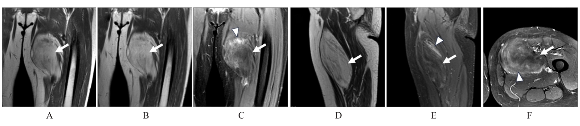
| [1] |
ANJU M S, CHANDRAMOHAN K, BHARGAVAN R V, et al. An overview on liposarcoma subtypes: genetic alterations and recent advances in therapeutic strategies[J]. J Mol Histol, 2024, 55(3): 227-240.
|
| [2] |
SZUHAI K, IJSZENGA M, KNIJNENBURG J, et al. Does parosteal liposarcoma differ from other atypical lipomatous tumors/well-differentiated liposarcomas? A molecular cytogenetic study using combined multicolor COBRA-FISH karyotyping and array-based comparative genomic hybridization[J]. Cancer Genet Cytogenet, 2007, 176(2): 115-120.
|
| [3] |
OLSON C R, SUAREZ-KELLY L P, ETHUN C G, et al. Resection status does not impact recurrence in well-differentiated liposarcoma of the extremity[J]. Am Surg, 2021, 87(11): 1752-1759.
|
| [4] |
LEE A T J, THWAY K, HUANG P H, et al. Clinical and molecular spectrum of liposarcoma[J]. J Clin Oncol, 2018, 36(2): 151-159.
|
| [5] |
杨 俊, 赵 敏, 周江军. 大腿巨大高分化脂肪肉瘤1例[J]. 临床骨科杂志, 2021, 24(5): 727.
|
| [6] |
赵康艳, 李新春, 陈镜聪, 等. 下肢高分化及黏液型脂肪肉瘤的影像表现与病理分析[J]. 影像诊断与介入放射学, 2012, 21(2): 133-136.
|
| [7] |
陈 华, 沙 娜, 刘 宁, 等. 人骨髓间充质干细胞通过YAP影响人脂肪肉瘤SW872细胞的生物学行为[J]. 吉林大学学报(医学版), 2024, 50(4): 1000-1008.
|
| [8] |
李婧婧, 热孜宛古丽·艾斯凯尔, 邢婉怡, 等. 单侧卵巢原发性去分化脂肪肉瘤1例报告及文献复习[J]. 吉林大学学报(医学版), 2023, 49(4): 1040-1045.
|
| [9] |
WAKELY P E JR. Atypical spindle cell/pleomorphic lipomatous tumour (ASPLT): a report of three FNA cases and comparison with spindle cell/pleomorphic lipoma cytopathology[J]. Cytopathology, 2023, 34(4): 346-352.
|
| [10] |
OHLIGER M A. Hyperpolarized 13C pyruvate MRI: an important window into tumor metabolism[J]. Radiol Imaging Cancer, 2024, 6(2): e240004.
|
| [11] |
邢汝维, 方志伟, 宋金纲, 等. 透明细胞肉瘤的治疗及疗效分析[J]. 中华骨科杂志, 2005, 25(8): 485-490.
|
| [12] |
YANG L G, CHEN S Q, LUO P, et al. Liposarcoma: advances in cellular and molecular genetics alterations and corresponding clinical treatment[J]. J Cancer, 2020, 11(1): 100-107.
|
| [13] |
DEI TOS A P. Liposarcoma: new entities and evolving concepts[J]. Ann Diagn Pathol, 2000, 4(4): 252-266.
|
| [14] |
DE VITA A, MERCATALI L, RECINE F, et al. Current classification, treatment options, and new perspectives in the management of adipocytic sarcomas[J]. Onco Targets Ther, 2016, 9: 6233-6246.
|
| [15] |
THWAY K. Well-differentiated liposarcoma and dedifferentiated liposarcoma: an updated review[J]. Semin Diagn Pathol, 2019, 36(2): 112-121.
|
| [16] |
COINDRE J M, PÉDEUTOUR F, AURIAS A. Well-differentiated and dedifferentiated liposarcomas[J]. Virchows Arch, 2010, 456(2): 167-179.
|
| [17] |
HÉLIAS-RODZEWICZ Z, PÉDEUTOUR F, COINDRE J M, et al. Selective elimination of amplified CDK4 sequences correlates with spontaneous adipocytic differentiation in liposarcoma[J]. Genes Chromosomes Cancer, 2009, 48(11): 943-952.
|
| [18] |
GEORGANTZOGLOU N, GREEN D, LEFFERTS J A, et al. A rare case of low-grade dedifferentiated liposarcoma presenting as a pharyngeal polyp: avoiding a pitfall with significant repercussions[J]. Int J Surg Pathol, 2022, 30(4): 405-412.
|
| [19] |
DEMICCO E G. Molecular updates in adipocytic neoplasms[J]. Semin Diagn Pathol, 2019, 36(2): 85-94.
|
| [20] |
GAMBELLA A, BERTERO L, RONDÓN-LAGOS M, et al. FISH diagnostic assessment of MDM2 amplification in liposarcoma: potential pitfalls and troubleshooting recommendations[J]. Int J Mol Sci, 2023, 24(2): 1342.
|
| [21] |
周炜洵, 刘彤华. 不典型脂肪瘤样肿瘤/分化好的脂肪肉瘤的分子遗传学进展及临床应用[J]. 中华病理学杂志, 2005, 34(3): 179-181.
|
| [22] |
朱 刚, 孙海斌, 汪 浒, 等. MRI、CT和病理检查对肢体脂肪肉瘤诊断价值的比较[J]. 吉林大学学报(医学版), 2017, 43(6): 1215-1219.
|
| [23] |
ZHANG T W, LIU B. MRI differential diagnosis and guidance for puncture biopsy of musculoskeletal dedifferentiated liposarcoma and well differentiated liposarcoma[J]. Cancer Manag Res, 2024, 16: 455-463.
|
| [24] |
SHIM E J, YOON M A, YOO H J, et al. An MRI-based decision tree to distinguish lipomas and lipoma variants from well-differentiated liposarcoma of the extremity and superficial trunk: Classification and regression tree (CART) analysis[J]. Eur J Radiol, 2020, 127: 109012.
|
| [25] |
KAWAGUCHI M, KATO H, KOBAYASHI K, et al. Differences in MRI findings of superficial spindle cell lipoma and atypical lipomatous tumor/well-differentiated liposarcoma[J]. Br J Radiol, 2023, 96(1143): 20220743.
|
| [26] |
CHOI K Y, JOST E, MACK L, et al. Surgical management of truncal and extremities atypical lipomatous tumors/well-differentiated liposarcoma: a systematic review of the literature[J]. Am J Surg, 2020, 219(5): 823-827.
|
 ),Qinghua LUO1,Hongguang JIN2,Liang HAN3