Journal of Jilin University(Medicine Edition) ›› 2026, Vol. 52 ›› Issue (1): 26-34.doi: 10.13481/j.1671-587X.20260104
• Research in basic medicine • Previous Articles Next Articles
Effect of iprodione on ferroptosis in spermatocytes GC-2 of mice
Xiaoxue HU,Xiaowen AI,Anna YANG,Yonglan ZHANG( )
)
- Department of Pharmaceutical Engineering,School of Pharmacy and Bioengineering,Chongqing University of Technology,Chongqing 400054,China
-
Received:2025-01-17Accepted:2025-03-01Online:2026-01-28Published:2026-02-24 -
Contact:Yonglan ZHANG E-mail:lanzy2015@cqut.edu.cn
CLC Number:
- R966
Cite this article
Xiaoxue HU,Xiaowen AI,Anna YANG,Yonglan ZHANG. Effect of iprodione on ferroptosis in spermatocytes GC-2 of mice[J].Journal of Jilin University(Medicine Edition), 2026, 52(1): 26-34.
share this article
Tab.1
Cell activities of GC-2 cells in various groups after treated with different doses of Ipr"
| Concentration of Ipr (μmol·L-1) | Cell activity |
|---|---|
| 0 | 100.00±2.75 |
| 0.001 | 99.29±4.43 |
| 0.010 | 99.04±3.13 |
| 0.100 | 97.49±4.41 |
| 1.000 | 85.64±3.00* |
| 10.000 | 67.70±3.82* |
| 100.000 | 31.08±4.68* |
Tab.2
Activities of SOD, levels of MDA and ratios of GSH/GSSG in GC-2 cells in various groups"
| Group | SOD activity [λB/(U·mg-1)] | MDA level [λB/(mol·g-1)] | Ratio of GSH/GSSG |
|---|---|---|---|
| Black control | 74.6±2.8 | 5.82±0.55 | 1.35±0.44 |
| 1.0 μmol·L-1 Ipr | 71.8±5.2 | 7.07±1.24 | 1.10±0.02 |
| 2.5 μmol·L-1 Ipr | 56.9±3.8** | 9.73±1.31* | 0.45±0.05** |
| 5.0 μmol·L-1 Ipr | 46.3±1.2** | 12.91±2.79** | 0.09±0.05** |
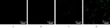
Fig. 1
Fluorescence microscope images of intracellular ROS levels in GC-2 cells in various groups"
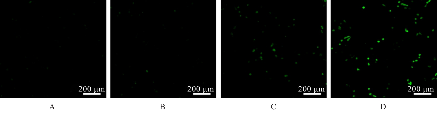
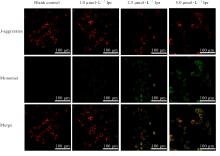
Fig. 2
Fluorescence microscope images of mitochondrial membrane potential in GC-2 cells in various groups"
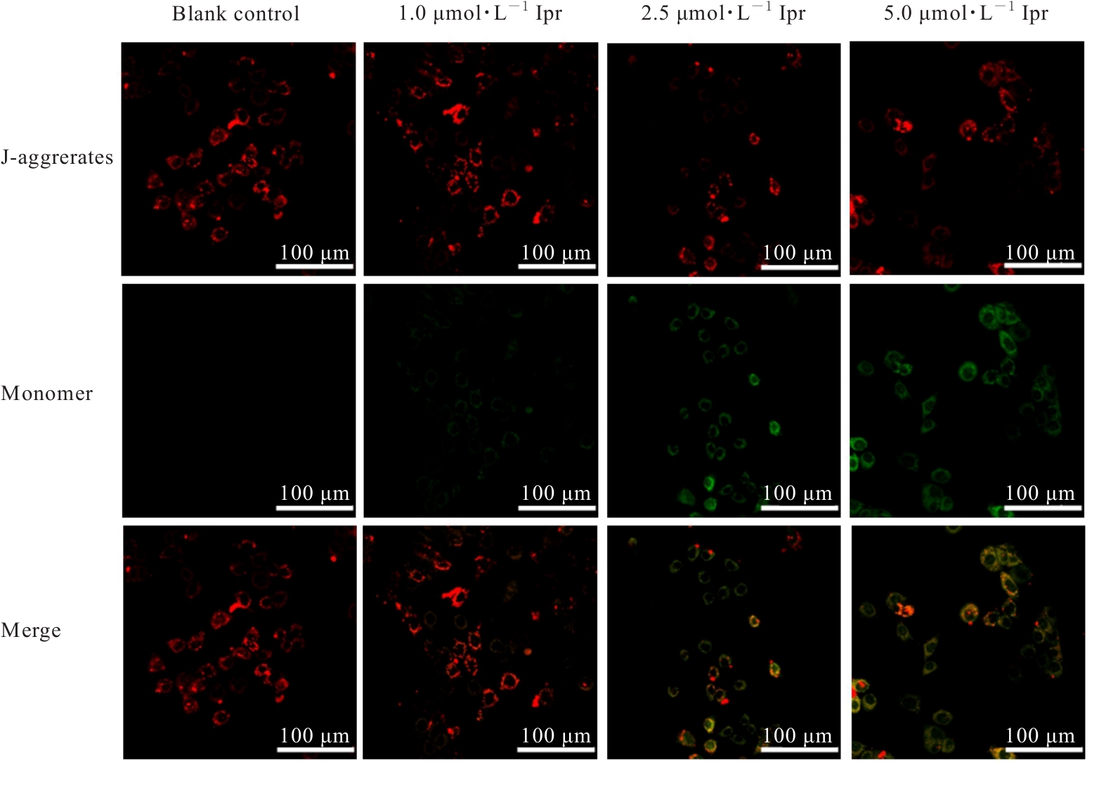

Fig. 3
Electrophoregram of expressions of HO-1,GPX4, and FTH1 proteins in GC-2 cells in various groups"
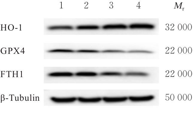
Tab.3
Expression levels of HO-1, GPX4, and FTH1 proteins in GC-2 cells in various groups"
| Group | HO-1 | GPX4 | FTH1 |
|---|---|---|---|
| Blank control | 1.00±0.00 | 1.00±0.00 | 1.00±0.00 |
| 1.0 μmol·L-1 Ipr | 1.22±0.04** | 0.83±0.21 | 0.98±0.12 |
| 2.5 μmol·L-1 Ipr | 1.37±0.06** | 0.62±0.07** | 0.71±0.22* |
| 5.0 μmol·L-1 Ipr | 1.53±0.12** | 0.55±0.07** | 0.49±0.07** |

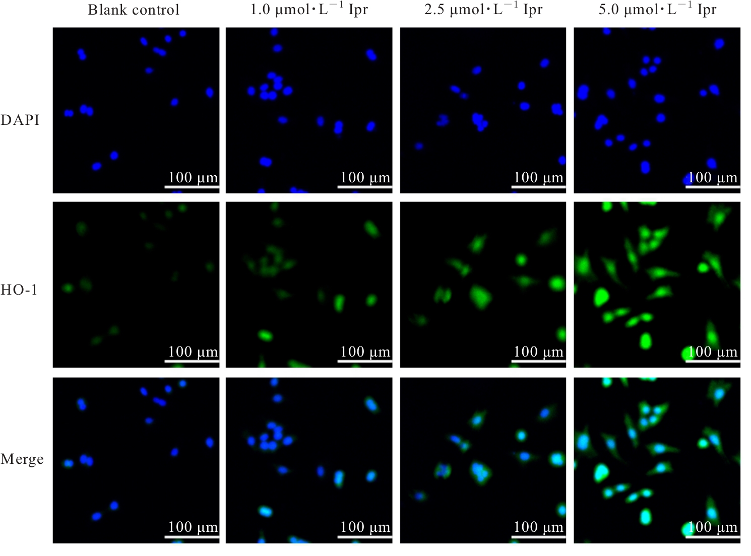
Fig. 4
Fluorescence intensities of HO-1 protein in GC-2 cells in various groups detected of immunofluorescence method"
| [1] | JENARDHANAN P, PANNEERSELVAM M, MATHUR P P. Effect of environmental contaminants on spermatogenesis[J]. Semin Cell Dev Biol, 2016, 59: 126-140. |
| [2] | CORPUZ-HILSABECK M, CULTY M. Impact of endocrine disrupting chemicals and pharmaceuticals on sertoli cell development and functions[J]. Front Endocrinol, 2023, 14: 1095894. |
| [3] | TVRDA E, PEER R, SIKKA S C, et al. Iron and copper in male reproduction: a double-edged sword[J]. J Assist Reprod Genet, 2015, 32(1): 3-16. |
| [4] | XIONG L, BIN Z, YOUNG J L, et al. Exposure to low-dose cadmium induces testicular ferroptosis[J]. Ecotoxicol Environ Saf, 2022, 234: 113373. |
| [5] | ZHANG X H, JIANG L P, CHEN H B, et al. Resveratrol protected acrolein-induced ferroptosis and insulin secretion dysfunction via ER-stress- related PERK pathway in MIN6 cells[J]. Toxicology, 2022, 465: 153048. |
| [6] | YANG L, JIANG L P, SUN X C, et al. DEHP induces ferroptosis in testes via p38α-lipid ROS circulation and destroys the BTB integrity[J]. Food Chem Toxicol, 2022, 164: 113046. |
| [7] | SU Y J, LIU Z L, XIE K Y, et al. Ferroptosis: a novel type of cell death in male reproduction[J]. Genes, 2022, 14(1): 43. |
| [8] | STOCKWELL B R, FRIEDMANN ANGELI J P, BAYIR H, et al. Ferroptosis: a regulated cell death nexus linking metabolism, redox biology, and disease[J]. Cell, 2017, 171(2): 273-285. |
| [9] | WANG R K, DENG L G, WANG Y H, et al. Synergistic effects of combined lead and iprodione exposure on P53 signaling-mediated hepatotoxicity, enterotoxicity and transgenerational toxicity in zebrafish[J]. Sci Total Environ, 2025, 958: 178127. |
| [10] | HASSAN M A, BOHY K M E L, SHARKAWY N I E L, et al. Iprodione and chlorpyrifos induce testicular damage, oxidative stress, apoptosis and suppression of steroidogenic- and spermatogenic-related genes in immature male albino rats[J]. Andrologia, 2021, 53(4): e13978. |
| [11] | ABD-ELHAKIM Y M, SHARKAWY N I E L, BOHY K M E L, et al. Iprodione and/or chlorpyrifos exposure induced testicular toxicity in adult rats by suppression of steroidogenic genes and SIRT1/TERT/PGC-1α pathway[J]. Environ Sci Pollut Res Int, 2021, 28(40): 56491-56506. |
| [12] | SHARMA P, HUQ A U, SINGH R. Cypermethrin-induced reproductive toxicity in the rat is prevented by resveratrol[J]. J Hum Reprod Sci, 2014, 7(2): 99-106. |
| [13] | ARTINI P G, CASAROSA E, CARLETTI E, et al. In vitro effect of myo-inositol on sperm motility in normal and oligoasthenospermia patients undergoing in vitro fertilization[J]. Gynecol Endocrinol, 2017, 33(2): 109-112. |
| [14] | ARMSTRONG J S, RAJASEKARAN M, CHAMULITRAT W, et al. Characterization of reactive oxygen species induced effects on human spermatozoa movement and energy metabolism[J]. Free Radic Biol Med, 1999, 26(7/8): 869-880. |
| [15] | LIU T, HOU B L, WANG Z P, et al. Polystyrene microplastics induce mitochondrial damage in mouse GC-2 cells[J]. Ecotoxicol Environ Saf, 2022, 237: 113520. |
| [16] | MAIORINO M, CONRAD M, URSINI F. GPx4, lipid peroxidation, and cell death: discoveries, rediscoveries, and open issues[J]. Antioxid Redox Signal, 2018, 29(1): 61-74. |
| [17] | GROSSHANS K, CALVIN H I. Estimation of glutathione in purified populations of mouse testis germ cells[J]. Biol Reprod, 1985, 33(5): 1197-1205. |
| [18] | ABDULLAH F, KHAN NOR-ASHIKIN M N, AGARWAL R, et al. Glutathione (GSH) improves sperm quality and testicular morphology in streptozotocin-induced diabetic mice[J]. Asian J Androl, 2021, 23(3): 281-287. |
| [19] | JIANG X J, STOCKWELL B R, CONRAD M. Ferroptosis: mechanisms, biology and role in disease[J]. Nat Rev Mol Cell Biol, 2021, 22(4): 266-282. |
| [20] | DIXON S J, OLZMANN J A. The cell biology of ferroptosis[J]. Nat Rev Mol Cell Biol, 2024, 25(6): 424-442. |
| [21] | TANG D L, CHEN X, KANG R, et al. Ferroptosis: molecular mechanisms and health implications[J]. Cell Res, 2021, 31(2): 107-125. |
| [22] | WANG X L, YIN L S, WEN Y J, et al. Mitochondrial regulation during male germ cell development[J]. Cell Mol Life Sci, 2022, 79(2): 91. |
| [23] | DAVIES R, MINHAS S, JAYASENA C N. The role of seminal reactive oxygen species assessment in the setting of infertility and early pregnancy loss[J]. World J Urol, 2023, 41(11): 3257-3265. |
| [24] | LIU J, KANG R, TANG D. Signaling pathways and defense mechanisms of ferroptosis[J]. FEBS J, 2022, 289(22): 7038-7050. |
| [25] | ZHAO X, LIU Z H, GAO J, et al. Inhibition of ferroptosis attenuates busulfan-induced oligospermia in mice[J]. Toxicology, 2020, 440: 152489. |
| [26] | ADEDOYIN O, BODDU R, TRAYLOR A, et al. Heme oxygenase-1 mitigates ferroptosis in renal proximal tubule cells[J]. Am J Physiol Renal Physiol, 2018, 314(5): F702-F714. |
| [27] | WU Y H, WANG J K, ZHAO T X, et al. Di-(2-ethylhexyl) phthalate exposure leads to ferroptosis via the HIF-1α/HO-1 signaling pathway in mouse testes[J]. J Hazard Mater, 2022, 426: 127807. |
| [28] | TORTI F M, TORTI S V. Regulation of ferritin genes and protein[J]. Blood, 2002, 99(10): 3505-3516. |
| [29] | SAMMARCO M C, DITCH S, BANERJEE A, et al. Ferritin L and H subunits are differentially regulated on a post-transcriptional level[J]. J Biol Chem, 2008, 283(8): 4578-4587. |
| [30] | ZHUGE R, ZHANG L, XUE Q, et al. Ferritinophagy is involved in hexavalent chromium-induced ferroptosis in Sertoli cells[J]. Toxicol Appl Pharmacol, 2024, 492: 117139. |
| [1] | Ziyi TANG,Shiying YANG,Tianzhen YANG,Wenqiang LIU,Jiangxue ZHONG,Li YIN. Induction effect of pesticide pyraclostrobin on ferroptosis of spermatocytes GC-2 of mice [J]. Journal of Jilin University(Medicine Edition), 2026, 52(1): 18-25. |
| [2] | Huiyuan YU,Ling JIN,Ying YU,Xue WANG,Bing WANG. Improvement effect of epicatechin on liver injury in mice induced by acetaminophen and its mechanism [J]. Journal of Jilin University(Medicine Edition), 2025, 51(6): 1498-1507. |
| [3] | Han LIN,Qiuyan YANG,Jieyue ZHONG,Bolun CHEN,Wangxia TONG. Improvement effect of cordycepin on ferroptosis in HepG2 cells induced by RSL3 and its mechanism [J]. Journal of Jilin University(Medicine Edition), 2025, 51(3): 576-589. |
| [4] | Kaiqi NIU,He CHANG,Guangfu LYU,Pengyu ZHENG,Xueting CHI,Jia ZHOU,Yuchen WANG,Xiaowei HUANG. Inhibitory effect of astragaloside Ⅳ on cisplatin-induced liver injury in mice and its mechanism [J]. Journal of Jilin University(Medicine Edition), 2025, 51(2): 370-377. |
| [5] | Liang LI,Xiangdong ZHOU,Jie WANG,Chaoqun XU,Mengxia ZHU,Shanjun YU,Qi LI. Effect of bitter-taste receptor T2R38 activation on ferroptosis of human airway epithelium NuLi-1 cells induced by cigarette smoke exposure and its mechanism [J]. Journal of Jilin University(Medicine Edition), 2025, 51(2): 333-340. |
| [6] | Yanyan BAI,Yutong ZHOU,Haijuan SUI,Zhuo LIU. Improvement effect of asiatic acid on damage of lipopolysaccharide-induced hippocampum neuron in rats through Nrf2/HO-1 signaling pathway [J]. Journal of Jilin University(Medicine Edition), 2025, 51(1): 85-95. |
| [7] | Guobin HE,Huan WANG. Effect of knockdown of RIP3 on autophagy, pyroptosis, and ferroptosis of hypoxia/reoxygenation-induced human renal tubular epithelial HK2 cells [J]. Journal of Jilin University(Medicine Edition), 2024, 50(6): 1644-1653. |
| [8] | Yi LONG,Ziyi YOU,Xiuying TAN,Rou ZHANG,Yuhan ZHANG,Lina YANG. Protective effect of sodium butyrate on acute liver injury in mice induced by lipopolysaccharide combined with D-galactosamine and its mechanism [J]. Journal of Jilin University(Medicine Edition), 2024, 50(6): 1614-1620. |
| [9] | Baolian MA,Xiaoxue HU,Xiaowen AI,Yonglan ZHANG. Inhibitory effect of diosmetin on ferroptosis of GC-2 spermatocytes induced by RSL3 in mice and its mechanism [J]. Journal of Jilin University(Medicine Edition), 2024, 50(6): 1481-1490. |
| [10] | Xiaoyong PENG,Yu ZHU,Shuangbo ZHANG,Yingguo ZHU,Tao LI,Liangming LIU,Jianmin WANG,Guangming YANG. Alleviative effect of fluid resuscitation on damage of structure injury of vascular cells after blast injury complicated with hemorrhagic shock in rats by inhibiting ferroptosis of vascular tissue [J]. Journal of Jilin University(Medicine Edition), 2024, 50(5): 1227-1234. |
| [11] | Fangyang JIANG,Jing XIAO,He CHANG,Mingyang SUN,Wenjing ZHANG,Guangfu LYU,He LIN,Zhe LIN,Xiaowei HUANG,Yuchen WANG. Effect of polygonatum odoratum polysaccharide on acute kidney injury in mice induced by cisplatin and its ferroptosis mechanism [J]. Journal of Jilin University(Medicine Edition), 2024, 50(5): 1235-1242. |
| [12] | Junping WEI,Dajia FU,Qingwen MENG,Daofei LIN,Yanzai LIN. Effect of bone marrow mesenchymal stem cell-derived exosomes on myocardial fibrosis in rats induced by isoproterenol and its mechanism [J]. Journal of Jilin University(Medicine Edition), 2024, 50(5): 1348-1357. |
| [13] | Yanjue YE,Ziyi TANG,Yupei TAN,Shiying YANG,Yong LIU,Li YIN. Effect of azathioprine on ferroptosis in spermatocytes of mice induced by RSL3 [J]. Journal of Jilin University(Medicine Edition), 2024, 50(5): 1217-1226. |
| [14] | Yingqun NI,Mao YANG,Di YANG,Chenglin GUO,Wenjun ZHU,Yaqin YU,Qin LU,Jinzhi LUO,Chunqin WU,Zhaohui FANG. Screening of key differentially expressed genes involved in osteogenic differentiation of lower limb vascular smooth muscle cells and validation [J]. Journal of Jilin University(Medicine Edition), 2024, 50(3): 620-627. |
| [15] | Ruipeng ZHANG,Jie LI. Resistance and regeneration effects of lncRNA GPRC5D-AS1 on muscle atrophy of myocytes in mice induced by dexamethasone and its mechanism [J]. Journal of Jilin University(Medicine Edition), 2023, 49(6): 1457-1465. |
|
||