Journal of Jilin University(Medicine Edition) ›› 2022, Vol. 48 ›› Issue (6): 1462-1473.doi: 10.13481/j.1671-587X.20220612
• Research in basic medicine • Previous Articles Next Articles
Effect of lncRNA-MIAT on M2-type polarization of tumor-associated macrophages and its mechanism
Jing XU1( ),Jian GUO1,Xingwei PU2,Daxing LI1
- 1.Department of Orthopedics, Guizhou Provincial Orthopedic Hospital, Guiyang 550000, China
2.Department of Spine, Guizhou Orthopedic Hospital, Guiyang 550000, China
-
Received:2022-01-31Online:2022-11-28Published:2022-12-07 -
Contact:Jing XU E-mail:xuj1101@163.com
CLC Number:
- R33
Cite this article
Jing XU,Jian GUO,Xingwei PU,Daxing LI. Effect of lncRNA-MIAT on M2-type polarization of tumor-associated macrophages and its mechanism[J].Journal of Jilin University(Medicine Edition), 2022, 48(6): 1462-1473.
share this article
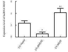
Fig. 1
Expression levels of lncRNA-MIAT in THP-1 cells in various groups detected by RT-qPCR method"


Fig. 2
Percentages of M2-type macrophages in THP-1 cells in various groups detected by flow cytometry method"


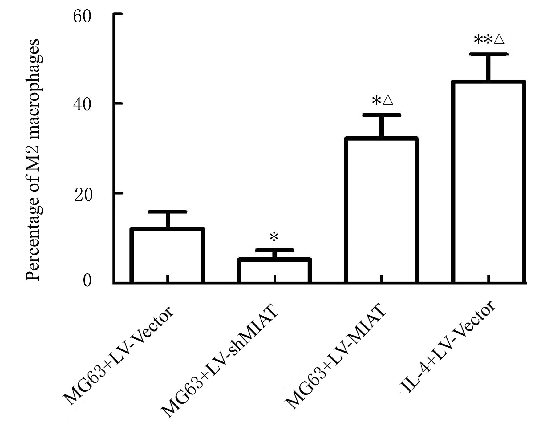
Fig. 3
Percentages of polarized M2 macrophages in THP-1 cells in various groups"
Tab. 1
Levels of VEGF, IL-10,and TGF-β1 in supernatant of THP-1 cells in various groups [n=5,x±s,ρB /(ng·L-1)]"
| Group | VEGF | IL-10 | TGF-β1 |
|---|---|---|---|
| MG63+LV-Vector | 3.15±0.93 | 2.07±0.48 | 2.53±0.61 |
| MG63+LV-shMIAT | 1.91±0.16* | 0.82±0.15* | 1.46±0.28* |
| MG63+LV-MIAT | 6.44±1.05*△ | 4.82±0.71*△ | 5.17±0.92*△ |
| IL-4+LV-Vector | 8.21±1.19*△ | 9.74±0.83*△ | 11.05±1.66*△ |
Fig. 4
Morphology of proliferation of HUVEC in various groups detected by EdU staining method (Bar=100 μm)"
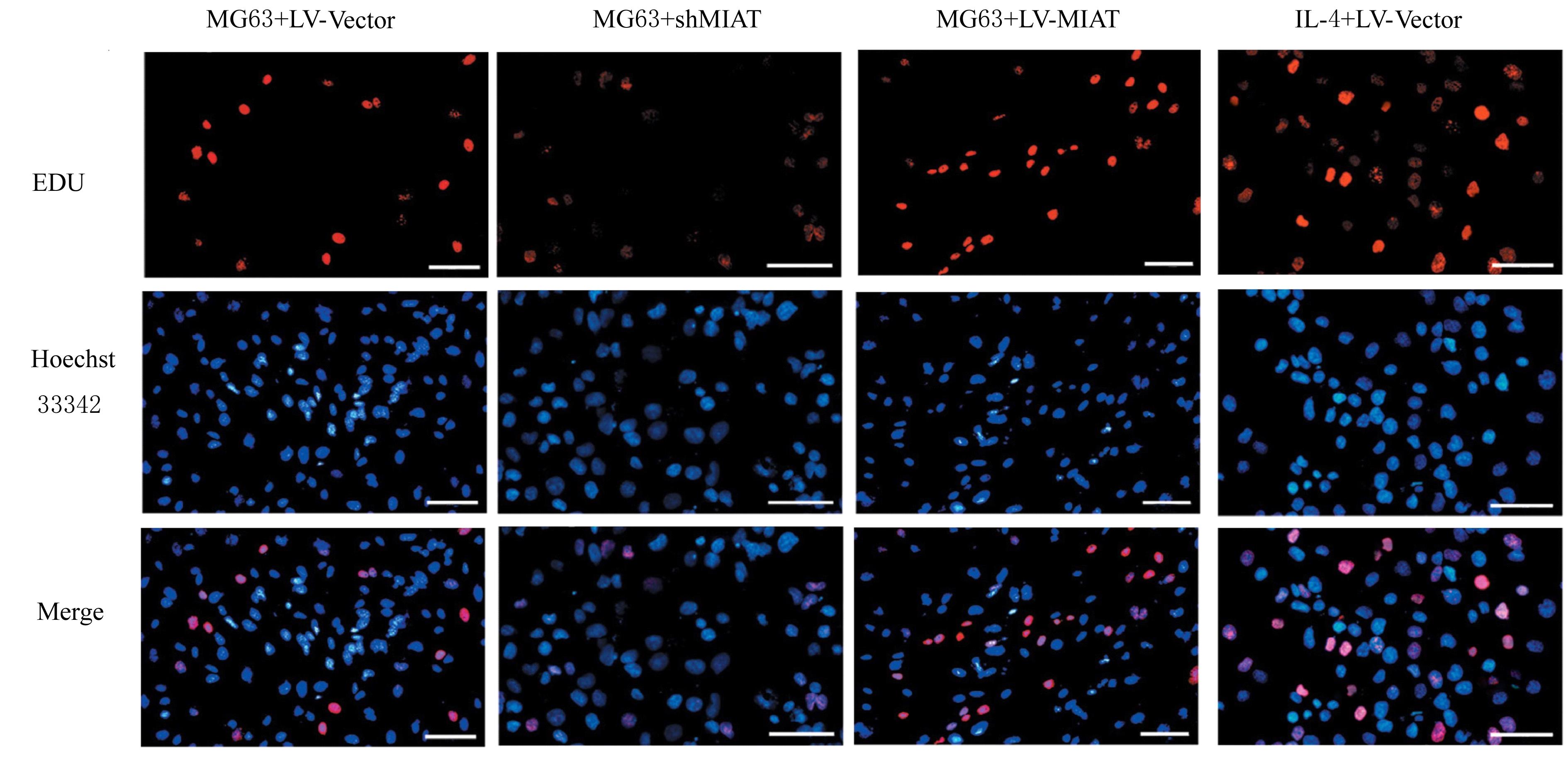

Fig. 5
Positive rates of EdU in HUVEC in various groups"

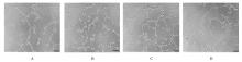
Fig. 6
Angiogenesis of HUVEC in various groups detected by tube formation method (Bar=100 μm)"
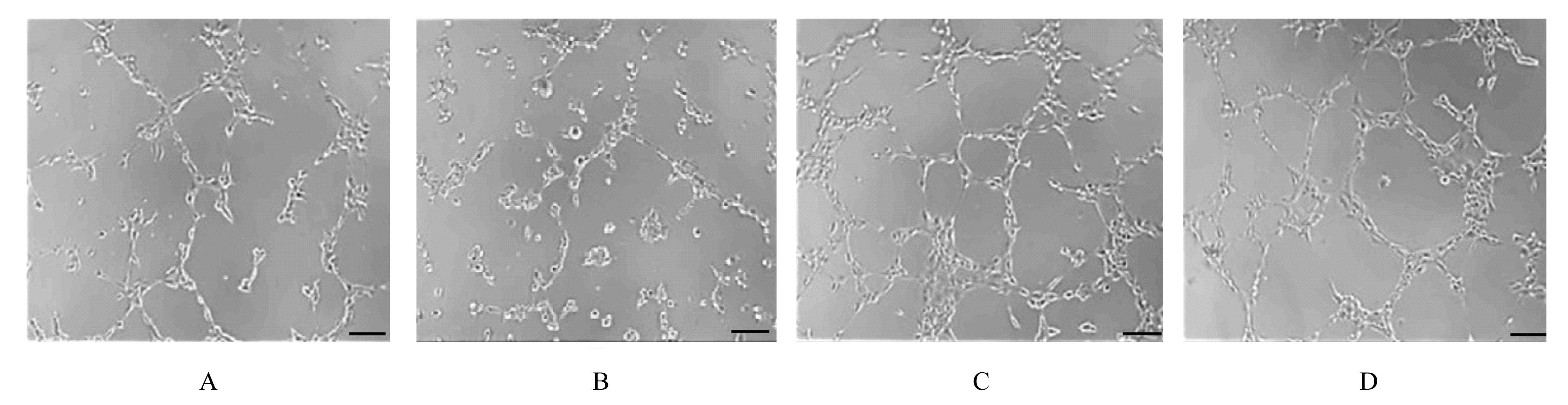
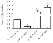
Fig. 7
Number of tube formation of HUVEC in various groups"


Fig. 8
Electrophoregram(A)and histogram (B) of expressions levels of STAT6, p-STAT6,and JAK1 proteins in THP-1 cells in various groups detected by Western blotting method"
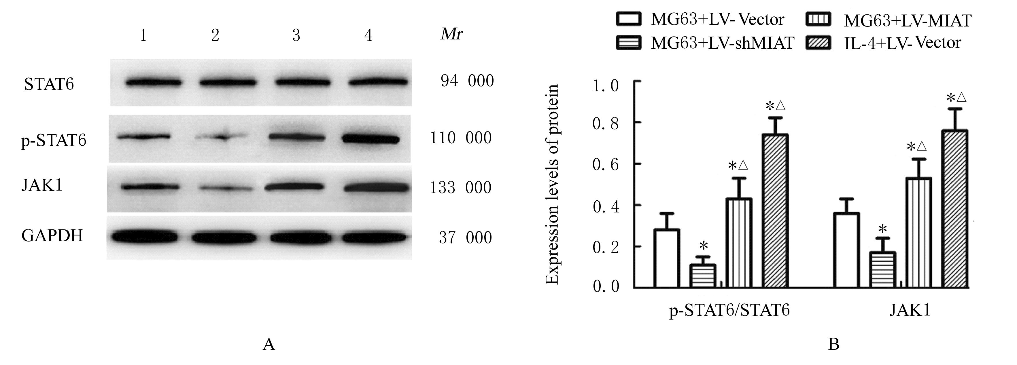

Fig. 9
Electrophoregram( A)and histogram (B) of expressions of VEGFR2,Notch1,and DLL4 proteins in HUVEC in various groups detected by Western blotting method"

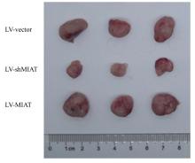
Fig. 10
Gross morphology of transplanted tumor of nude mice in various groups"
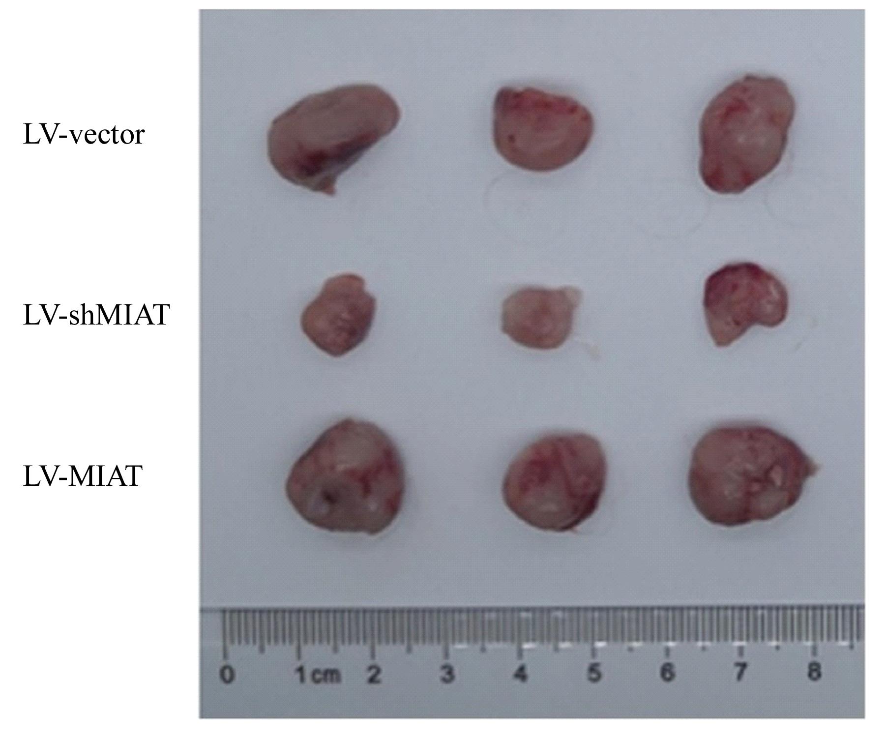

Fig. 11
Volumes of transplanted tumor of nude mice in various groups"


Fig. 12
Weights of transplanted tumor of nude mice in various groups"
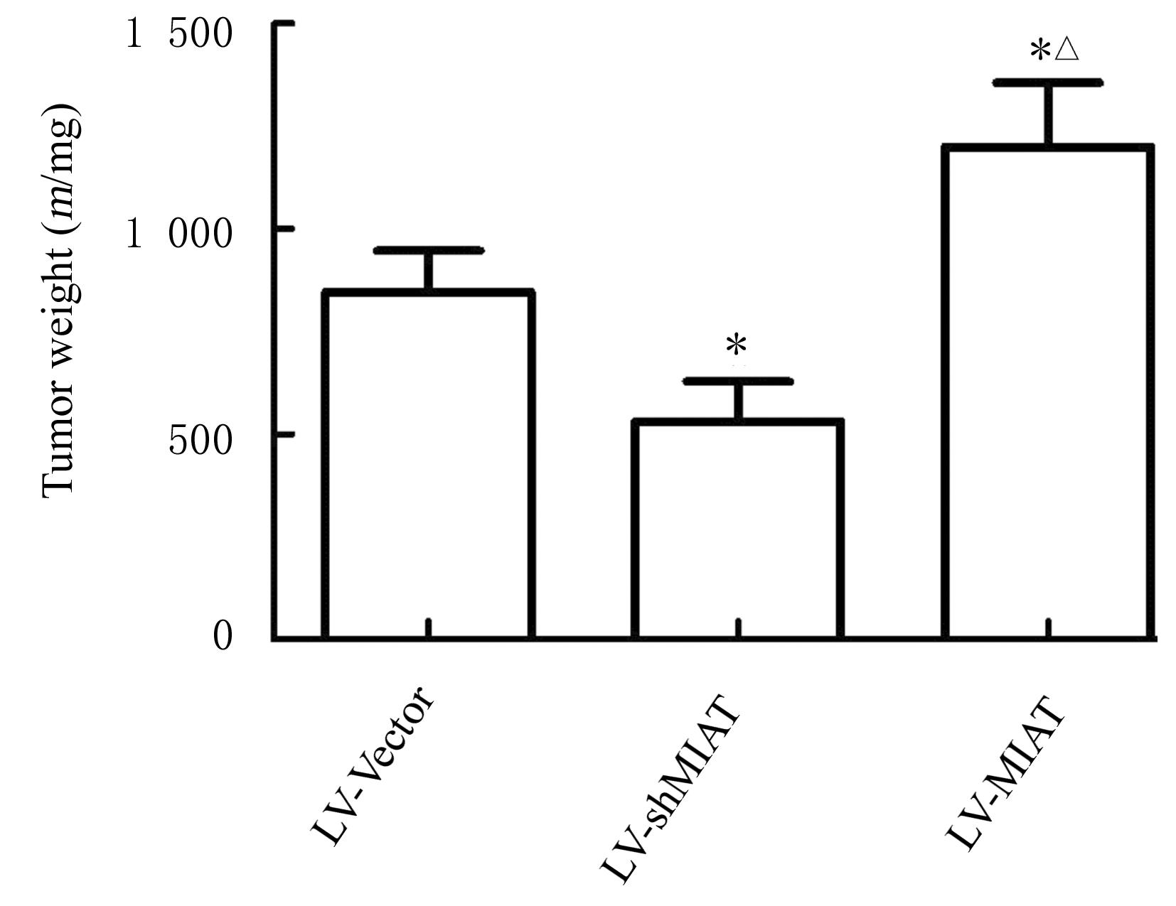
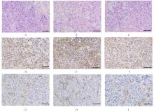
Fig. 13
Expressions of CD163 and CD31 in transplanted tumor tissue of nude mice in various groups (Bar=100 μm)"

| 1 | TAHIR I, ANDREI V, POLLOCK R, et al. Malignant giant cell tumour of bone: a review of clinical, pathological and imaging features[J]. Skeletal Radiol, 2022, 51(5): 957-970. |
| 2 | LIN Z L, WU Z Y, LUO W. Chimeric antigen receptor T-cell therapy: the light of day for osteosarcoma[J]. Cancers, 2021, 13(17): 4469. |
| 3 | LOCATI M, CURTALE G, MANTOVANI A, Diversity,mechanisms, and significance of macrophage plasticity [J]. Annu Rev Pathol, 2020, 15: 123-147. |
| 4 | WU K Y, LIN K J, LI X Y, et al. Redefining tumor-associated macrophage subpopulations and functions in the tumor microenvironment[J]. Front Immunol, 2020, 11: 1731. |
| 5 | MOHAPATRA S, PIOPPINI C, OZPOLAT B, et al. Non-coding RNAs regulation of macrophage polarization in cancer[J]. Mol Cancer, 2021, 20(1): 24. |
| 6 | WINKLER L, DIMITROVA N. A mechanistic view of long noncoding RNAs in cancer[J]. Wiley Interdiscip Rev RNA, 2022, 13(3): e1699. |
| 7 | ZHANG Y Y, MAO Q J, XIA Q M, et al. Noncoding RNAs link metabolic reprogramming to immune microenvironment in cancers[J]. J Hematol Oncol, 2021, 14(1): 169. |
| 8 | WANG Y F, FU L Y, LU T T, et al. Clinicopathological and prognostic significance of long non-coding RNA MIAT in human cancers: a review and meta-analysis[J]. Front Genet, 2021, 12: 729768. |
| 9 | ZHANG C Y, XIE L S, LIANG H L, et al. LncRNA MIAT facilitates osteosarcoma progression by regulating mir-128-3p/VEGFC axis[J].IUBMB Life,2019,71(7): 845-853. |
| 10 | JI Q, ZHU J, FANG C L, et al. Down-regulation of MIAT suppresses osteosarcoma progression by acting as a ceRNA for miR-141-3p to regulate SIX1-mediated PI3K/AKT pathway[J]. Eur Rev Med Pharmacol Sci, 2020, 24(5): 2218-2228. |
| 11 | WANG Z Y, YANG K, ZHAO L, et al. LncRNA MIAT downregulates IL-1β, TNF-α to suppress macrophage inflammation but is suppressed by ATP-induced NLRP3 inflammasome activation[J]. Cell Cycle, 2021, 20(2): 194-203. |
| 12 | LI P F, HAO Z F, WU J Y, et al. Comparative proteomic analysis of polarized human THP-1 and mouse RAW264.7 macrophages[J]. Front Immunol, 2021, 12: 700009. |
| 13 | HUANG Y F, LU L, SHEN H L, et al. LncRNA SNHG4 promotes osteosarcoma proliferation and migration by sponging miR-377-3p[J]. Mol Genet Genomic Med, 2020, 8(8): e1349. |
| 14 | ZHOU S Y, XU A L, SONG T F, et al. lncRNA MIAT regulates cell growth, migration, and invasion through sponging miR-150-5p in ovarian cancer[J]. Cancer Biother Radiopharm, 2020, 35(9): 650-660. |
| 15 | SONG F C, YANG Y, LIU J X. Long non-coding RNA MIAT promotes the proliferation and invasion of laryngeal squamous cell carcinoma cells by sponging microRNA-613[J]. Exp Ther Med, 2021, 21(3): 232. |
| 16 | WANG L X, ZHANG S X, WU H J, et al. M2b macrophage polarization and its roles in diseases[J]. J Leukoc Biol, 2019, 106(2): 345-358. |
| 17 | ATRI C, GUERFALI F Z, LAOUINI D. Role of human macrophage polarization in inflammation during infectious diseases[J]. Int J Mol Sci, 2018, 19(6): 1801. |
| 18 | RHEE I. Diverse macrophages polarization in tumor microenvironment[J]. Arch Pharm Res, 2016, 39(11): 1588-1596. |
| 19 | HAO J, HU Y X, LI Y M, et al. Involvement of JNK signaling in IL4-induced M2 macrophage polarization[J]. Exp Cell Res, 2017, 357(2): 155-162. |
| 20 | CUI L Y, YANG G D, YE J N, et al. Dioscin elicits anti-tumour immunity by inhibiting macrophage M2 polarization via JNK and STAT3 pathways in lung cancer[J]. J Cell Mol Med, 2020, 24(16): 9217-9230. |
| 21 | HE Y, GAO Y, ZHANG Q, et al. IL-4 switches microglia/macrophage M1/M2 polarization and alleviates neurological damage by modulating the JAK1/STAT6 pathway following ICH[J]. Neuroscience, 2020, 437: 161-171. |
| 22 | PARK Y G, CHOI J, JUNG H K, et al. Baicalein inhibits tumor progression by inhibiting tumor cell growth and tumor angiogenesis[J]. Oncol Rep, 2017, 38(5): 3011-3018. |
| [1] | Junxiu LIU,Jia ZHOU,Guangfu LYU,Yuchen WANG,Xuefeng ZHUANG,Jiarui ZHAO,Xiaowei HUANG,Ruili LI. Protective effect of Shenhong Buxue Granule on vascular endothelium of mice with vascular endothelial dysfunction of Qi stagnation and blood stasis type and its mechanism [J]. Journal of Jilin University(Medicine Edition), 2022, 48(6): 1437-1447. |
| [2] | Chengyuan HE,Hongyu YANG,Yujing TAN,Hang SU,Hongshu LI,Chun LI. Expression of IL-17A in non-small cell lung cancer tissue and its regulation on VEGF expression via NF-κB signaling pathway [J]. Journal of Jilin University(Medicine Edition), 2022, 48(4): 1003-1009. |
| [3] | YUE Sheng, QIAO Guohua, YUE Lei, ZHU Ping. Inhibitory effect of berberine on Ang Ⅱ-induced apoptosis of human umbilical vein endothelial cells via ROS/JNK signaling pathway and its mechanism [J]. Journal of Jilin University(Medicine Edition), 2019, 45(04): 872-876. |
| [4] | LIU Lixing, MIN Xianhui, ZHANG Min, HU Jianzhang. Expression of ABCA1 in serum of patients with preeclampsia and its effect on injury of endothelial cells [J]. Journal of Jilin University Medicine Edition, 2015, 41(03): 448-453. |
| [5] | ZHONG Ling-zhi,LI Wen-xue,LI Xiu-ying,LI Xin-na,LI Yu-lin,LI Rong-gui. Construction of eukaryotic expression vector of human mitochondrial superoxide dismutase and its expression in HUVEC [J]. Journal of Jilin University Medicine Edition, 2014, 40(01): 10-14. |
| [6] | HAO Tie-zhu,SHEN Huan,TAO Zhi-kan,WANG Wei,ZHAO Dan-dan. Influence of lovastatin in expressions of MCP-1 and IL-10 of HUVECs treated with TNF-α [J]. Journal of Jilin University Medicine Edition, 2013, 39(5): 928-932. |
| [7] | TAO Tao,CHEN Hong,GU Xue-shuang,ZHOU Jun-hao,PENG Dan-yi,ZHANG Li-jun. [J]. Journal of Jilin University Medicine Edition, 2013, 39(3): 559-564. |
| [8] | TONG Guang-hui1|TONG Wei-wei1|MA Li2|LIU Yong1. Effects of glucose and insulin on |production of solubility endothelial cell protein C receptor in human umbilical vein endothelial cells [J]. J4, 2011, 37(5): 851-854. |
| [9] | GAO Ying,ZHANG Yun-jian,LI Yun,TAN Yan. Protective effects of radix astragali on ECV304 cells exposed to high level of glucose [J]. J4, 2008, 34(5): 810-813. |
|