Journal of Jilin University(Medicine Edition) ›› 2026, Vol. 52 ›› Issue (1): 125-134.doi: 10.13481/j.1671-587X.20260113
• Research in basic medicine • Previous Articles Next Articles
Therapeutic effect of prunetin on myocardial infarction model mice and its mechanism
Meng CAI,Yang GUO,Yingfang MA,Jinglei CUI,Jia LUO,Lili WEI,Yunhua ZHANG,Yang WANG( )
)
- Key Laboratory of Xinjiang Local and Ethnic High incidence Diseases,Ministry of Education,School of Medicine,Shihezi University,Shihezi 832000,China
-
Received:2025-01-09Accepted:2025-03-01Online:2026-01-28Published:2026-02-24 -
Contact:Yang WANG E-mail:wangyang_shz@foxmail.com
CLC Number:
- R542.22
Cite this article
Meng CAI,Yang GUO,Yingfang MA,Jinglei CUI,Jia LUO,Lili WEI,Yunhua ZHANG,Yang WANG. Therapeutic effect of prunetin on myocardial infarction model mice and its mechanism[J].Journal of Jilin University(Medicine Edition), 2026, 52(1): 125-134.
share this article
Tab.1
LVEF and LVFS of mice in various groups"
| Group | n | LVEF | LVFS |
|---|---|---|---|
| Sham operation | 9 | 75.76±3.34 | 43.72±3.09 |
| Model | 8 | 36.15±11.33* | 17.47±5.74* |
| Low dose of prunetin | 8 | 46.75±4.26△ | 23.20±4.08△ |
| High dose of prunetin | 8 | 52.65±4.97△ | 27.32±3.38△ |
| Enalaprilat | 8 | 59.42±3.52△ | 31.10±2.25△ |
Tab.2
Levels of serum CK-MB and cTn-Ⅰ of in mice in various groups"
| Group | CK-MB [ρB/(μg·L-1)] | cTn-Ⅰ [ρB/(ng·L-1)] |
|---|---|---|
| Sham operation | 4.53±2.12 | 33.33±13.41 |
| Model | 23.44±9.53* | 62.42±7.05* |
| Low dose of prunetin | 7.00±2.30△ | 32.46±14.85△ |
| High dose of prunetin | 5.83±1.38△ | 28.70±11.70△ |
| Enalaprilat | 6.27±1.37△ | 48.83±11.12△ |
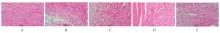
Fig. 1
Pathomorphology of myocardium tissue of mice in various groups(HE,×400)"
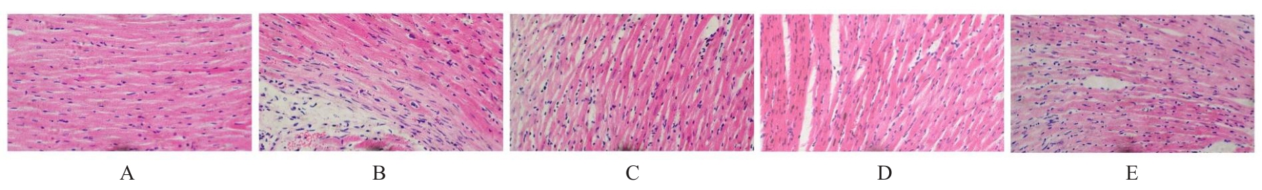
Fig. 2
Fibrotic degrees of myocardium tissue of mice in various groups (Masson,×150)"
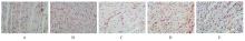
Fig. 3
Expressions of CD31 protein in myocardium tissue of MI border zone of mice in variou groups (Immunohistochemistry,×400)"
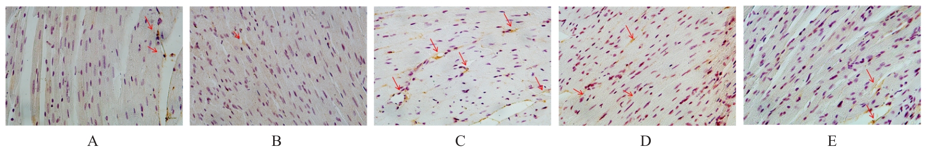
Fig. 4
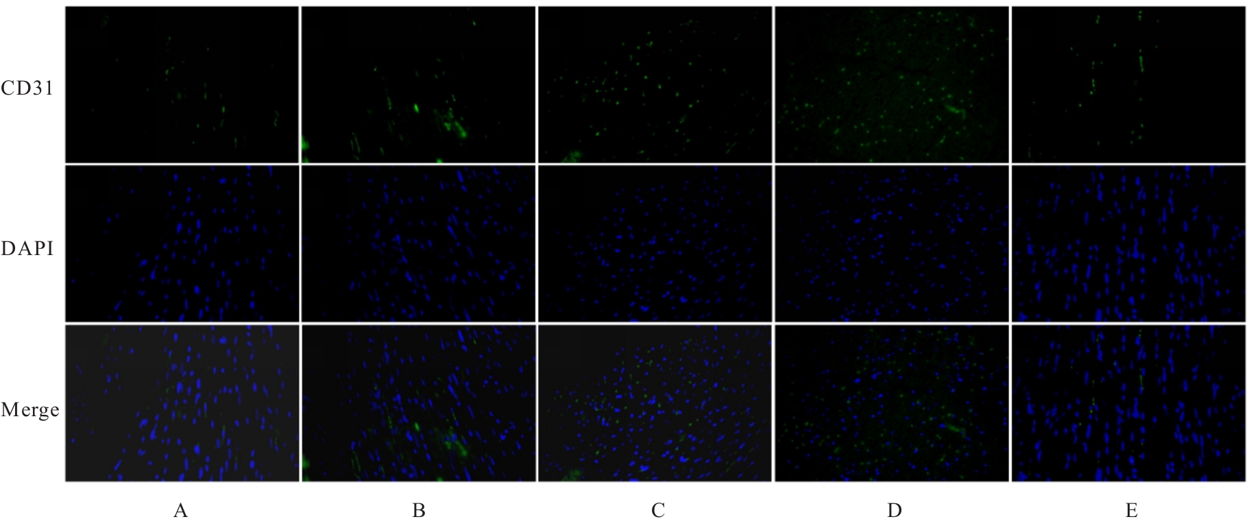
Expressions of CD31 protein in MI marginal zone of mice in various groups (Immunofluorecence,×400)"
Tab.3
Number of new vessels in MI marginal zone of mice in various groups"
| Group | Number of new vessels |
|---|---|
| Sham operation | 12.24±5.62 |
| Model | 10.67±3.74 |
| Low dose of prunetin | 18.22±5.26 |
| High dose of prunetin | 22.06±8.65* |
| Enalaprilat | 12.11±6.50 |
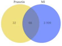
Fig. 5
Intersection diagram of target points of prunetin and MI"
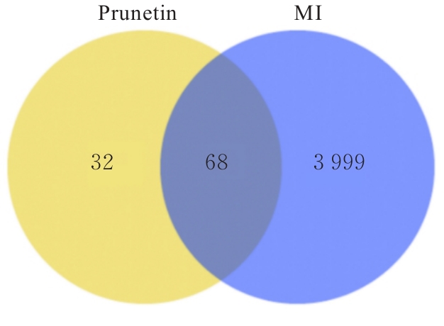
Fig. 6
PPI network diagram(A) and target network (B) of prunetin and MI"
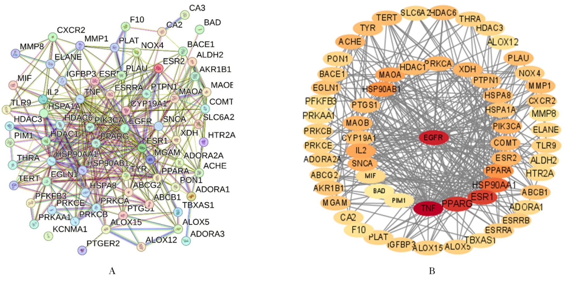
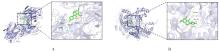
Fig. 7
Molecule docking diagram of prunetin with EGFR(A) and prunetin with PIK3CA(B)"

| [1] | O’DONNELL A, YUTZEY K E. Mechanisms of heart valve development and disease[J]. Development, 2020, 147(13): dev183020. |
| [2] | SU M X, CUI J M, ZHAO J, et al. Skimmin ameliorates cardiac function via the regulation of M2 macrophages in a myocardial infarction mouse model[J]. Perfusion, 2023, 38(6): 1298-1307. |
| [3] | ZHANG H, FABER J E. De-novo collateral formation following acute myocardial infarction: Dependence on CCR2⁺ bone marrow cells[J]. J Mol Cell Cardiol, 2015, 87: 4-16. |
| [4] | ZHANG H, PRABHAKAR P, SEALOCK R, et al. Wide genetic variation in the native pial collateral circulation is a major determinant of variation in severity of stroke[J]. J Cereb Blood Flow Metab, 2010, 30(5): 923-934. |
| [5] | 张 淳, 湛 疆. 敲除组蛋白脱乙酰酶6对心肌缺血再灌注损伤小鼠心脏功能的保护作用[J]. 郑州大学学报(医学版), 2025, 60(6): 785-788. |
| [6] | GOUIN K H 3rd, HELLSTROM S K, CLEGG L E, et al. Arterialized collateral capillaries progress from nonreactive to capable of increasing perfusion in an ischemic arteriolar tree[J]. Microcirculation, 2018, 25(3): e12438. |
| [7] | BRITTEN M B, ABOLMAALI N D, ASSMUS B, et al. Infarct remodeling after intracoronary progenitor cell treatment in patients with acute myocardial infarction (TOPCARE-AMI): mechanistic insights from serial contrast-enhanced magnetic resonance imaging[J]. Circulation, 2003, 108(18): 2212-2218. |
| [8] | AGHAJANIAN A, ZHANG H, BUCKLEY B K, et al. Decreased inspired oxygen stimulates de novo formation of coronary collaterals in adult heart[J]. J Mol Cell Cardiol, 2021, 150: 1-11. |
| [9] | WANG N, CHEN C Y, YANG D Z, et al. Mesenchymal stem cells-derived extracellular vesicles, via miR-210, improve infarcted cardiac function by promotion of angiogenesis[J]. Biochim Biophys Acta Mol Basis Dis, 2017, 1863(8): 2085-2092. |
| [10] | NASSER M I, MASOOD M, ADLAT S, et al. Mesenchymal stem cell-derived exosome microRNA as therapy for cardiac ischemic injury[J]. Biomed Pharmacother, 2021, 143: 112118. |
| [11] | GALLET R, DAWKINS J, VALLE J, et al. Exosomes secreted by cardiosphere-derived cells reduce scarring, attenuate adverse remodelling, and improve function in acute and chronic porcine myocardial infarction[J]. Eur Heart J, 2017, 38(3): 201-211. |
| [12] | KERVADEC A, BELLAMY V, HARANE N E L, et al. Cardiovascular progenitor-derived extracellular vesicles recapitulate the beneficial effects of their parent cells in the treatment of chronic heart failure[J]. J Heart Lung Transplant, 2016, 35(6): 795-807. |
| [13] | JUN P, RAHMAT E, HAN C H, et al. Traditional Chinese medicine and traditional Indonesian medicine: a comparative review of herbal medicines restricted in pregnancy[J]. Chin J Integr Med, 2021, 27(10): 794-800. |
| [14] | 杨清华, 王庆高, 潘朝锌, 等. 养心通脉方促心肌梗死后大鼠缺血心肌血管生成作用及对VEGF、bFGFmRNA表达的影响 [J]. 实用中医内科杂志, 2019, 33(12): 70-72. |
| [15] | 程嵩奕. 黄芪甲苷调控PTEN/PI3K/Akt信号通路介导心肌梗死后血管新生与心肌保护的实验研究[D]. 南京: 南京中医药大学, 2018. |
| [16] | 陈岩岩. 从“心主血脉” 思想探讨益气通阳逐瘀生新法组方促心肌缺血再灌注大鼠血管新生机理[D]. 长沙: 湖南中医药大学, 2021. |
| [17] | LIU R, MENG F, BAI H, et al. Inhibitory effect of quercetin, rutin and puerarin on LDL oxidation induced by Cu2+[J]. Zhongguo Zhong Yao Za Zhi, 2007, 32(19): 2058-2062. |
| [18] | AHN T G, YANG G, LEE H M, et al. Molecular mechanisms underlying the anti-obesity potential of prunetin, an O-methylated isoflavone[J]. Biochem Pharmacol, 2013, 85(10): 1525-1533. |
| [19] | WONG S L, CHANG H S, WANG G J, et al. Secondary metabolites from the roots of Neolitsea daibuensis and their anti-inflammatory activity[J]. J Nat Prod, 2011, 74(12): 2489-2496. |
| [20] | KHAN K, PAL S, YADAV M, et al. Prunetin signals via G-protein-coupled receptor, GPR30(GPER1): Stimulation of adenylyl cyclase and cAMP-mediated activation of MAPK signaling induces Runx2 expression in osteoblasts to promote bone regeneration[J]. J Nutr Biochem, 2015, 26(12): 1491-1501. |
| [21] | LIU R, HUSSAIN S, MADDU N, et al. Protective effect of prunetin on isoproterenol-induced myocardial infarction in wistar rats via biochemical characterization[J]. Comb Chem High Throughput Screen, 2025, 28(7):1170-1180. |
| [22] | HAN J X, ZHENG S T, JIN J, et al. Polydopamine-loaded prunetin nanomaterials activate DRD2 to reduce UV-induced inflammation by stabilizing and promoting Nrf2 nuclear translocation[J]. Acta Biomater, 2023, 169: 556-565. |
| [23] | MIRZA-AGHAZADEH-ATTARI M, EKRAMI E M, MOUSAVI AGHDAS S A L I, et al. Targeting PI3K/Akt/mTOR signaling pathway by polyphenols: Implication for cancer therapy[J]. Life Sci, 2020, 255: 117481. |
| [24] | BURKE J E, TRISCOTT J, EMERLING B M, et al. Beyond PI3Ks: targeting phosphoinositide kinases in disease[J]. Nat Rev Drug Discov, 2023, 22(5): 357-386. |
| [25] | 王鑫宇, 刘 滢, 隋 昕, 等. 短期饮食限制对小鼠肝脏胰岛素信号通路与线粒体的影响[J]. 中国兽医学报, 2024, 44(4): 719-727. |
| [26] | YAN C P, WANG X Z, WANG Q, et al. A novel conductive polypyrrole-chitosan hydrogel containing human endometrial mesenchymal stem cell-derived exosomes facilitated sustained release for cardiac repair[J]. Adv Healthc Mater, 2024, 13(10): e2304207. |
| [27] | 毛丽斯, 朱晓红. 网络药理学在中药领域的应用进展[J]. 中医药管理杂志, 2021, 29(13): 98-102. |