吉林大学学报(医学版) ›› 2023, Vol. 49 ›› Issue (1): 193-197.doi: 10.13481/j.1671-587X.20230125

• 临床医学 • 上一篇    

外阴血管肌纤维母细胞瘤复发1例报告及文献复习

贾荣霞,周旭,石贽堃,包美静,王冠群,褚雨晴,金洋,林杨()   

  1. 吉林大学第二医院妇产科,吉林 长春 130041
  • 收稿日期:2022-02-01 出版日期:2023-01-28 发布日期:2023-02-03
  • 通讯作者: 林杨 E-mail:linyang@jlu.edu.cn
  • 作者简介:贾荣霞(1995-),女,山西省临汾市人,在读硕士研究生,主要从事人乳头瘤病毒感染和宫颈肿瘤诊治方面的研究。
  • 基金资助:
    国家自然科学基金项目(81802613);吉林省卫健委科研项目(2020J039);吉林省财政厅科研项目(2019SCZT016)

Recurrent vulvar angiomyofibroblastoma :A case report and literature review

Rongxia JIA,Xu ZHOU,Zhikun SHI,Meijing BAO,Guanqun WANG,Yuqing CHU,Yang JIN,Yang LIN()   

  1. Department of Obstetrics and Gynecology,Second Hospital,Jilin University,Changchun 130041,China
  • Received:2022-02-01 Online:2023-01-28 Published:2023-02-03
  • Contact: Yang LIN E-mail:linyang@jlu.edu.cn

摘要:

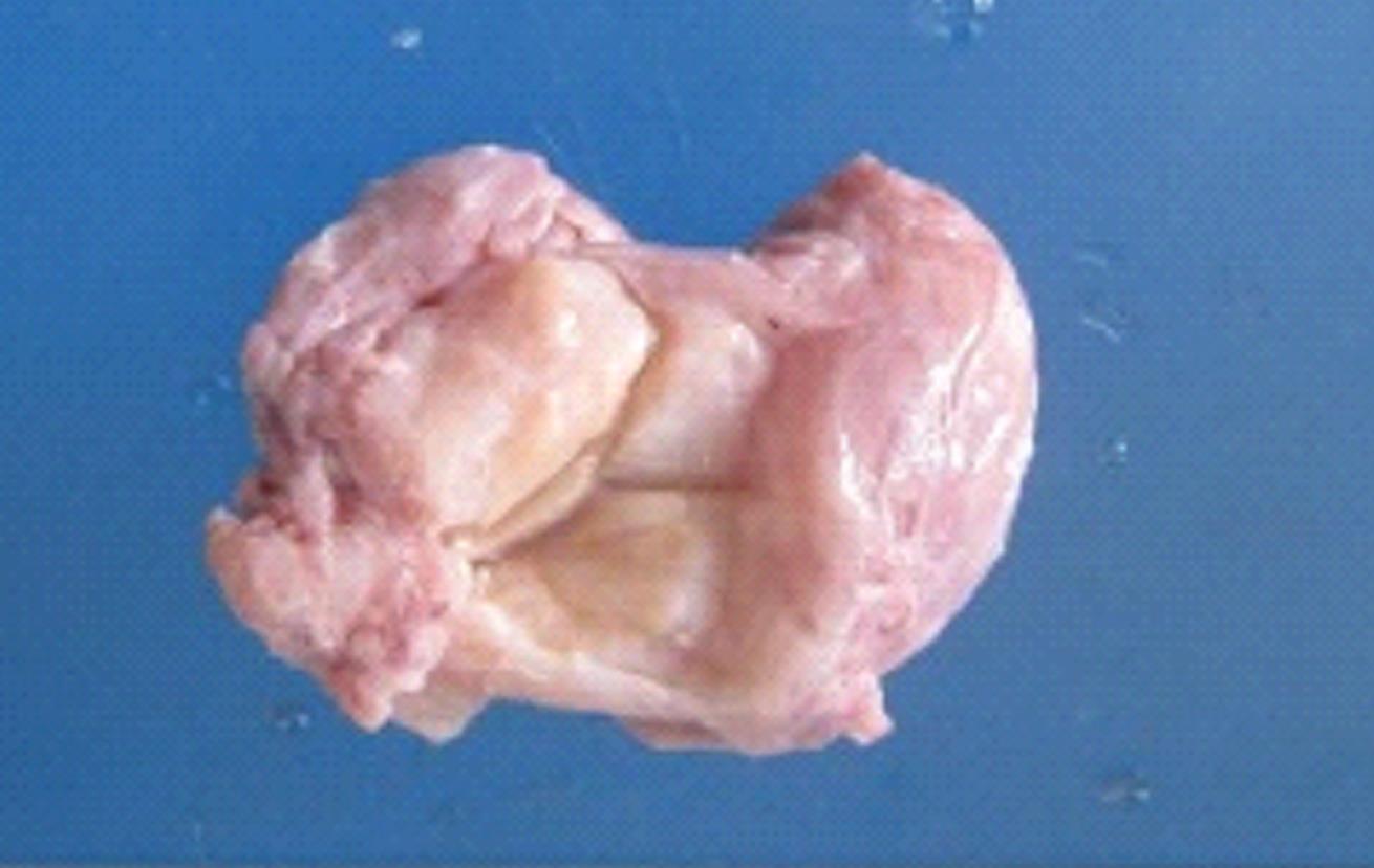
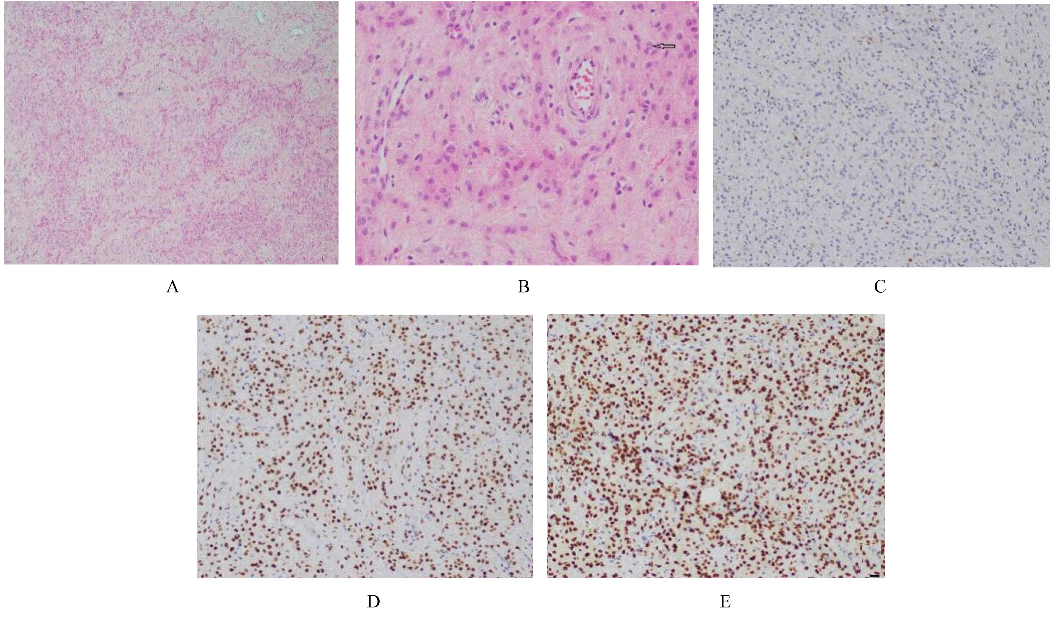
目的 探讨外阴血管肌纤维母细胞瘤(AMFB)复发患者的临床特征、诊断过程和治疗方法,旨在提高临床医生对该病的认识。 方法 收集1例复发性外阴AMFB患者的临床资料、影像学检查结果、病理检查结果和免疫组织化学检测结果,分析上述资料并进行相关文献复习。 结果 患者,女性,50岁,5年前因外阴肿物于当地医院行外阴肿物病灶切除术,术后病理提示AMFB。现因自觉外阴部肿物6个月,高度怀疑外阴AMFB复发入院。妇科检查,左侧大阴唇下方可见肿物凸起,表面皮肤完整,肿物深部凸向直肠,与肠管界限不清,大小约为5.0 cm×4.0 cm×4.0 cm,形态尚规则,界限尚清,活动性欠佳,无明显压痛,无溃破和皮损。浅表超声显示左侧大阴唇处可见范围约5.0 cm×4.0 cm×4.0 cm的稍低无回声光团,其内可见多处团状高回声;彩色多普勒血流显像(CDFI)示内部及周边可见血流信号。行外阴肿物切除术,结合术后病理和免疫组织化学诊断为外阴AMFB复发。 结论 AMFB虽较少见复发及恶变,但应高度重视AMFB的术后随访,以减少复发并提高治愈率。

关键词: 血管肌纤维母细胞瘤, 外阴, 免疫组织化学, 复发

Abstract:

Objective: To investigate the clinical characteristics, diagnosis procedure and treatment method of the patient with recurrent vulvar angiomyofibroblastoma (AMFB),and to improve the understanding of clinical doctors to this disease. Methods The clinical data, imaging examination results, pathological examination results and immunohistochemical examination results of 1 patient with recurrent vulvar AMFB were collected; the above clinical data were analyzed, and the relevant literatures were reviewed. Results A 50-year-old female patient underwent excision of vulvar mass 5 years ago in the local hospital for a vulvar mass,the postoperative pathology suggested AMFB.The patient was now admitted to the hospital due to a 6-month self-reported vaginal mass and a high degree of suspicion of recurrence of AMFB.The gynecological examination suggested a swelling under the labia major on the left side, with complete skin surface, deep protrusion towards the rectum and unclear boundary with the bowel;the size of tumor was about 5.0 cm×4.0 cm×4.0 cm,with regular shape, clear boundary,and poor activity,and there was no obvious tenderness,ulceration and skin lesion. Superficial ultrasound showed a slightly low anechoic light mass with a range of 5.0 cm×4.0 cm×4.0 cm at the left labia major, in which multiple mass hyperechoics were observed. Color Doppler flow imaging (CDFI) showed visible blood flow signals in and around the labia major. Conclusion Although recurrence and malignancy of AMFB are rare, postoperative follow-up of AMFB should be given high priority to reduce the recurrence and increase the cure rate.

Key words: Angiomyofibroblastoma, Vulva, Immunohistochemistry, Recurrence

中图分类号: 

  • R711.72