吉林大学学报(医学版) ›› 2022, Vol. 48 ›› Issue (6): 1510-1517.doi: 10.13481/j.1671-587X.20220617

• 临床研究 • 上一篇    下一篇

不同部位孤立性纤维性肿瘤患者临床病理特征和预后影响因素分析

高云鹤,姚佳楠,曹岚清,许传杰()   

  1. 吉林大学第二医院病理科, 吉林 长春 130041
  • 收稿日期:2022-01-22 出版日期:2022-11-28 发布日期:2022-12-07
  • 通讯作者: 许传杰 E-mail:xucj@jlu.edu.cn
  • 作者简介:高云鹤(1984-), 女,吉林省长春市人, 主治医师, 医学博士, 主要从事肿瘤诊治方面的研究。
  • 基金资助:
    国家自然科学基金项目(82002808);吉林省发改委产业技术研究与开发基金项目(2015Y031-7)

Clinicopathological characteristics and analysis on prognostic factors of patients with solitary fibrous tumors in different sites

Yunhe GAO,Jianan YAO,Lanqing CAO,Chuanjie XU()   

  1. Department of Pathology,Second Hospital,JilinUniversity,Changchun 130041,China
  • Received:2022-01-22 Online:2022-11-28 Published:2022-12-07
  • Contact: Chuanjie XU E-mail:xucj@jlu.edu.cn

摘要:

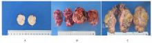
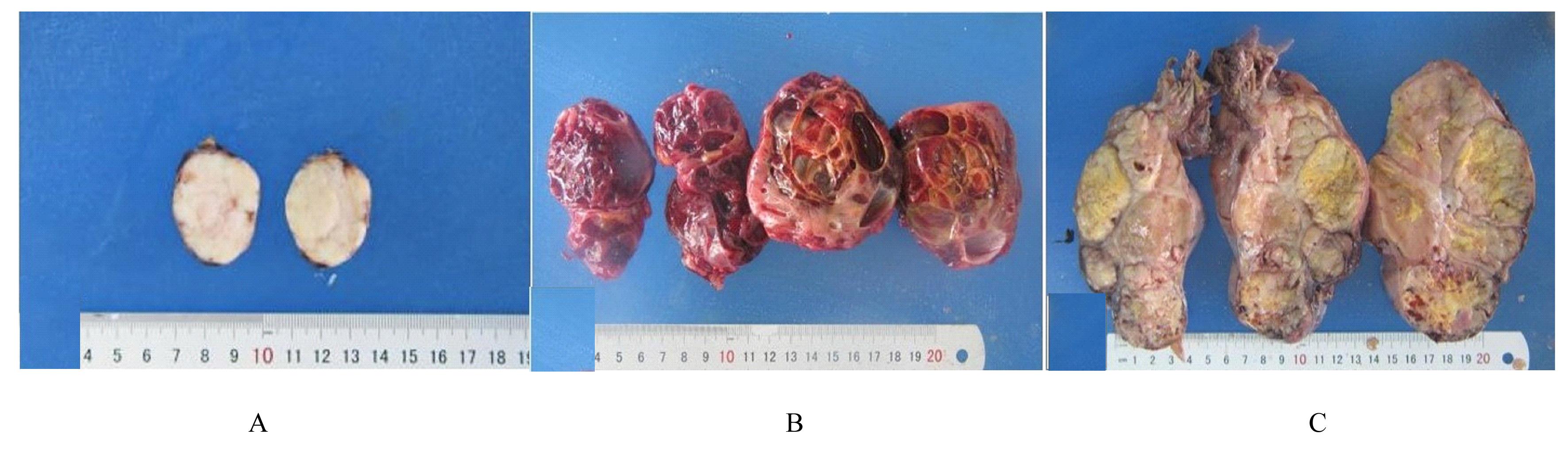
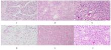
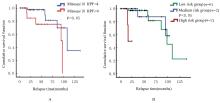
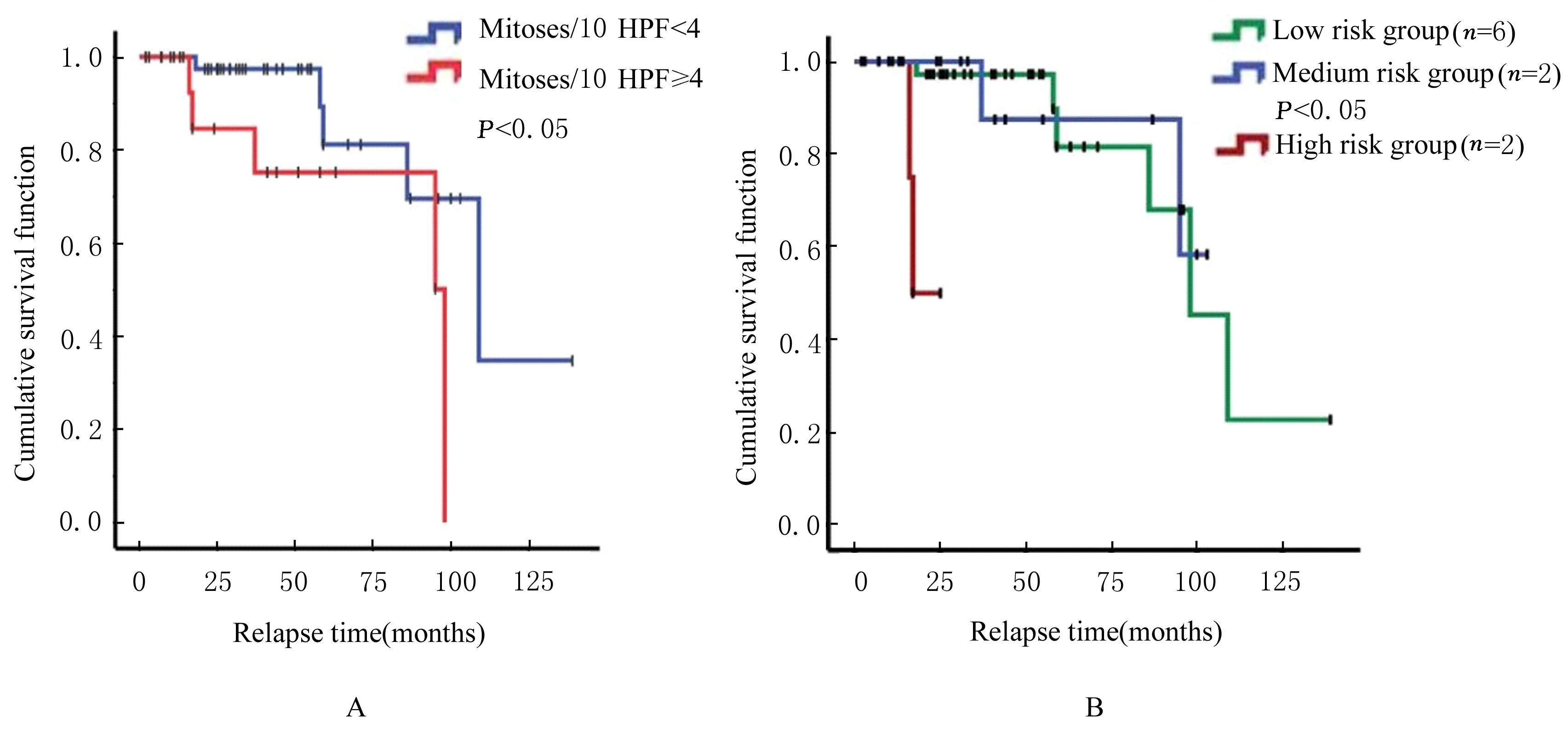
目的 探讨孤立性纤维性肿瘤(SFT)患者的临床病理特征和预后相关影响因素,为该病病理诊断、临床治疗及患者预后判断提供依据。 方法 收集经手术切除原发于不同系统的86例SFT患者临床病理资料,根据危险度分级标准分为低(n=65)、中(n=14)和高危险组(n=7)。观察肿瘤大体形态表现、HE染色和免疫组织化学染色检测SFT形态表现,获取患者随访资料行预后相关分析。采用Kaplan-Meier生存曲线法分析不同部位的单一临床病理因素与患者无进展生存期的关系,多因素Cox回归分析法进行多因素与患者无进展生存期关系的分析。 结果 86例SFT中,男性37例, 女性49例, 不同部位SFT的患者性别构成比比较差异无统计学意义(P>0.05)。不同部位SFT的患者年龄比较差异无统计学意义(P>0.05)。不同部位SFT在低、中、高危险组的分布情况比较差异均无统计学意义(P>0.05)。不同部位肿瘤直径比较差异有统计学意义(P<0.01)。光镜下,肿瘤细胞形态及排列多样,呈梭形或卵圆形细胞疏密不等地无结构排列,常见特征性鹿角样分枝状血管和粗细不等的胶原纤维,大部分肿瘤细胞形态温和、异型性和核分裂象不明显。免疫组织化学染色,STAT-6核弥漫强阳性;CD34、Bcl-2及CD99不同程度阳性表达。61例患者获随访信息,随访时间为2~139个月。其中10例患者出现复发,各组患者复发率分别为低危险组9%、中危险组14%、高危险组28%。单因素分析, 核分裂象<4个/10HPF与≥4个/10HPF患者无进展生存期比较差异有统计学意义(P<0.05);高危险组与低、中危险组患者无进展生存期比较差异有统计学意义(P<0.05)。多因素分析,性别、年龄、肿瘤直径和核分裂象计数均非患者无进展生存期的独立预测因素(P>0.05)。 结论 SFT可发生于人体多种器官和系统, 形态学表现多样。发生在中枢神经系统、上呼吸道及眼眶的肿瘤直径明显小于女性生殖道、腹腔、皮下软组织、肺及胸膜。STAT-6是SFT诊断特异且敏感指标;核分裂象≥4个/10HPF和高危险分级是患者无进展生存期缩短的危险因素;多因素综合分析肿瘤危险度分级不能完全反映患者预后。

关键词: 孤立性纤维性肿瘤, 临床病理特征, 免疫组织化学, 危险度分级, 预后

Abstract:

Objective: To investigate the clinicopathological features and prognosis-related factors of the patients with solitary fibrous tumor (SFT), and to provide the evidence for its pathological diagnosis, clinical treatment and prognosis judgment. Methods The clinicopathological data of 86 patients with SFT who had undergone surgical resection from different systems were collected and divided into low (n=65), medium (n=14) and high risk groups (n=7) according to the risk classification criteria. The general morphology of tumor was observed,HE staining and immunohistochemical staining were used to detect the morphology of SFT,and the follow-up data of the patients were obtained for the prognosis-related correlation analysis. Kaplan-Meier survival curve method was used to analyze the relationship between single clinicopathological factor in different parts and progression free survival of the patients, and Cox regression analysis was used to analyze the relationships between multiple factors and progression free survival of the patients. Results Of the 86 cases of SFT, 37 were male and 49 were female. There was no significant difference in the gender constituent ratio of the patients with SFT at different sites (P>0.05). There was no significant difference in the age of patients with SFT at different sites (P>0.05). There was no significant difference in the distribution of SFT at different sites among low,medium, and high risk groups (P>0.05). There was significant statistical difference in the tumor diameter at different sites(P<0.01). The microscope results showed that the shape and arrangement of tumor cells were diverse, and the spindle or oval cells were not arranged structurally in varying density; the characteristic antler like branching vessels and collagen fibers of varying thickness were common;most of the tumor cells were mild in shape and heterotypic, and the mitotic image was not obvious.The immunohistochemiscal staining results showed that the STAT-6 nucleus was diffusely and strongly positive; CD34, Bcl-2 and CD99 were positive in different degrees. A total of 61 cases were followed up for 2-139 months. Among them, 10 cases recurred, and the recurrence rates were 9% in low risk group, 14% in medium risk group, and 28% in high risk group, respectively. The univariate analysis results showed that there was a significant difference in the progression free survival between the patients with mitotic images<4/10 HPF and those with mitotic images ≥ 4/10 HPF (P<0.05); there were significant differences in the progression free survival between high risk group and low,medium risk groups(P<0.05). The multivariate analysis results showed that gender, age, tumor diameter and mitotic count were not the independent predictors of progression free survival of the patients (P>0.05). Conclusion SFT can occur in many organs and systems of human body, and its morphology is diverse. The diameters of tumors in the central nervous system, upper respiratory tract and orbit are significantly smaller than those in female genital tract, abdominal cavity, subcutaneous soft tissue, lung and pleura. STAT-6 is a specific and sensitive index for SFT diagnosis; mitotic images ≥ 4/10 HPF and high risk classification are the risk factors for the progression free survival shortening; multivariate comprehensive analysis of tumor risk classification can not fully reflect the prognosis of the patients.

Key words: Solitary fibrous tumor, Clinicopathological characteristics, Immunohistochemistry, Risk models, Recurrence

中图分类号: 

  • R73