吉林大学学报(医学版) ›› 2023, Vol. 49 ›› Issue (4): 896-904.doi: 10.13481/j.1671-587X.20230410

• 基础研究 • 上一篇    下一篇

莫特塞尼与EZH2抑制剂GSK126联合应用对肝癌Huh7细胞增殖和凋亡的影响及其机制

梁媛媛1,2,赵颂1,胡静1,安妮1,魏验芦1,苏荣健1()   

  1. 1.锦州医科大学基础医学院细胞生物学教研室,辽宁 锦州 121000
    2.锦州医科大学附属第一医院 风湿免疫科,辽宁 锦州 121000
  • 收稿日期:2022-06-18 出版日期:2023-07-28 发布日期:2023-07-26
  • 通讯作者: 苏荣健 E-mail:srjsrj186@163.com
  • 作者简介:梁媛媛(1990-),女,辽宁省凌海市人,在读硕士研究生,主要从事肝癌治疗及其分子机制方面的研究。
  • 基金资助:
    国家自然科学基金项目(81903109);辽宁省教育厅自然科学基金项目(2020-MS-299)

Effect of motesanib combined with EZH2 inhibitor GSK126 on proliferation and apoptosis of liver cancer Huh7 cells and its mechanism

Yuanyuan LIANG1,2,Song ZHAO1,Jing HU1,Ni AN1,Yanlu WEI1,Rongjian SU1()   

  1. 1.Department of Cell Biology,School of Basic Medical Sciences,Jinzhou Medical University,Jinzhou 121001,China
    2.Department of Rheumatoid Immunity,First Affiliated Hospital,Jinzhou Medical University,Jinzhou 121000,China
  • Received:2022-06-18 Online:2023-07-28 Published:2023-07-26
  • Contact: Rongjian SU E-mail:srjsrj186@163.com

摘要:

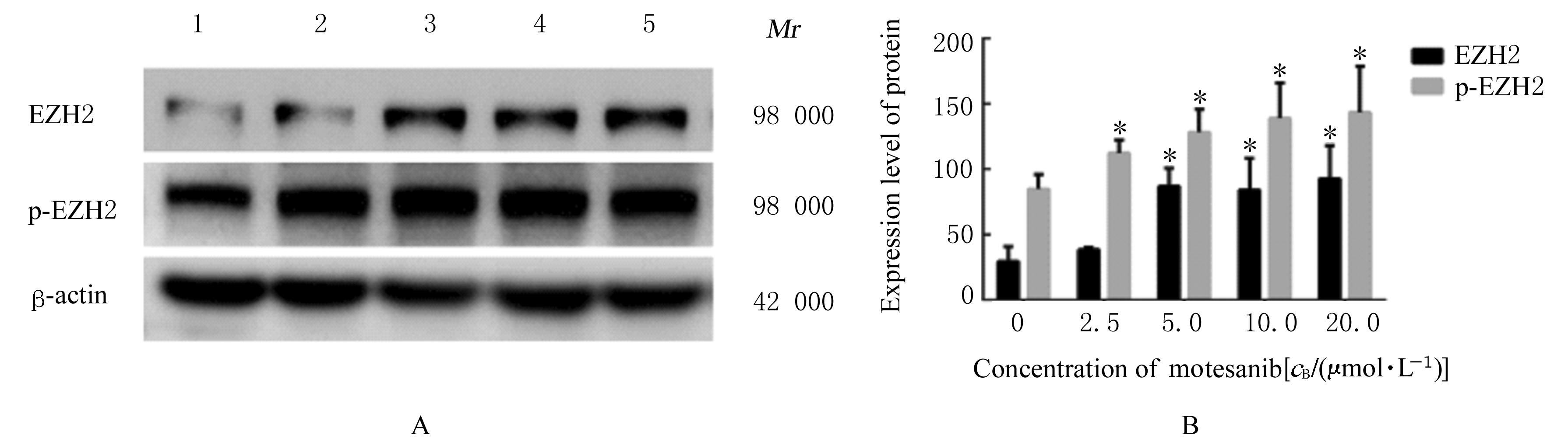
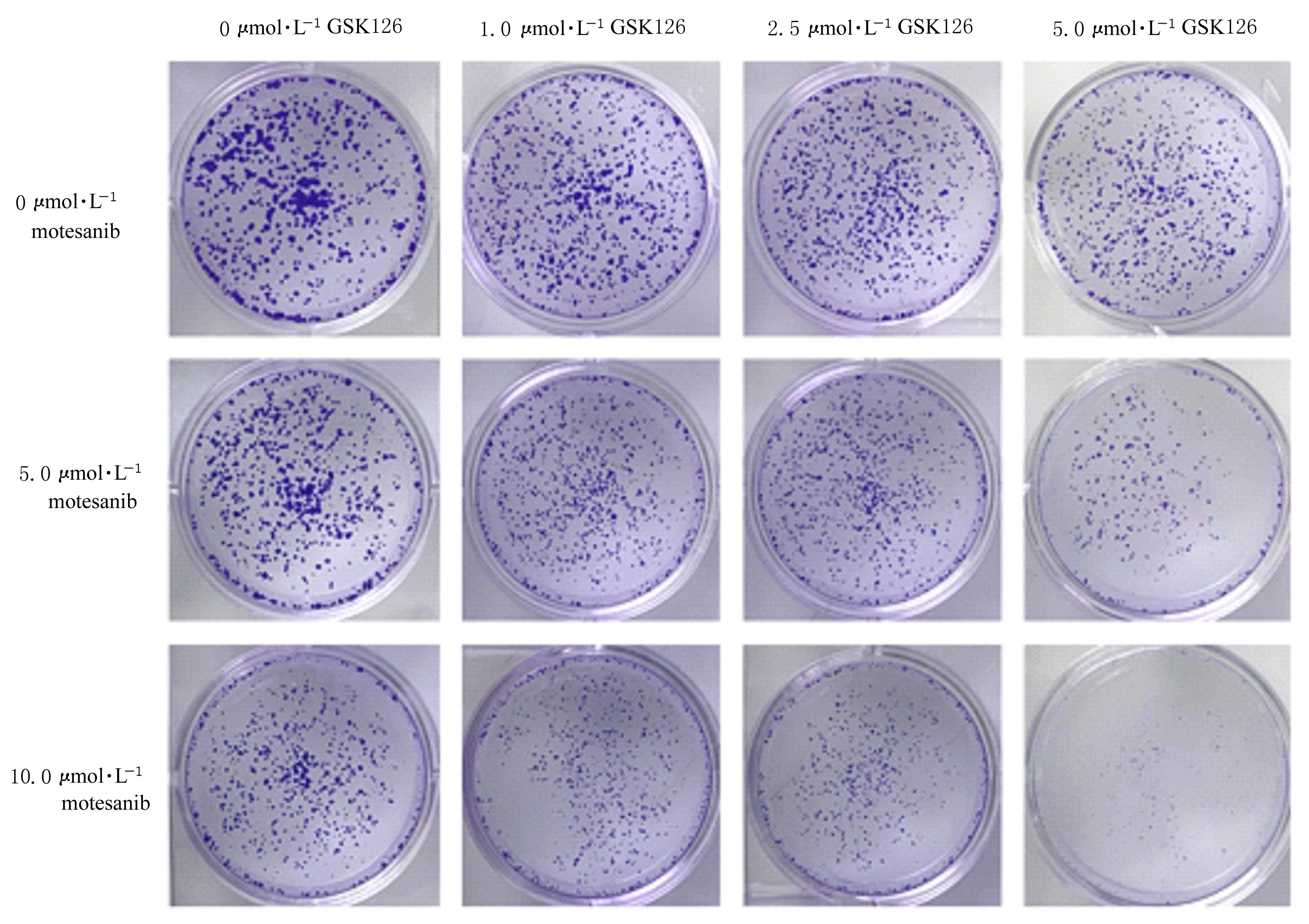
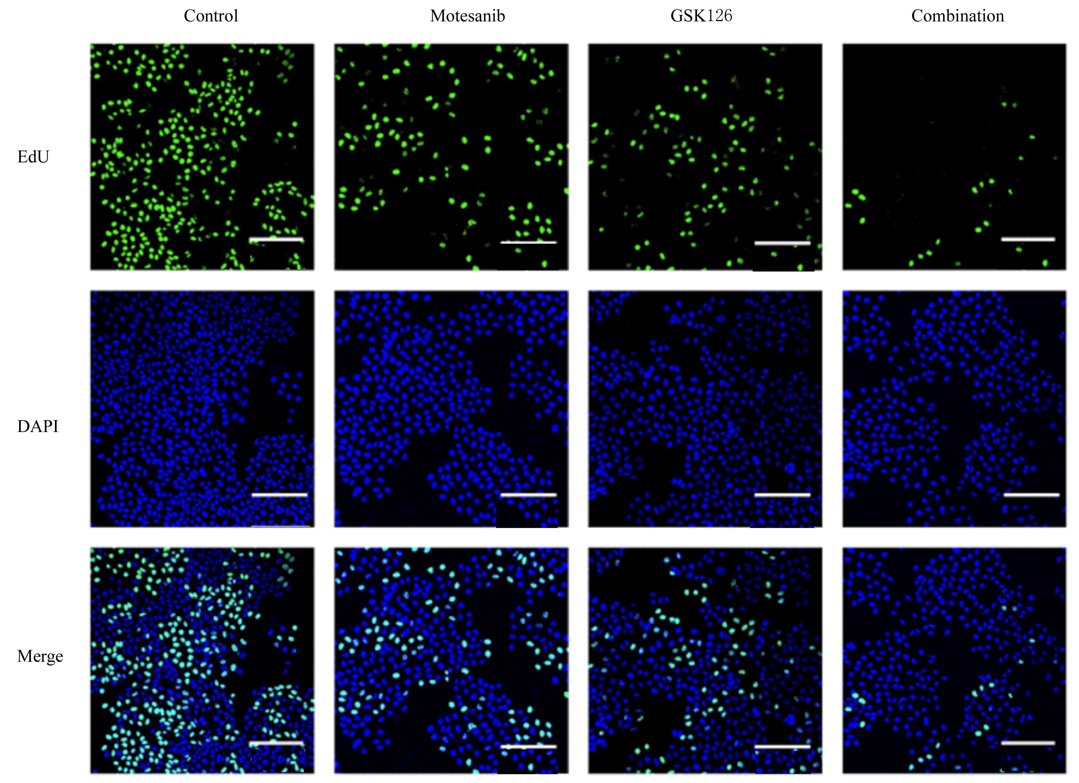
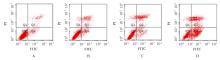
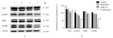
目的 探讨多激酶抑制剂莫特塞尼联合果蝇zeste基因增强子的人类同源物2(EZH2)抑制剂GSK126对肝癌Huh7细胞体外增殖和凋亡的影响,初步阐明其相关机制。 方法 不同浓度(0、1、5、10、20、40和60 μmol·L-1)莫特塞尼处理Huh7细胞,采用CCK-8法检测各组Huh7细胞增殖率,采用Western blotting法检测不同浓度(0、2.5、5.0、10.0和20.0 μmol·L-1)莫特塞尼组Huh7细胞中EZH2和磷酸化EZH2(p-EZH2)蛋白表达水平。莫特塞尼和GSK126联合作用于Huh7细胞,采用CCK-8法和克隆形成实验确定后续实验合适的药物浓度。Huh7细胞分为对照组、莫特塞尼(10 μmol·L-1)组、GSK126(5 μmol·L-1)组和联合组(莫特塞尼+GSK126),采用5-乙炔基-2'脱氧尿嘧啶核苷(EdU)荧光染色法检测各组Huh7细胞增殖率,流式细胞术检测各组Huh7细胞凋亡率,Western blotting法检测各组Huh7细胞中细胞外信号调节激酶(ERK)、磷酸化ERK(p-ERK)、蛋白激酶B(AKT)和磷酸化AKT(p-AKT)蛋白表达水平。 结果 CCK-8法检测,不同浓度莫特塞尼组Huh7细胞存活率随着药物浓度升高逐渐降低,与0 μmol·L-1 莫特塞尼组比较,20、40和60 μmol·L-1莫特塞尼组Huh7细胞增殖率明显降低(P<0.05或P<0.01);Western blotting法检测,与0 μmol·L-1莫特塞尼组比较,5.0、10.0和20.0 μmol·L-1莫特塞尼组Huh7细胞中EZH2和p-EZH2蛋白表达水平明显升高(P<0.01)。通过CCK-8法和克隆形成实验确定10 μmol·L-1莫特塞尼和5 μmol·L-1 GSK126用于后续实验。与对照组比较,莫特塞尼组、GSK126组和联合组Huh7细胞增殖率明显降低(P<0.01),细胞凋亡率明显升高(P<0.05或P<0.01);与莫特塞尼组和GSK126组比较,联合组Huh7细胞增殖率明显降低(P<0.01),细胞凋亡率明显升高(P<0.01)。与对照组、莫特塞尼组和GSK126组比较,联合组Huh7细胞中p-AKT和p-ERK蛋白表达水平明显降低(P<0.05或P<0.01)。 结论 单独应用莫特塞尼抑制肝癌Huh7细胞增殖和促进凋亡效果不明显,可能与EZH2升高导致细胞耐药有关。莫特塞尼与GSK126联合应用可通过抑制AKT和ERK信号通路,增强莫特塞尼对肝癌Huh7细胞的增殖抑制和促凋亡作用。

关键词: 莫特塞尼, 肝肿瘤, 细胞增殖, 细胞凋亡, 果蝇zeste基因增强子的人类同源物2, GSK126

Abstract:

Objective To discuss the effect of multi-kinase inhibitor motesanib combined with enhancer of zeste homolog 2(EZH2)inhibitor GSK126 on the proliferation and apoptosis of the liver cancer Huh7 cells in vitro, and to clarify its related mechanism. Methods The Huh7 cells were treated with different concentrations (0,1,5,10,20,40,and 60 μmol·L-1)of motesanib.The proliferation rates of Huh7 cells in various groups were detected by CCK-8 assay;Western blotting method was used to detect the expression levels of EZH2 and phosphorylated EZH2(p-EZH2) proteins in the Huh7 cells in different concentrations(0,2.5,5.0,10.0,20.0 μmol·L-1) of motesanib groups.The Huh7 cells were treated with motesanib and GSK126 in various groups;the appropriate drug concentrations were selected by CCK-8 assay and colony formation assay.The Huh7 cells were divided into control group,motesanib(10 μmol·L-1) group, GSK126( 5 μmol·L-1)group, and combination (motesanib+GSK126) group. The 5-ethynyl-2'-deoxyu ridine(EdU) fluorescence staining assay was used to detect the proliferation rates of Huh7 cells in various groups;the apoptotic rates of Huh7 cells in various groups were detected by flow cytometry; Western blotting method was used to detect the expression levels of extracellular regulated protein kinases(ERK), phosphorylated ERK (p-ERK),protein kinase B(AKT),and phosphorylated AKT(p-AKT) proteins in the Huh7 cells in various groups. Results The CCK-8 assay results showed that proliferation rate of Huh7 cells in different concentrations of motesenib groups were decreased gradually with the increasing of drug concentrations; compared with motesenib (0 μmol·L-1) group, the proliferation rates of the Huh7 cells in 20, 40, and 60 μmol·L-1 motesenib groups were decreased significantly (P<0.05 or P<0.01).The Western blotting results showed that compared with 0 μmol·L-1 motesanib group, the expression levels of EZH2 and p-EZH proteins in the Huh7 cells in 5,10, and 20 μmol·L-1 motesanib groups were increased (P<0.05 or P<0.01).The CCK-8 results and colony formation experiment results showed that 10 μmol·L-1 motesanib and 5 μmol·L-1 GSK126 could be used for the following experiment.Compared with control group,the proliferation rates of the Huh7 cells in motesanib group,GSK126 group,and combination group were significantly decreased(P<0.05 or P<0.01); compared with motesanib group and GSK126 group, the proliferation rate of the cells in combination group was decreased (P<0.01),and the apoptotic rate was increased (P<0.01).Compared with control group,motesanib group and GSK126 group, the expression levels of p-AKT and p-ERK proteins in the Huh7 cells in combination group were significantly decreased (P<0.05 or P<0.01). Conclusion Motesanib alone has no significantly inhibitory effect on the proliferation and promoting effect on the apoptosis of the liver cancer Huh7 cells, which may be related to the increasing of EZH2 leading to the cell drug resistance. The combination of motesanib and GSK126 enhances the proliferation inhibition and pro-apoptotic effects of motesanib on the Huh7 cells by inhibiting AKT and ERK signaling pathways.

Key words: Motesanib, Hepatocellular carcinoma, Cell proliferation, Apoptosis, Enhancer of zeste homolog 2, GSK126

中图分类号: 

  • R735.7