吉林大学学报(医学版) ›› 2023, Vol. 49 ›› Issue (3): 691-696.doi: 10.13481/j.1671-587X.20230318

• 基础研究 • 上一篇    下一篇

circ_EFCAB2在癫痫细胞模型中的表达及其作用机制

张舒雅1,2,孙洪英1(),毛戬1,孟晨曦1,巴格隆3   

  1. 1.内蒙古科技大学包头医学院第一附属医院神经内科,内蒙古 包头 014010
    2.内蒙古科技大学包头医学院第三附属医院老年病科,内蒙古 包头 014030
    3.内蒙古科技大学包头医学院第三附属医院 功能科,内蒙古 包头 014030
  • 收稿日期:2022-08-19 出版日期:2023-05-28 发布日期:2023-06-20
  • 通讯作者: 孙洪英 E-mail:sunhongying2004@sina.com
  • 作者简介:张舒雅(1989-),女,内蒙古自治区鄂尔多斯市人,在读硕士研究生,主要从事癫痫的基础和临床方面的研究。
  • 基金资助:
    内蒙古自治区科技厅自然科学基金项目(2020MS08073);内蒙古自治区卫健委卫生计生科研计划项目(201702130)

Expression of circ_EFCAB2 in epileptic cell model and its mechanism

Shuya ZHANG1,2,Hongying SUN1(),Jian MAO1,Chengxi MENG1,Gelong BA3   

  1. 1.Department of Neurology,First Affiliated Hospital,Baotou Medical College,Inner Mongolia University of Science and Technology,Baotou 014010,China
    2.Department of Geriatrics,Third Affiliated Hospital,Baotou Medical College,Inner Mongolia University of Science and Technology,Baotou 014030,China
    3.Department of Ultrasound,Third Affiliated Hospital,Baotou Medical College,Inner Mongolia University of Science and Technology,Baotou 014030,China
  • Received:2022-08-19 Online:2023-05-28 Published:2023-06-20
  • Contact: Hongying SUN E-mail:sunhongying2004@sina.com

摘要:

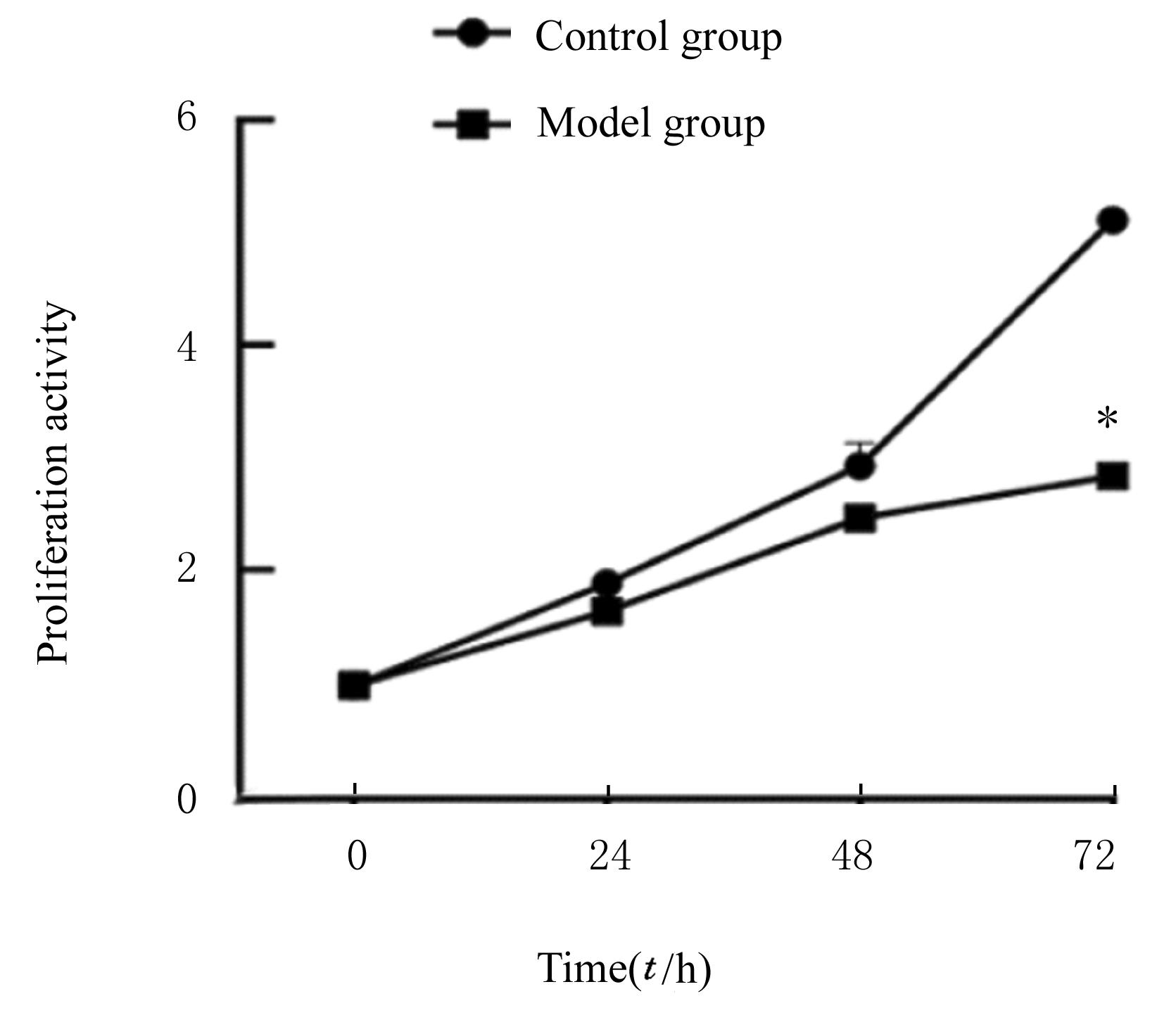
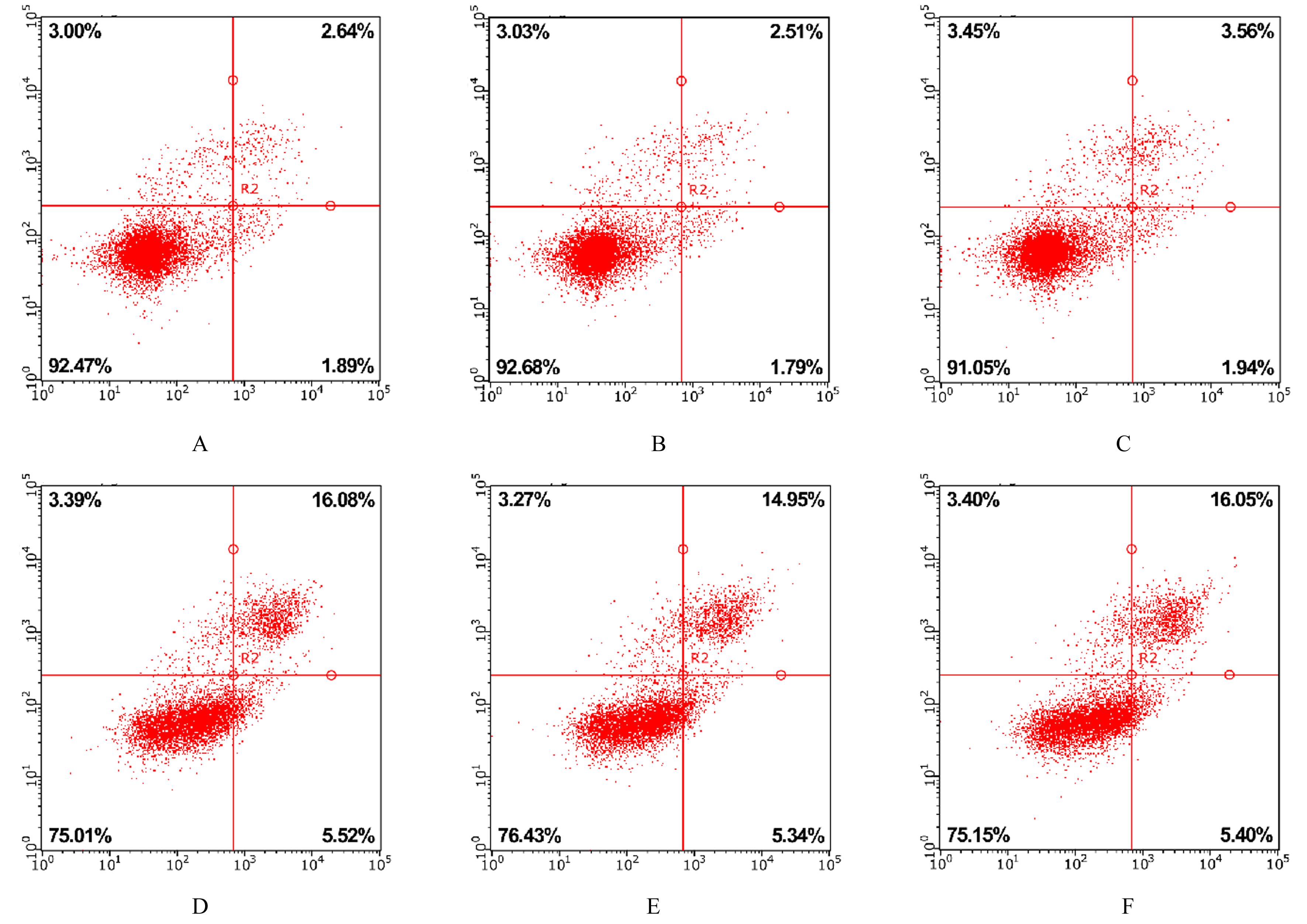
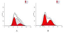
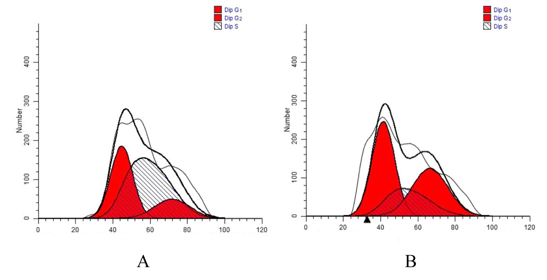
目的 探讨circ_EFCAB2在体外癫痫细胞模型中的表达,阐明其可能的作用机制。 方法 人神经母细胞瘤LA-N-5细胞中构建无镁癫痫细胞模型(模型组),未经无镁细胞外液处理的细胞作为对照组。实时荧光定量PCR(RT-qPCR)法检测2组细胞中circ_EFCAB2 mRNA表达水平。将细胞分为核糖核酸酶(RNase)R消化组和RNase R未消化组,RNA酶消化实验和RT-qPCR法检测2组细胞中circ_EFCAB2和线性EFCAB2 mRNA表达水平。核质分离实验检测癫痫细胞中circ_EFCAB2 mRNA表达水平,CCK-8法检测2组细胞增殖能力,流式细胞术检测2组细胞凋亡率和不同细胞周期细胞百分率。 结果 模型组细胞有自发高频率的痫样放电,提示癫痫细胞模型建立成功。RT-qPCR法检测,与对照组比较,模型组细胞中circ_EFCAB2 mRNA表达水平升高(P<0.05)。RNA酶消化实验和RT-qPCR法检测,与RNase R未消化组比较,RNase R消化组细胞中线性EFCAB2 mRNA表达水平明显降低(P<0.01)。核质分离实验检测,癫痫细胞的细胞质和细胞核中circ_EFCAB2 mRNA表达水平比较差异无统计学意义(P>0.05)。CCK-8 法检测,转染72 h时,与对照组比较,模型组细胞增殖能力明显降低(P<0.01)。流式细胞术检测,与对照组比较,模型组细胞凋亡率明显升高(P<0.01);与对照组比较,模型组S期细胞百分率明显降低(P<0.01),G1+G2期细胞百分率明显升高(P<0.01)。 结论 上调的circ_EFCAB2可能通过抑制细胞增殖、促进细胞凋亡和阻滞细胞周期等途径在癫痫的发病过程中发挥作用。

关键词: 癫痫, circ_EFCAB2, 癫痫细胞模型, 细胞凋亡, 细胞增殖

Abstract:

Objective To discuss the expression of circs_EFCAB2 in epileptic cell model in vitro,and to clarify its possible mechanism. Methods The magnesium-free epilepsy cell model (model group) was constructed in the human neuroblastoma LA-N-5 cells, and the cells untreated with magnesium-free extracellular fluid were regarded as control group. Real-time fluorescence quantitative PCR (RT-qPCR) method was used to detect the expression level of circ_EFCAB2 mRNA in the cells in two groups.The cells were didived into ribonuclease(RNase) R digestion group and RNase R undigestion group. RNA enzyme digestion experiment and RT-qPCR method were used to detect the expression levels of circ_ EFCAB2 and linear EFCAB2 mRNA in the cells in two groups; nuclear cytoplasmic separation experiment was used to detect the expression levels of circle_EFCAB2 mRNA in the epileptic cells; CCK-8 method was used to detect the proliferation activities of the cells in two groups; flow cytometry was used to detect the apoptotic rate and percentages of the cells at different cell cycles in two groups. Results The cells in model group showed spontaneous high-frequency epileptiform discharges, indicating the epilepsy cell model was successfully established. The RT-qPCR results showed that compared with control group, the expression level of circ_ EFCAB2 mRNA in the cells in model group was increased (P<0.05). The RNA enzyme digestion experiment and RT-qPCR results showed that compared with RNase R undigestion group, the expression level of linear EFCAB2 mRNA in the cells in RNase R digestion group was decreased (P<0.01). The nuclear cytoplasmic separation experiment results showed that there was no statistically significant difference in the expression level of circ_ EFCAB2 mRNA in the epileptic cells between the cytoplasm and nucleus of the epileptic cells(P>0.05). The CCK-8 results showed that at 72 h after transfection, compared with control group, the proliferation activity of the cells in model group was significantly decreased (P<0.01). The flow cytometry analysis results showed that compared with control group, the apoptotic rate of the cells in model group was significantly increased (P<0.01);compared with control group, the percentage of the cells at S phase in model group was significantly decreased (P<0.01), and the percentage of the cells at G1+G2 phase was significantly increased (P<0.01). Conclusion The upregulated circ_ EFCAB2 may play a role in the pathogenesis of epilepsy by inhibiting the cell proliferation, promoting the apoptosis, and blocking the cell cycles.

Key words: Epilepsy, Circ_EFCAB2, Epilepsy cell model, Apoptosis, Cell proliferation

中图分类号: 

  • R742.1