吉林大学学报(医学版) ›› 2023, Vol. 49 ›› Issue (3): 656-664.doi: 10.13481/j.1671-587X.20230314

• 基础研究 • 上一篇    下一篇

lncRNA PAX8-AS1对结直肠癌细胞增殖、凋亡和侵袭的作用及其机制

闫圣玉1,刘昌化2(),许志杰1,丁雅婷1,谢亚锋1,张侨1,刘菀莹1   

  1. 1.南华大学衡阳医学院附属第二医院肛肠科,湖南 衡阳 421000
    2.南华大学衡阳医学院附属 第二医院胃肠外科,湖南 衡阳 421001
  • 收稿日期:2022-06-02 出版日期:2023-05-28 发布日期:2023-06-20
  • 通讯作者: 刘昌化 E-mail:344792164@qq.com
  • 作者简介:闫圣玉(1985-),男,湖南省衡阳市人,副主任医师,医学硕士,主要从事肛肠疾病基础和临床方面的研究。
  • 基金资助:
    湖南省卫健委科研项目(202104010708)

Effect of lncRNA PAX8-AS1 on proliferation, apoptosis and invasion of colorectal cancer cells and its mechanism

Shengyu YAN1,Changhua LIU2(),Zhijie XU1,Yating DING1,Yafeng XIE1,Qiao ZHANG1,Wanying LIU1   

  1. 1.Department of Proctology,Second Affiliated Hospital,Hengyang Medical School,University of South China,Hengyang 421001,China
    2.Department of Gastrointestinal Surgery,Second Affiliated Hospital,Hengyang Medical School,University of South China,Hengyang 421001,China
  • Received:2022-06-02 Online:2023-05-28 Published:2023-06-20
  • Contact: Changhua LIU E-mail:344792164@qq.com

摘要:

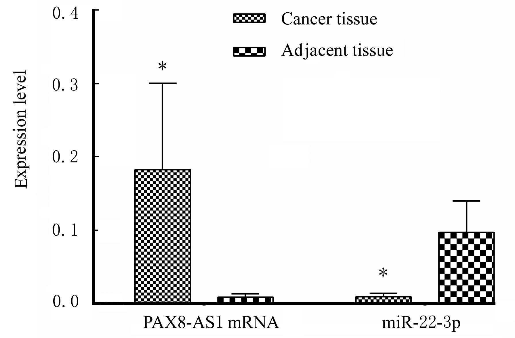
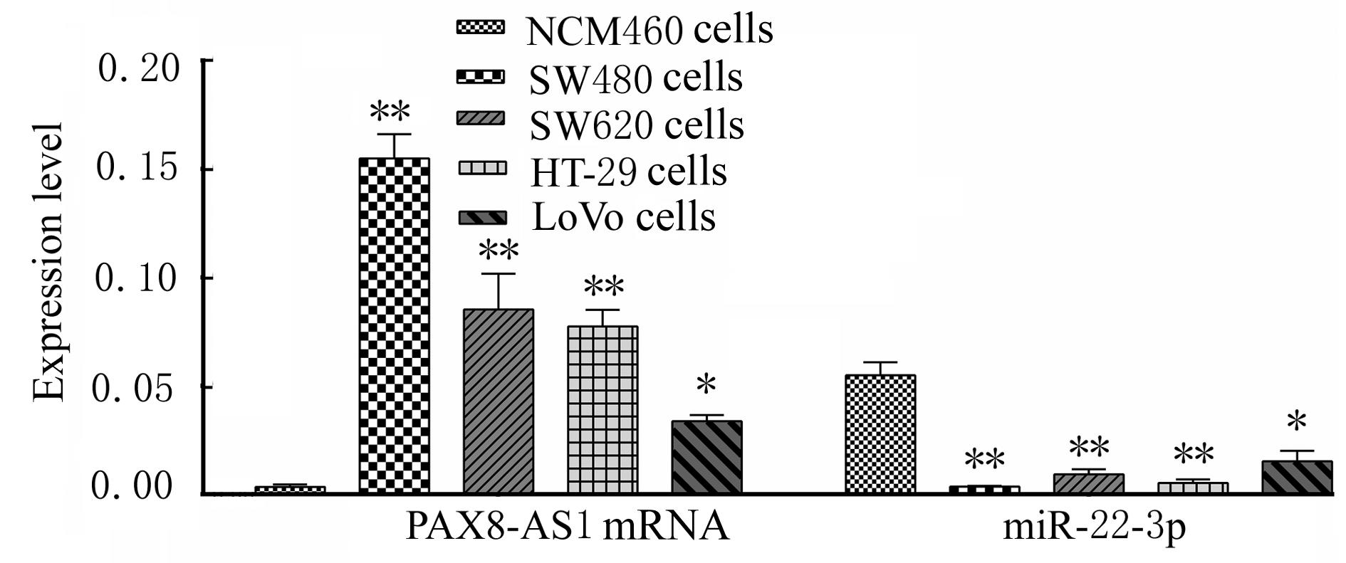
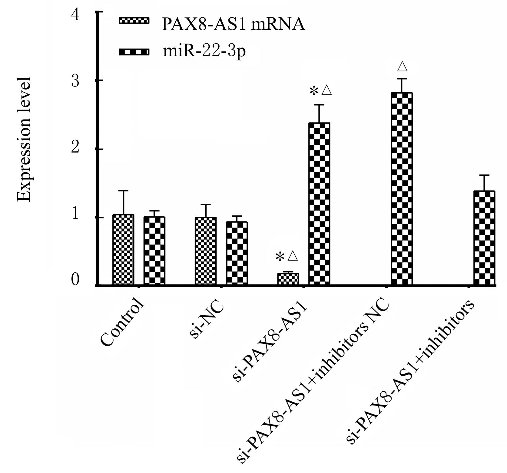
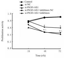
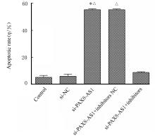
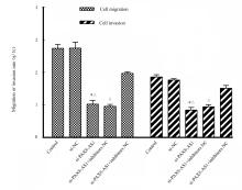
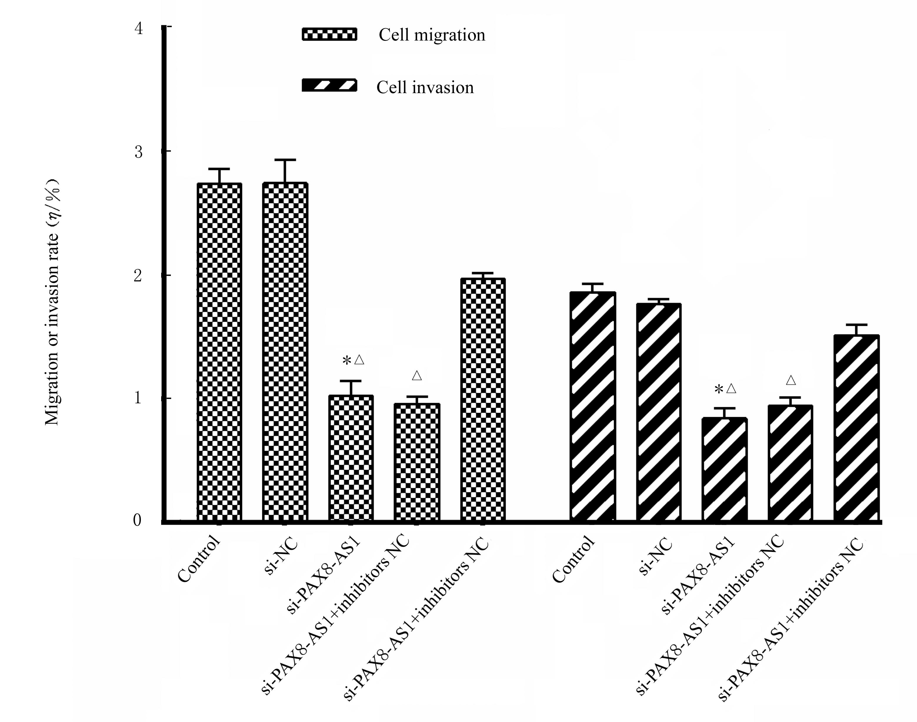
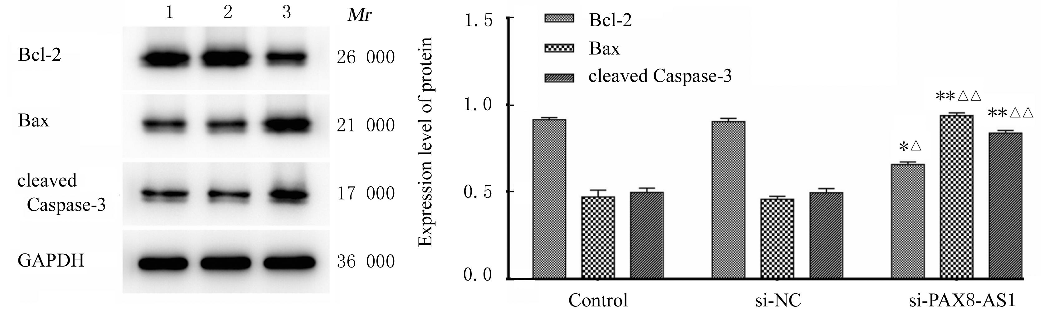
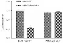
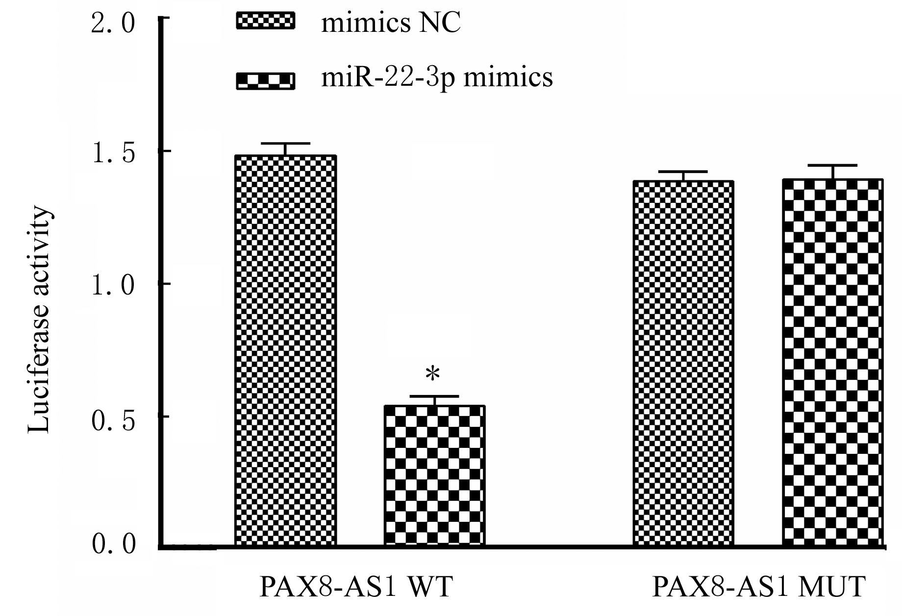
目的 探讨长链非编码RNA配对盒8反义RNA 1(PAX8-AS1)在人结直肠癌(CRC)组织中的表达水平及其对CRC细胞增殖、凋亡及侵袭的作用,并阐明其作用机制。 方法 采用实时荧光定量PCR(RT-qPCR)法检测94例CRC患者癌组织和癌细胞中PAX8-AS1 mRNA和miR-22-3p表达水平。将PAX8-AS10 siRNA小分子序列与miR-22-3p inhibitors分别转染或共转染至CRC细胞中,转染后细胞分为si-NC组(转染阴性序列)、si-PAX8-AS1组(转染PAX8-AS1 siRNA)、si-PAX8-AS1+inhibitors NC组(共转染PAX8-AS1 siRNA和inhibitors NC)和si-PAX8-AS1+inhibitors组(共转染PAX8-AS1 siRNA和miR-22-3p inhibitors),另取未经任何转染的SW480细胞作为对照组。RT-qPCR法检测转染后各组细胞中PAX8-AS1 mRNA和miR-22-3p表达水平,MTT法检测各组细胞增殖活性,流式细胞术检测各组细胞凋亡率,Transwell小室实验检测各组细胞迁移和侵袭率,Western blotting法检测各组细胞中B细胞淋巴瘤2(Bcl-2)、Bcl-2相关X蛋白(Bax)和cleaved Caspase-3蛋白表达水平,双荧光素酶报告基因实验验证PAX8-AS1与miR-22-3p的靶向关系。 结果 RT-qPCR法检测,与癌旁组织比较,癌组织中PAX8-AS1 mRNA表达水平明显升高(P<0.01),miR-22-3p表达水平明显降低(P<0.01);与人正常结肠上皮细胞NCM460比较, SW480、SW620、HT-29和LoVo细胞中PAX8-AS1 mRNA表达水平均明显升高(P<0.05或P<0.01),miR-22-3p表达水平均明显降低(P<0.05或P<0.01)。与对照组和si-NC组比较,si-PAX8-AS1组细胞中PAX8-AS1 mRNA表达水平明显降低(P<0.01),miR-22-3p表达水平明显升高(P<0.01);与si-NC组比较,si-PAX8-AS1+ inhibitors NC组细胞中miR-22-3p表达水平明显升高(P<0.01)。MTT法检测,与对照组比较,si-PAX8-AS1组、si-PAX8-AS1+inhibitors NC组和si-PAX8-AS1+ inhibitors组细胞培养48和72 h时细胞增殖活性均明显降低(P<0.01);与si-NC组比较, si-PAX8-AS1+ inhibitors NC组细胞培养48和72 h时细胞增殖活性均明显降低(P<0.01)。流式细胞术检测,与对照组比较,si-PAX8-AS1组细胞凋亡率明显升高(P<0.01);与si-NC组比较,si-PAX8-AS1组和si-PAX8-AS1+ inhibitors NC组细胞凋亡率明显升高(P<0.01)。Transwell小室实验检测,与对照组比较,si-PAX8-AS1组细胞迁移和侵袭率明显降低(P<0.01);与si-NC组比较,si-PAX8-AS1组和si-PAX8-AS1+ inhibitors NC组细胞迁移和侵袭率明显降低(P<0.01)。Western blotting法检测,与对照组和si-NC组比较,si-PAX8-AS1组细胞中Bax和cleaved Caspase-3蛋白表达水平明显升高(P<0.01),Bcl-2蛋白表达水平降低(P<0.05)。TargetScan预测PAX8-AS1与miR22-3p存在靶向结合位点。双荧光素酶报告基因实验,与共转染mimics NC的细胞比较,miR-22-3p mimics共转染后PAX8-AS1-WT细胞中荧光素酶活性明显降低(P<0.01)。 结论 PAX8-AS1在人CRC组织中高表达,沉默PAX8-AS1可抑制CRC细胞增殖、迁移和侵袭,并诱导细胞凋亡,其机制可能与上调miR22-3p表达有关。

关键词: 长链非编码RNA, 配对盒8反义RNA 1, 微小RNA-22-3p, 结直肠肿瘤, 细胞凋亡

Abstract:

Objective To discuss the expression levels of long non-coding RNA paired box 8 antisense RNA 1 (PAX8-AS1) in the human colorectal cancer (CRC) tissue and its effects on the proliferation, apoptosis and invasion of the CRC cells,and to charify its mechanism. Methods The expression levels of PAX8-AS1 mRNA and miR-22-3p in cancer tissue and cancer cells of the CRC patients were detected by real-time fluorescence quantitative PCR (RT-qPCR) method.The PAX8-AS10 siRNA small molecule sequences and miR-22-3p inhibitors were transfected alone or co-transfected into the CRC cells, and the cells after transfection were divided into si-NC group (transfected with negative sequences), si-PAX8-AS1 group (transfected with PAX8-AS1 siRNA), si-PAX8-AS1+inhibitors NC group (co-transfected with PAX8-AS1 siRNA and inhibitors NC), and si-PAX8-AS1+ inhibitors group (co-transfected with PAX8-AS1 siRNA and miR-22-3p inhibitors), and the SW480 cells without any transfection were regarded as control group. The expression levels of PAX8-AS1 mRNA and miR-22-3p in the cells in various groups after transfection were measured by RT-qPCR method; the proliferation activities of the cells in various groups were detected by MTT method; the migration and invasion rates of the cells in various groups were measured by Transwell chamber assay; the apoptotic rates of cells in various groups were measured by flow cytometry; the expression levels of B-cell lymphoma-2(Bcl-2), Bcl-2 associated X protein(Bax), and cleaved Caspase-3 proteins in the cells in various groups were measured by Western blotting method; the target relationship between PAX8-AS1 and miR-22-3p was verified by dual luciferase reporter gene assay. Results The RT-qPCR results showed that compared with adjacent tissue,the expression level of PAX8-AS1 mRNA in cancer tissue was significantly increased(P<0.01), and the expression level of miR-22-3p was significantly decreased(P<0.01); compared with the human normal colonic epithelial cells NCM460,the PAX8-AS1 mRNA expression levels in the SW480, SW620, HT-29 and LoVo cells were all significantly increased (P<0.01), while the miR-22-3p expression levels were all significantly decreased (P<0.05 or P<0.01). Compared with control group and si-NC group, the expression level of PAX8-AS1 mRNA in the cells in si-PAX8-AS1 group was decreased(P<0.01), and the expression level of miR-22-3p was significantly increased(P<0.01); compared with si-NC group,the expression level of miR-22-3p in the cells in si-PAX8-AS1+inhibitors group was increased(P<0.01).The MTT assay results showed that compared with control group,the proliferation activities of the cells in si-PAX8-AS1 group,si-PAX8-AS1+inhibitors NC group,and si-PAX8-AS1+inhibitors group were significantly decreased after culture for 48 and 72 h (P<0.01); compared with si-NC group, the proliferation activities of the cells in si-PAX8-AS1+ inhibitors NC group were significantly decreased after culture for 48 and 72 h (P<0.01).The flow cytometry results showed that compared with control group, the apoptotic rate of cells in si-PAX8-AS1 group was significantly increased (P<0.01);compared with si-NC group,the apoptotic rate of cells in si-PAX8-AS1+ inhibitors NC group was significantly increased (P<0.01).The Transwell chamber assay results showed that compared with control group, the rates of migration and invasion cells in si-PAX8-AS1 group were significantly decreased(P<0.01); compared with si-NC group,the rates of migration and invasion cells in si-PAX8-AS1 group and si-PAX8-AS1+ inhibitors NC group were significantly decreased(P<0.01).The Western blotting results showed that compared with control group and NC group, the expression levels of Bax and cleaved Caspase-3 proteins in the cells in si-PAX8-AS1 group were significantly increased(P<0.01), and expression level of the Bcl-2 protein was significantly decreased(P<0.05).The TargetScan predicted that there were target binding sites between PAX8-AS1 and miR22-3p.The dual luciferase reporter gene assay results showed that compared with the cells co-transfected with mimics NC, the luciferase activity in the PAX8-AS1-WT cells was significantly decreased after co-transfected with miR-22-3p mimics (P<0.01). Conclusion The PAX8-AS1 is highly expressed in the human CRC tissue, and silencing the PAX8-AS1 expression can inhibit the proliferation, migration and invasion of the CRC cells and induce the apoptosis,and its mechanism may be ralated to up-regulation the miR-22-3p expression.

Key words: Long non-coding RNA, Paired box 8 antisense RNA 1, Micro RNA, Colorectal neoplasm, Apoptosis

中图分类号: 

  • R735.35