吉林大学学报(医学版) ›› 2023, Vol. 49 ›› Issue (3): 714-721.doi: 10.13481/j.1671-587X.20230321

• 基础研究 • 上一篇    下一篇

槐耳清膏对乳腺癌他莫昔芬耐药细胞的影响及其作用机制

薛姣姣1,2,郝磊3,张毓秀1,2,代贺阳4,张丽霞1,郭少伟1,张晶晶5,李阳1,李庆霞1,2()   

  1. 1.河北省人民医院肿瘤四科,河北 石家庄 050000
    2.河北医科大学研究生院,河北 石家庄 050000
    3.南方医科大学第七附属医院肿瘤防治科,广东 佛山 528200
    4.华北理工大学研究生院,河北 唐山 063000
    5.河北省人民医院保健处,河北 石家庄 050000
  • 收稿日期:2022-06-22 出版日期:2023-05-28 发布日期:2023-06-20
  • 通讯作者: 李庆霞 E-mail:liqingxia@hebmu.edu.cn
  • 作者简介:薛姣姣(1992-),女,陕西省延安市人,在读硕士研究生,主要从事乳腺癌内分泌治疗方面的研究。
  • 基金资助:
    河北省科技厅自然科学基金精准医学联合培育项目(H2022307024);河北省卫健委医学科学研究课题计划项目(20210441)

Effect of Huaier aqueous extract on breast cancer tamoxifen-resistant LCC2 cells and its mechanism

Jiaojiao XUE1,2,Lei HAO3,Yuxiu ZHANG1,2,Heyang DAI4,Lixia ZHANG1,Shaowei GUO1,Jingjing ZHANG5,Yang LI1,Qingxia LI1,2()   

  1. 1.Fourth Department of Oncology, People’s Hospital, Hebei Province, Shijiazhuang 05000, China
    2.Graduate School, Hebei Medical University, Shijiazhuang 05000, China
    3.Department of Oncology Prevention and Control, Seventh Affiliated Hospital, Southern Medical University, Foshan 528200, China
    4.Graduate School, North China University of Science and Technology, Tangshan 063000, China
    5.Department of Health Service, People’s Hospital, Hebei Province, Shijiazhuang 05000, China
  • Received:2022-06-22 Online:2023-05-28 Published:2023-06-20
  • Contact: Qingxia LI E-mail:liqingxia@hebmu.edu.cn

摘要:

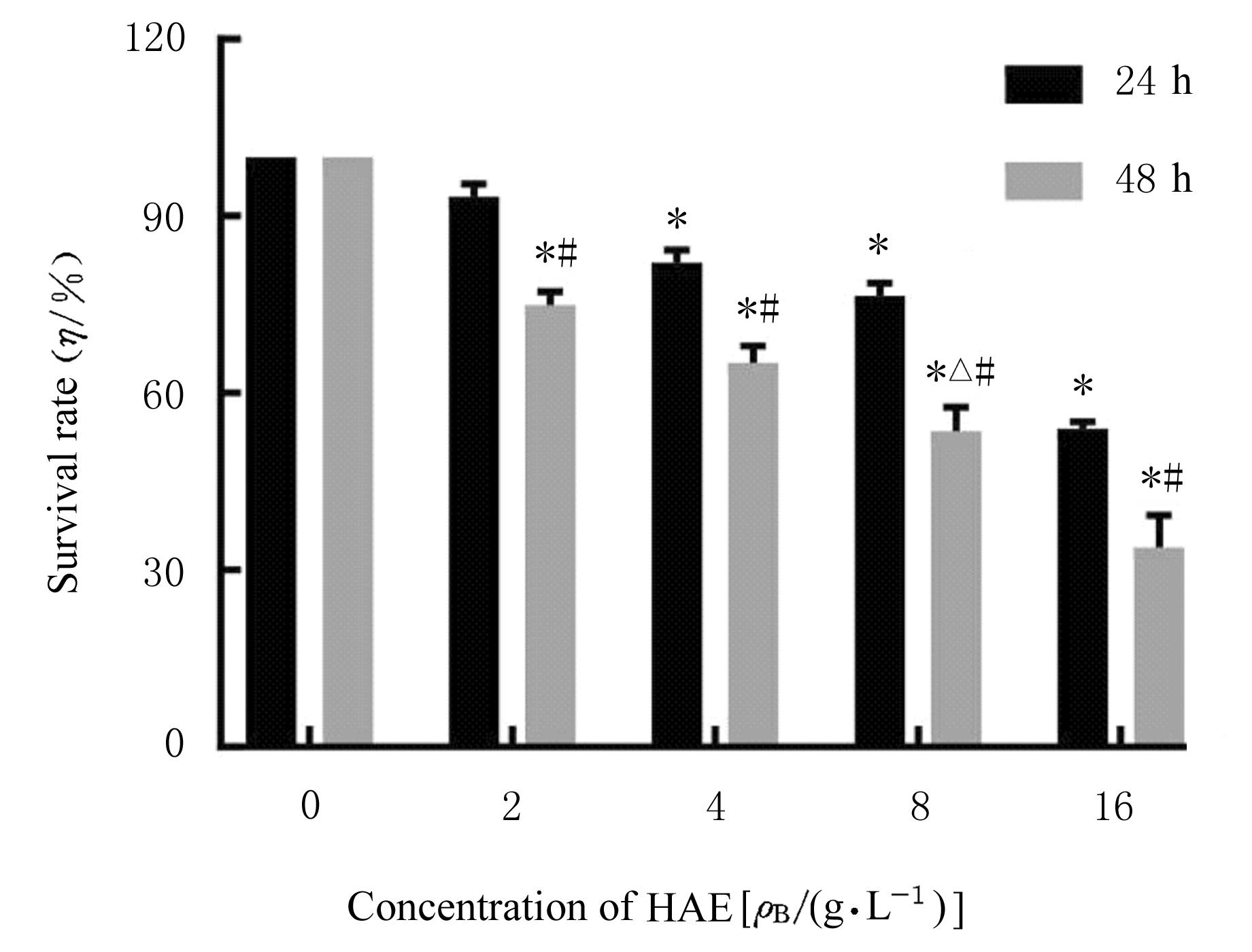
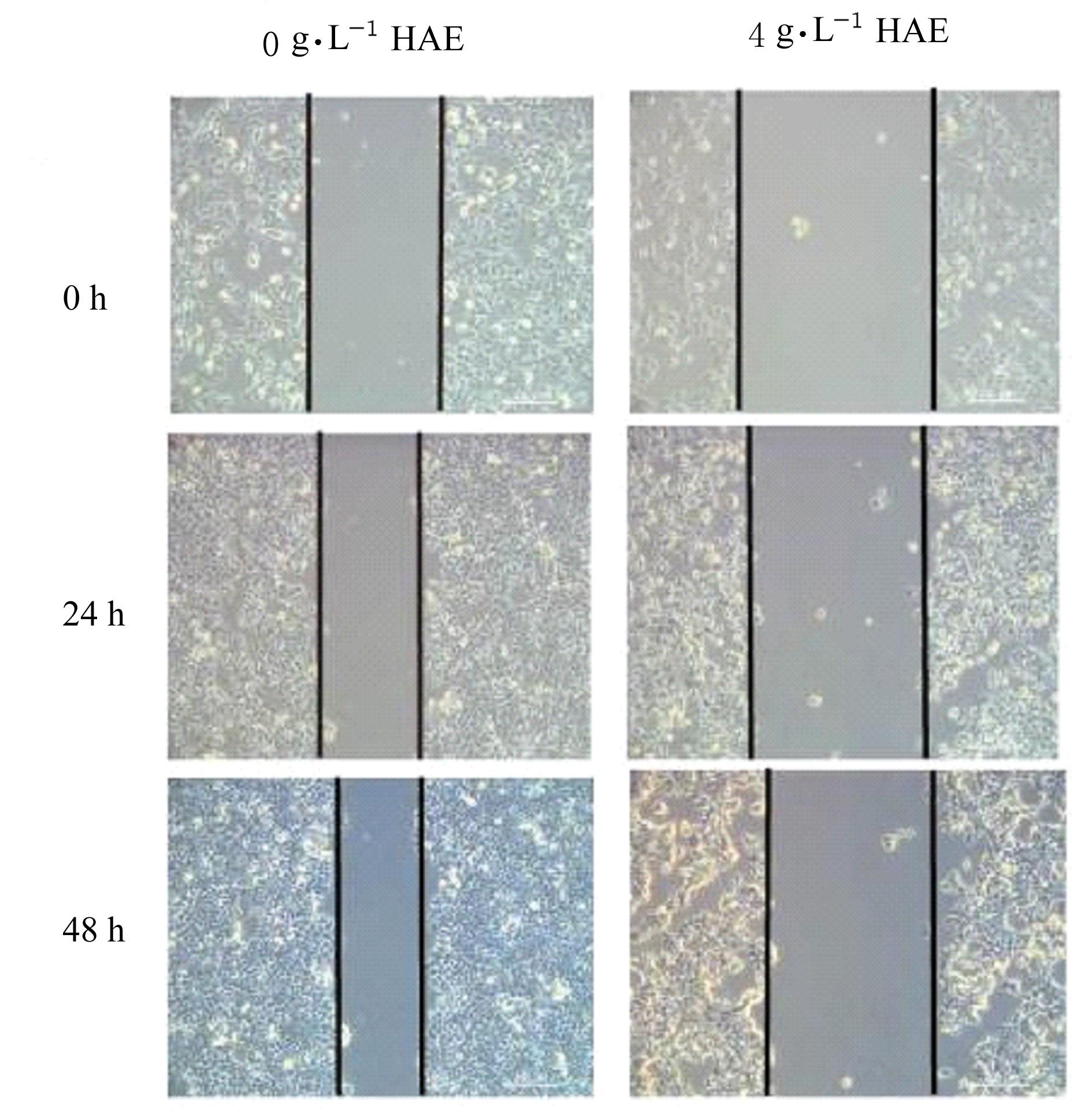
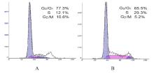
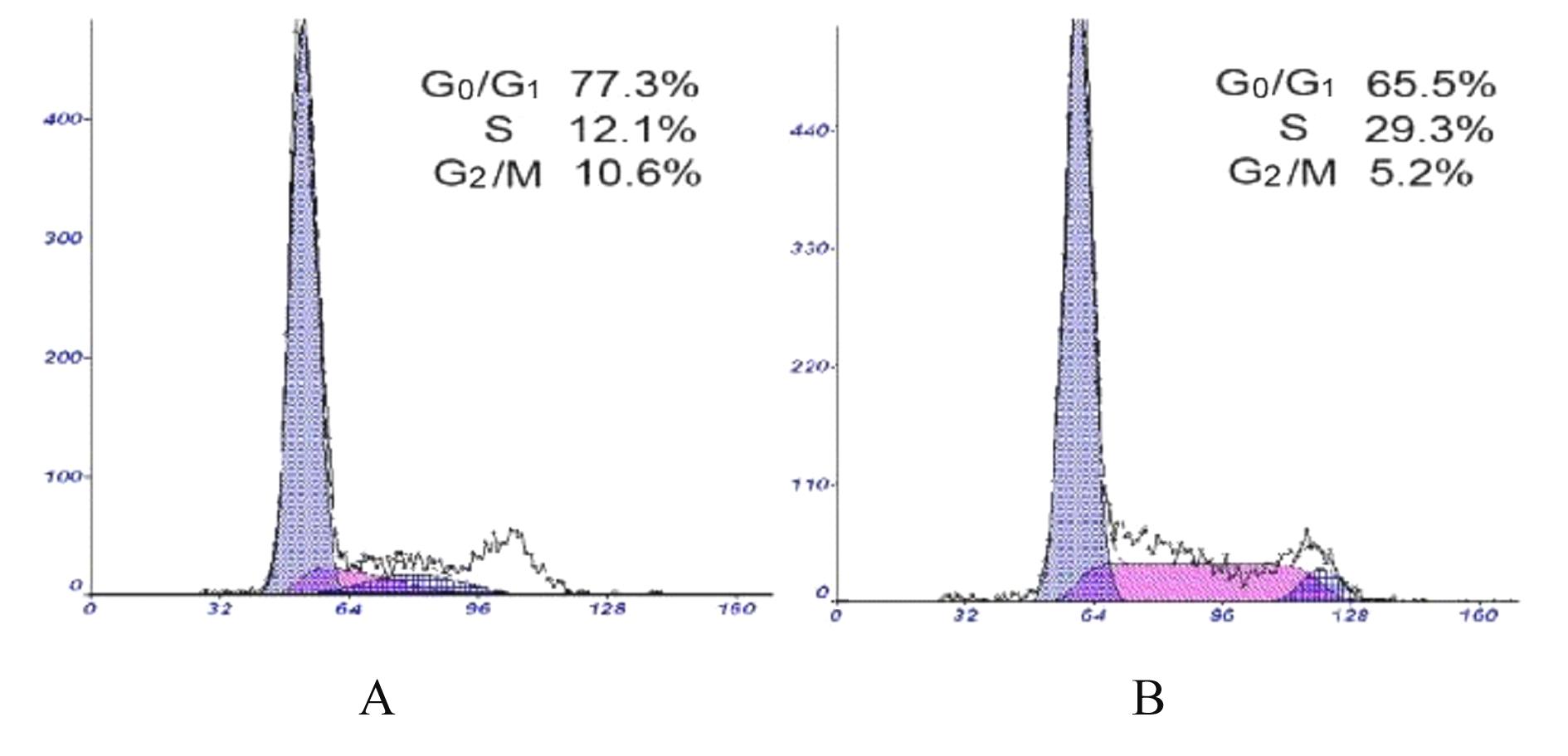
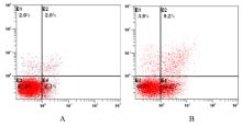
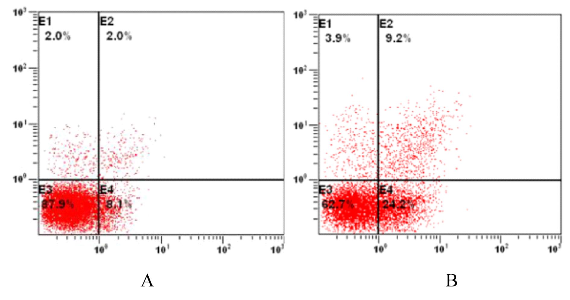
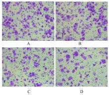
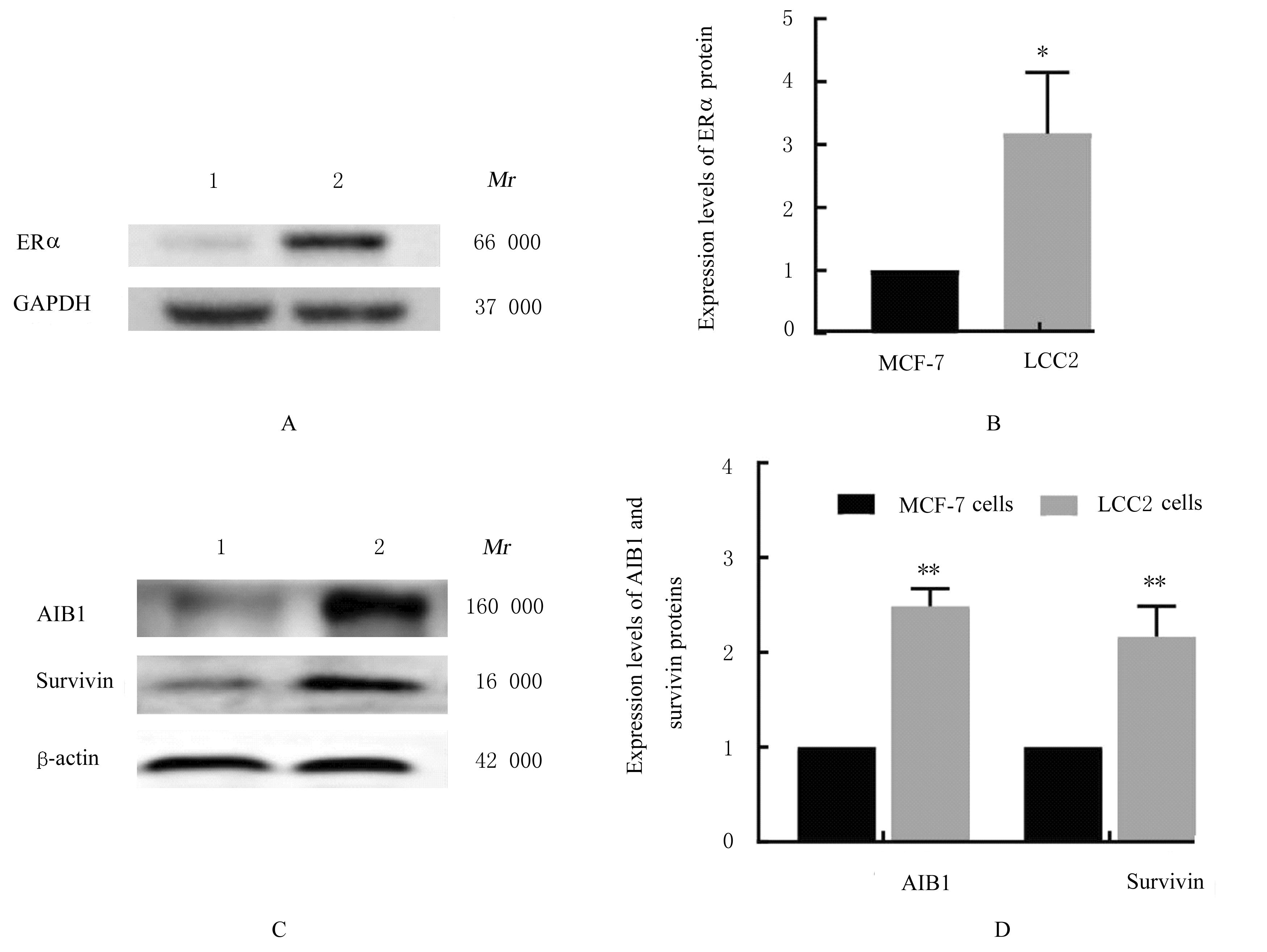
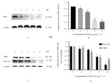
目的 探讨槐耳清膏(HAE)对乳腺癌他莫昔芬(TAM)耐药LCC2细胞增殖、迁移、细胞周期和凋亡的影响,并阐明其作用机制。 方法 细胞分为对照组(给予DMEM基础培养基)、TAM组(给予2 μmol·L-1 TAM)、TAM+HAE组(给予2 μmol·L-1 TAM+4 g·L-1 HAE)和0、2、4、8及16 g·L-1 HAE组。MTT法检测各组细胞存活率,细胞划痕实验检测各组细胞迁移率,流式细胞术检测各组不同细胞周期细胞百分率和细胞凋亡率, Transwell小室实验检测各组迁移细胞数,Western blotting法检测各组细胞中雌激素受体α(ERα)、乳腺癌扩增基因1(AIB1)和survivin蛋白表达水平。 结果 MTT法检测,各浓度HAE组作用细胞24 h,与0 g·L-1 HAE组比较,4、8和16 g·L-1 HAE组细胞存活率明显降低(P<0.01)。作用48 h时,与0 g·L-1 HAE组比较,2、4、8和16 g·L-1 HAE组细胞存活率明显降低(P<0.01);与4 g·L-1 HAE组比较,8 g·L-1 HAE组细胞存活率降低(P<0.05);与作用24 h时比较,作用48 h时各浓度HAE组细胞存活率明显降低(P<0.01)。作用48 h时,与TAM组和4 g·L-1 HAE组比较,TAM+HAE组细胞存活率明显降低(P<0.01)。细胞划痕实验,作用细胞48 h时,与0 g·L-1 HAE组比较,4 g·L-1 HAE组细胞迁移率降低(P<0.05)。流式细胞术检测,作用48 h时,与0 g·L-1 HAE组比较,4 g·L-1 HAE组S期细胞百分率和细胞凋亡率明显升高(P<0.05或P<0.01)。Transwell小室实验,作用48 h时,与TAM组和4 g·L-1 HAE组比较,TAM+HAE组迁移细胞数明显减少(P<0.01)。Western blotting法检测,与MCF-7细胞比较,LCC2细胞中ERα、AIB1和survivin蛋白表达水平明显升高(P<0.05或P<0.01);作用48 h时,与0 g·L-1 HAE组比较,2、4、8和16 g·L-1 HAE组细胞中ERα、AIB1及survivin蛋白表达水平明显降低(P<0.01)。 结论 HAE可抑制LCC2细胞的增殖和迁移,阻滞细胞周期于S期并促进细胞凋亡,同时可恢复LCC2细胞对TAM的敏感性,其作用机制可能与下调LCC2细胞中ERα、AIB1和survivin蛋白表达有关。

关键词: 乳腺癌, 槐耳清膏, 他莫昔芬, 细胞增殖, 耐药

Abstract:

Objective: To discuss the effect of Huaier aqueous extract (HAE) on the proliferation, migration, cell cycle,and apoptosis of tamoxifen (TAM) resistant breast cancer LCC2 cells, and to clarify its mechanism. Methods The cells were divided into control group (given DMEM basal medium) and TAM group (given 2 μmol·L-1 TAM), TAM+HAE group (given 2 μmol·L-1 TAM+4 g·L-1 HAE) and 0, 2, 4, 8, and 16 g·L-1 HAE groups. MTT method was used to detect the survival rates and the proliferation rates of the cells in various groups; cell scratch test was used to detect the migration rates of the cells in various groups;flow cytometry was used to detect the percentages of the cells at different cell cycles and apoptotic rates of the cells in various groups;Transwell chamber test was used to detect the migration number of the cells in various groups; Western blotting method was used to detect the expression levels of estrogen receptor α (ER α) in each group,amplified in breast cancer 1(AIB1),and survivin proteins in the cells in various groups. Results The MTT assay showed that after treated for 24 h, compared with 0 g·L-1 HAE group, the survival rates of the HAE cells in 4,8,and 16 g·L-1 HAE groups were significantly decreased(P<0.01). At 48 h after treatment, compared with 0 g·L-1 HAE group, the survival rates of the cells in 2,4,8, and 16 g·L-1 HAE groups were significantly decreased (P<0.01);compared with 4 g·L-1 HAE group,the survival rate of the cells in 8 g·L-1 HAE groups was decreased(P<0.05);compared with 24 h after treatment, the surival rates of the cells in different concentrations of HAE groups were significantly decreased (P<0.01) after treated for 48 h. At 48 treatment,compared with TAM and 4 g·L-1 HAE group,the proliferation rate of the cells in TAM+HAE group was significantly decreased(P<0.01).The cell scratch test results showed that the cell migration rate of the cells in 4 g·L-1 HAE group was lower than that in 0 g·L-1 HAE group after treated for 48 h (P<0.05). The flow cytometry results showed that after treated for 48 h, compared with 0 g·L-1 HAE group, the percentage of the cells at S phase and the apoptotic rate of the cells in 4 g·L-1HAE group were significantly increased (P<0.05 or P<0.01). The Transwell chamber experiment results showed that after treated for 48 h, compared with TAM group and 4 g·L-1 HAE group, the migration number of the cells in TAM+HAE group was significantly decreased (P<0.01). The Western blotting results showed that compared with MCF-7 cells,the expression levels of ERα,AIB1, and survivin proteins in the LCC2 cells were increased (P<0.05 or P<0.01);at 48 h after treatment, compared with 0 g·L-1 HAE group,the expression levels of ERα,AIB1, and survivin proteins in the LCC2 cells in 2,4,8, and 16 g·L-1 HAE groups were significantly decreased (P<0.01). Conclusion HAE can inhibit the proliferation and migration, arrest the cell cycle at S phase, and promote the apoptosis of the LCC2 cells,and it can also restore the sensitivity of the LCC2 cells to TAM, and its mechanism may be related to down-regulation of the expression levels of ER α, AIB1,and survivin proteins in the LCC2 cells.

Key words: Breast cancer, Huaier aqueous extract, Tamoxifen, Cell proliferation, Drug resistance

中图分类号: 

  • R737.9