吉林大学学报(医学版) ›› 2023, Vol. 49 ›› Issue (4): 931-940.doi: 10.13481/j.1671-587X.20230414

• 基础研究 • 上一篇    下一篇

PMS2通过ERK/ERCC1通路对结肠癌SW480细胞生物学行为的影响

黄雪茹1,丁绪浩1,陈素贤2,谭琦2,吴月明3,牛晓敏1,王亚帝4(),佟青1()   

  1. 1.锦州医科大学附属第三医院临床检验诊断学教研室,辽宁 锦州 121000
    2.锦州医科大学附属 第三医院病理科,辽宁 锦州 121000
    3.锦州医科大学附属第三医院肿瘤二科,辽宁 锦州 121000
    4.锦州医科大学附属第三医院精准医学中心,辽宁 锦州 121000
  • 收稿日期:2022-07-20 出版日期:2023-07-28 发布日期:2023-07-26
  • 通讯作者: 王亚帝,佟青 E-mail:15084126408@qq.com;2294305399@qq.com
  • 作者简介:黄雪茹(1992-),女,辽宁省丹东市人,在读硕士研究生,主要从事临床检验方面的研究。
  • 基金资助:
    辽宁省教育厅科学研究经费项目(JYTJCZR2020071)

Effect of PMS2 on biological behaviors of colon cancer SW480 cells through ERK/ERCC1 pathway

Xueru HUANG1,Xuhao DING1,Suxian CHEN2,Qi TAN2,Yueming WU3,Xiaomin NIU1,Yadi WANG4(),Qing TONG1()   

  1. 1.Department of Clinical Laboratory Diagnostics,Third Affiliated Hospital,Jinzhou Medical University,Jinzhou 120001,China
    2.Department of Pathology,Third Affiliated Hospital,Jinzhou Medical University,Jinzhou 120001,China
    3.Department of Oncology,Third Affiliated Hospital,Jinzhou Medical University,Jinzhou 120001,China
    4.Precision Medicine Center,Third Affiliated Hospital,Jinzhou Medical University,Jinzhou 120001,China
  • Received:2022-07-20 Online:2023-07-28 Published:2023-07-26
  • Contact: Yadi WANG,Qing TONG E-mail:15084126408@qq.com;2294305399@qq.com

摘要:

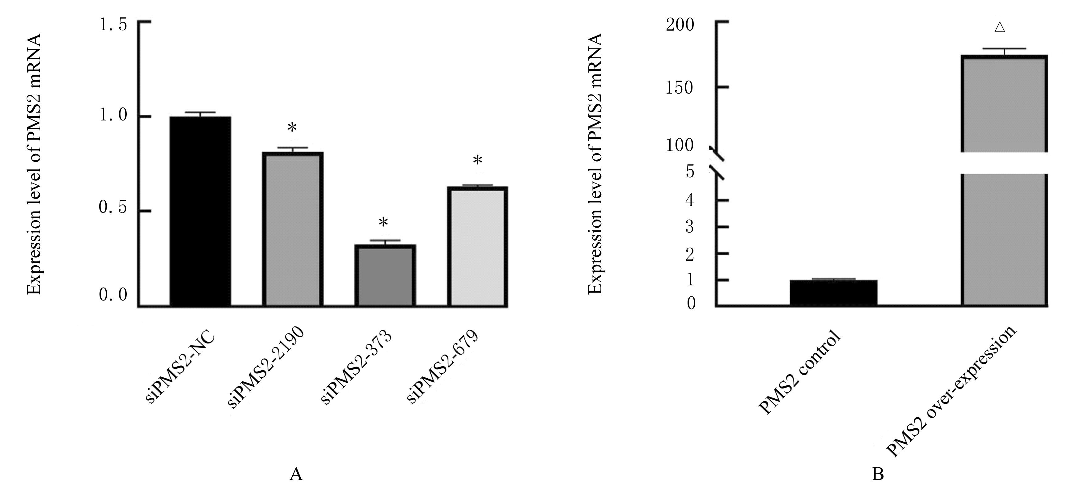
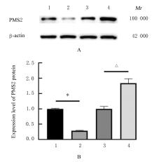
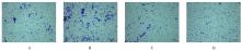
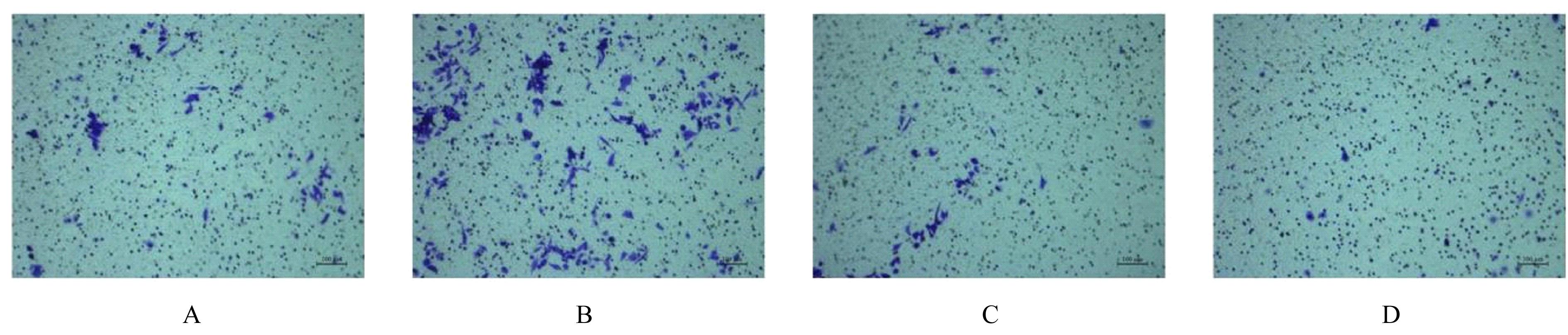
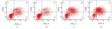
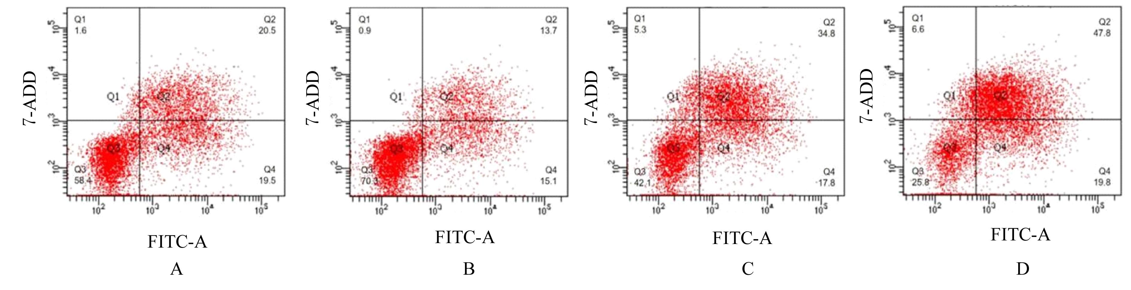
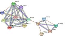
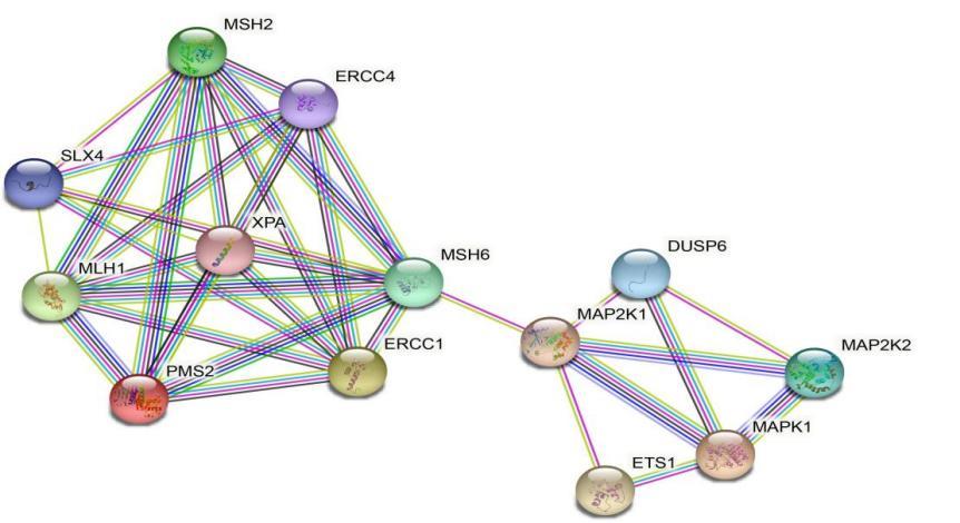
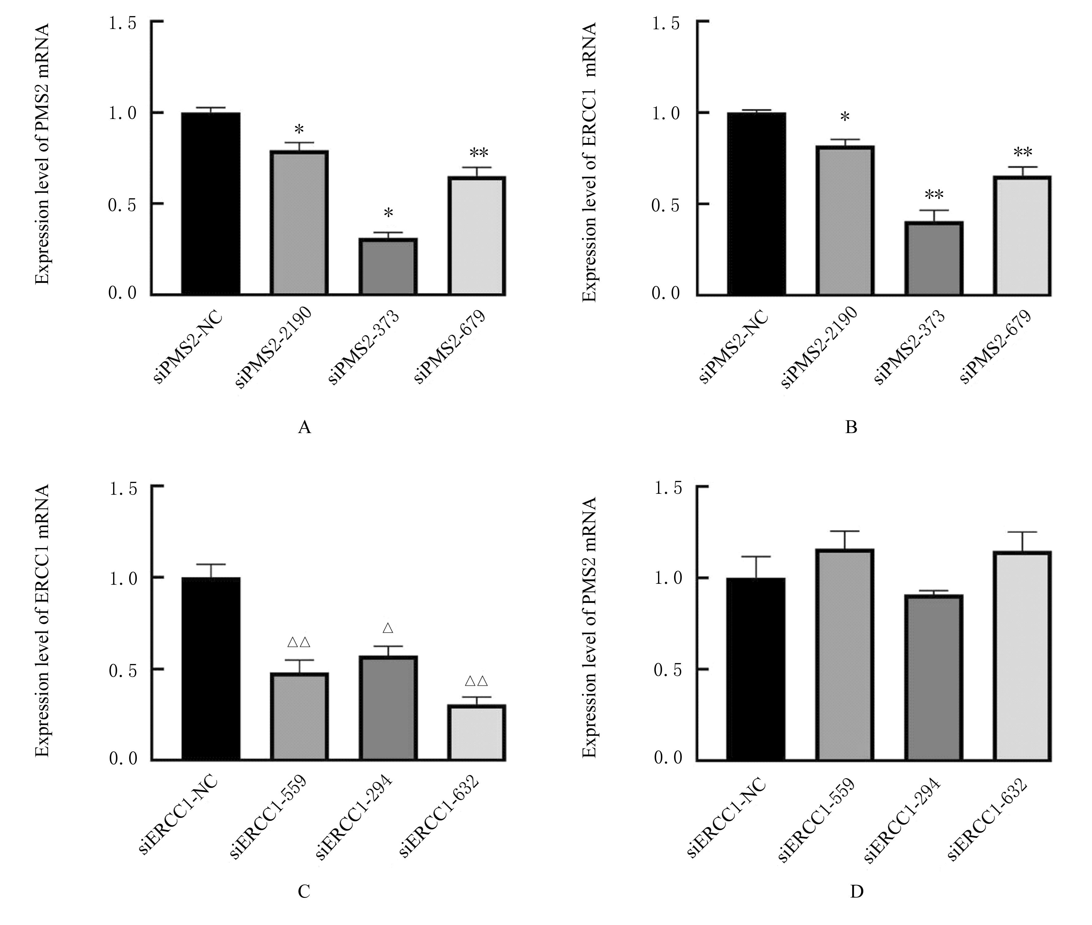
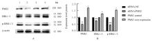
目的 探讨减数分裂后分离蛋白2(PMS2)表达对结肠癌SW480细胞生物学行为的影响,阐明PMS2与切除修复交叉互补组1(ERCC1)和细胞外调节蛋白激酶(ERK)信号转导通路的关系。 方法 将PMS2 siRNA质粒和PMS2过表达质粒分别转染入结肠癌SW480细胞(分别为PMS2敲减组和PMS2过表达组),同时设PMS2敲减对照组(siRNA-NC组)和PMS2过表达对照组(PMS2 control组)。采用实时荧光定量PCR(RT-qPCR)法检测各组细胞中PMS2 mRNA表达水平,Western blotting法检测各组细胞中PMS2蛋白表达水平,CCK-8法检测各组细胞增殖活性,细胞划痕实验检测各组细胞迁移率,Transwell小室实验检测各组细胞中侵袭细胞数,流式细胞术检测顺铂作用后各组细胞凋亡率。通过String数据库,对PMS2、ERCC1和ERK上下游蛋白的关系进行生物信息学分析。SW480细胞分别采用3条siRNA进行PMS2和ERCC1敲减,采用RT-qPCR法验证PMS2与ERCC1的相互作用,采用Western blotting法检测各组细胞中PMS2、细胞外调节蛋白激酶1/2(ERK1/2)和磷酸化ERK1/2 (p-ERK1/2)蛋白表达水平。 结果 RT-qPCR法和Western blotting法检测,PMS2基因敲减和过表达细胞模型构建成功。与siRNA-NC组比较,PMS2 敲减组细胞增殖活性和细胞迁移率明显升高(P<0.05或P<0.01),侵袭细胞数明显增加(P<0.01),顺铂作用后细胞凋亡率明显降低(P<0.01);与PMS2 control组比较,PMS2过表达组细胞增殖活性和细胞迁移率明显降低(P<0.01),侵袭细胞数明显减少(P<0.01),顺铂作用后细胞凋亡率明显升高(P<0.01)。 蛋白-蛋白互作(PPI)富集P值为2.09e-07,包含ERCC1和ERK1/2等相互作用节点数共有13个,提示PMS2、ERCC1和ERK1/2之间可能存在调控作用。与siRNA-NC组比较,各PMS2敲减组细胞中ERCC1 mRNA表达水平明显降低(P<0.05或P<0.01);与siERCC1-NC组比较,各ERCC1敲减组细胞中PMS2 mRNA表达水平差异无统计学意义(P>0.05)。与siRNA-NC组比较,PMS2敲减组细胞中PMS2、ERK1/2和p-ERK1/2蛋白表达水平均明显降低(P<0.05或P<0.01);与PMS2 control组比较,PMS2过表达组细胞中PMS2、ERK1/2和p-ERK1/2蛋白表达水平均明显升高(P<0.01)。 结论 PMS2表达可影响结肠癌SW480细胞增殖、迁移、侵袭和抗凋亡能力。PMS2与ERCC1存在互相作用关系,并可通过调节ERCC1参与ERK信号转导通路。

关键词: 结肠肿瘤, SW480细胞, 减数分裂后分离蛋白2, 切除修复交叉互补组1, 细胞外调节蛋白激酶1/2, 信号通路

Abstract:

Objective To discuss the effect of the expression of post-meiotic segregated protein 2 (PMS2) on the biological behaviors of colon cancer SW480 cells, and to clarify the relationships between PMS2 and excision repair cross-complementation group 1(ERCC1) and extracellular regulated protein kinase (ERK) signal transduction pathway. Methods The PMS2 siRNA and PMS2 over-expression plasmids were respectively transfected into the colon cancer SW480 cells (knockdown group and PMS2 over-expression group). The PMS2 knockdown control group (siRNA-NC group) and PMS2 over-expression control group (PMS2 control group) were wet up.Real-time fluorescence quantitative PCR(RT-qPCR) method was used to detect the expression levels of PMS2 mRNA in cells in various groups; the expression levels of PMS2 protein in the cells in various groups were detected by Western blotting method; the proliferation activities of cells in various groups were detected by CCK-8 assay; cell scratch test was used to detect the migration rates of cells in various groups;the numbers of invasion cells in various groups were detected by Transwell chamber assay; the apoptotic rates of cells in various groups after cisplatin treatment were detected by flow cytometry;bioinformatics analysis was performed on the relationship between upstream and downstream proteins of PMS2, ERCC1, and ERK by using String Database;the PMS2 and ERCC1 knockdown in the SW480 cells were treated with 3 siRNA.The RT-qPCR method was used to verify the reaction between PMS2 and ERCC1;the expression levels of PMS2,extracellular regulated protein kinases 1/2(ERK1/2), and phosphorylated ERK1/2(p-ERK1/2) proteins in the cells in various groups were detected by Western blotting method. Results The RT-qPCR and Western blotting results showed that the cell model of PMS2 gene knockdown and over-expression was successfully established. Compared with siRNA-NC group, the cell proliferative activity and cell migration rate in PMS2 knockdown group were increased(P<0.05 or P<0.01), the number of invasion cells was increased(P<0.01),the apoptotic rate was decreased after cisplatin treatment (P<0.05); compared with PMS2 control group, the cell proliferative activity and cell migration rate in PMS2 over-expression group were decreased(P<0.05), the number of invasion cells was decreased(P<0.01), and the apoptotic rate was increased after cisplatin treatment (P<0.05).The protein-protein interaction (PPI) enrichment P value was 2.09e-07, including a total of 13 interaction nodes such as ERCC1 and ERK1/2, suggesting that there might be a regulatory effect between PMS2, ERCC1 and ERK1/2. Compared with siRNA-NC group, the expression levels of ERCC1 mRNA in the cells in various PMS2-knockout groups were significantly decreased (P<0.05 or P<0.01). There was no significant difference in the expression of PMS2 mRNA in the cells between various ERCC1 knockdown groups and siERCC1-NC group (P>0.05). Compared with siRNA-NC group, the expression levels of PMS2, ERK1/2, and p-ERK1/2 proteins in the cells in PMS2 knockdown group were decreased (P<0.05 or P<0.01); compared with PMS2 control group, the expression levels of PMS2, ERK1/2,and p-ERK1/2 proteins in the cells in PMS2 over-expression group were increased (P<0.05). Conclusion PMS2 expression can affect the proliferation, migration, invasion,and anti-apoptotic abilities of the colon cancer SW480 cells.PMS2 interacts with ERCC1 and participates in ERK signaling transduction pathway by regulating ERCC1.

Key words: Colon neoplasms, SW480 cells, Posteiotic segregation increased 2, Excision repair cross-complementation group 1, Extracellular regulated protein kinases 1/2, Signaling pathway

中图分类号: 

  • R735.35