吉林大学学报(医学版) ›› 2024, Vol. 50 ›› Issue (2): 523-528.doi: 10.13481/j.1671-587X.20240227

• 临床研究 • 上一篇    

APRI、AAR和FIB-4等预测模型对自身免疫性肝硬化伴食管胃底静脉曲张的诊断价值

王素梅1,王楠2,于珍1,张金卷1,张健东1()   

  1. 1.天津市第三中心医院检验科 天津市重症疾病体外生命支持重点实验室 天津市人工细胞工程技术研究中心 天津市肝胆疾病研究所,天津 300170
    2.天津医科大学检验医学院,天津 300070
  • 收稿日期:2023-05-11 出版日期:2024-03-28 发布日期:2024-04-28
  • 通讯作者: 张健东 E-mail:zhangjiandongth@sina.com
  • 作者简介:王素梅(1983-),女,河北省廊坊市人,副主任技师,医学硕士,主要从事临床疾病检验方面的研究。
  • 基金资助:
    天津市科技局应用基础研究项目(22JCQNJC01590);天津市卫健委医学重点学科(专科)建设项目(TJYXZDXK-047A)

Diagnostic values of APRI, AAR, and FIB-4 predictive models in autoimmune cirrhosis combined with esophagogastric fundal varices

Sumei WANG1,Nan WANG2,Zhen YU1,Jinjuan ZHANG1,Jiandong ZHANG1()   

  1. 1.Department of Clinical Laboratory, Third Central Hospital, Tianjin City, Key Laboratory of Extracorporeal Life Support for Critical Diseases of Tianjin City, Artificial Cell Engineering Technology Research Center of Tianjin City, Tianjin Institute of Hepatobiliary Disease, Tianjin 300170, China
    2.School of Medical Laboratory, Tianjin Medical University, Tianjin 300070, China
  • Received:2023-05-11 Online:2024-03-28 Published:2024-04-28
  • Contact: Jiandong ZHANG E-mail:zhangjiandongth@sina.com

摘要:

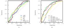
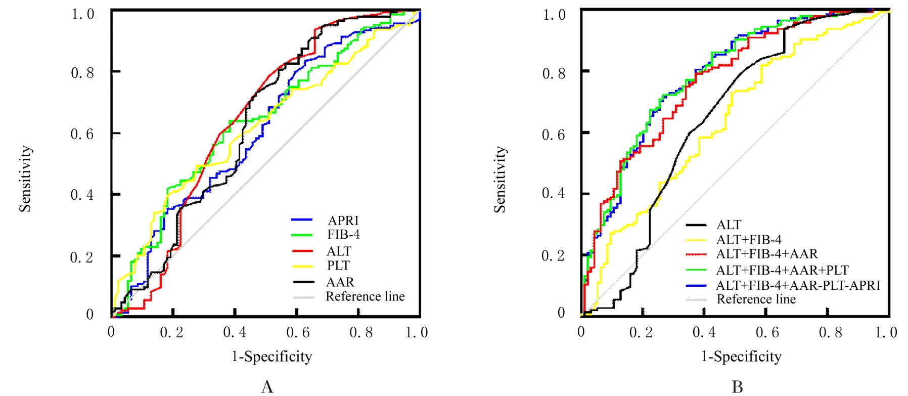
目的 评价无创模型对自身免疫性肝硬化伴食管胃底静脉曲张(EGV)的诊断价值,为早期诊断自身免疫性肝硬化伴EGV提供依据。 方法 回顾性收集238例诊断为自身免疫性肝硬化患者的临床资料,根据是否伴发EGV分为有EGV组和无EGV组。比较2组患者的谷氨酸氨基转移酶(ALT)、天门冬氨酸氨基转移酶(AST)、γ-谷氨酰转肽酶(γ-GT)、血小板(PLT)、AST/PLT指数(APRI)、纤维化-4指数(FIB-4)和AST/ALT比值(AAR)水平并绘制受试者工作特征(ROC)曲线,计算ROC曲线下面积(AUC)、灵敏度、特异度、阳性预测值和阴性预测值,评价各模型对自身免疫性肝硬化伴EGV的诊断价值。 结果 有EGV组患者的APRI、FIB-4和ALT明显高于无EGV组(P<0.01),而PLT水平和AAR则明显低于无EGV组(P<0.01)。单指标诊断模型以ALT的AUC最大,AUC为0.645(95%CI: 0.580~0.705,P<0.001),其灵敏度、特异度、阳性预测值和阴性预测值分别为93.75%、34.04%、68.53%及78.05%。多指标联合模型中以ALT+FIB-4+AAR+PLT联合模型的AUC最大,AUC为0.787(95%CI:0.730~0.838,P<0.001),其灵敏度、特异度、阳性预测值和阴性预测值分别为72.22%、73.40%、80.62%及63.30%。ALT+FIB-4+AAR、ALT+FIB-4+AAR+PLT和ALT+FIB-4+AAR+PLT+APRI联合模型的AUC与ALT和ALT+FIB-4模型比较差异均有统计学意义(P<0.05)。而上述3种模型AUC比较差异均无统计学意义(P>0.05)。 结论 APRI、FIB-4、ALT、PLT和AAR联合检测可提高自身免疫性肝硬化伴EGV的早期诊断效能。

关键词: 自身免疫, 肝硬化, 食管胃底静脉曲张, 预测模型, 诊断价值

Abstract:

Objective To evaluate the diagnostic value of non-invasive models for autoimmune cirrhosis with esophagogastric varices (EGV),and to provide the basis for the early diagnosis of autoimmune cirrhosis with EGV. Methods The retrospective collection of clinical data from 238 patients diagnosed with autoimmune cirrhosis was performed, and the patients divided into EGV group and non-EGV group according to whether complicated with EGV. The levels of alanine aminotransferase (ALT), aspartate aminotransferase (AST), γ-glutamyltransferase (γ-GT), platelet (PLT), AST / PLT index (APRI), fibrosis-4 index (FIB-4), and AST/ALT ratio (AAR) of the patients in both groups were compared and the receiver operating characteristic (ROC) curve was plotted to calculate the area under the curve (AUC), sensitivity, specificity, positive predictive value, and negative predictive value to evaluate the diagnostic values of various models for autoimmune cirrhosis with EGV. Results The levels of APRI, FIB-4, and ALT of the patients in EGV group were significantly higher than those in non-EGV group (P<0.01), while the levels of PLT and AAR were significantly lower than those in non-EGV group (P<0.01). In single indicator diagnostic model, ALT had the largest AUC of 0.645 (95% CI: 0.580-0.705, P<0.001), the sensitivity was 93.75%, the specificity was 34.04%, the positive predictive value was 68.53%, and the negative predictive value was 78.05%. In multiple indicator combined models, the combined model of ALT+FIB-4+AAR+PLT had the largest AUC of 0.787 (95% CI: 0.730-0.838, P<0.001), and the sensitivity was72.22%, the specificity was 73.40%, the positive predictive value was 80.62%, and the negative predictive value was 63.30%. Compared with ALT and ALT+FIB-4 models,the AUC of the combined models of ALT+FIB-4+AAR, ALT+FIB-4+AAR+PLT, and ALT+FIB-4+AAR+PLT+APRI had statistically significant differences (P<0.05). However, there were no significant differences in AUC among the above three models (P>0.05). Conclusion The combined detection of APRI, FIB-4, ALT, PLT, and AAR can improve the early diagnostic efficacy of autoimmune cirrhosis with EGV.

Key words: Autoimmunity, Liver cirrhosis, Esophagogastric fundus varices, Predictive modeling, Diagnostic value

中图分类号: 

  • R575.2