吉林大学学报(医学版) ›› 2025, Vol. 51 ›› Issue (1): 222-227.doi: 10.13481/j.1671-587X.20250127

• 临床医学 • 上一篇    

具有特殊影像学表现的富于巨细胞的骨肉瘤1例报告及文献复习

高子龙1,刘飙1,祁乐2,郎靖宇1,刘燕1()   

  1. 1.吉林大学中日联谊医院手足外科, 吉林 长春 130033
    2.吉林大学中日联谊医院创面修复科·整形重建显微外科, 吉林 长春 130033
  • 收稿日期:2024-11-19 接受日期:2024-12-11 出版日期:2025-01-28 发布日期:2025-03-06
  • 通讯作者: 刘燕 E-mail:lyan2009@jlu.edu.cn
  • 作者简介:高子龙(2000-),男,辽宁省沈阳市人,住院医师,在读硕士研究生,主要从事手足外科临床疾病方面的研究。
  • 基金资助:
    吉林省科技厅自然科学基金项目(YDZJ202301ZYTS044)

Giant cell-rich osteosarcoma with special imaging findings: A case report and literature review

Zilong GAO1,Biao LIU1,Le QI2,Jingyu LANG1,Yan LIU1()   

  1. 1.Department of Hand and Foot Surgery,China-Japan Union Hospital,Jilin University,Changchun 130033,China
    2.Department of Wound Repair,Plastic and Reconstructive Microsurgery,China-Japan Union Hospital Jilin University,Changchun 130033,China
  • Received:2024-11-19 Accepted:2024-12-11 Online:2025-01-28 Published:2025-03-06
  • Contact: Yan LIU E-mail:lyan2009@jlu.edu.cn

摘要:

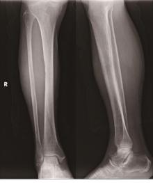
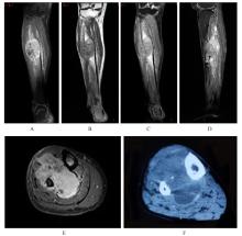
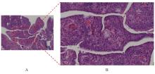
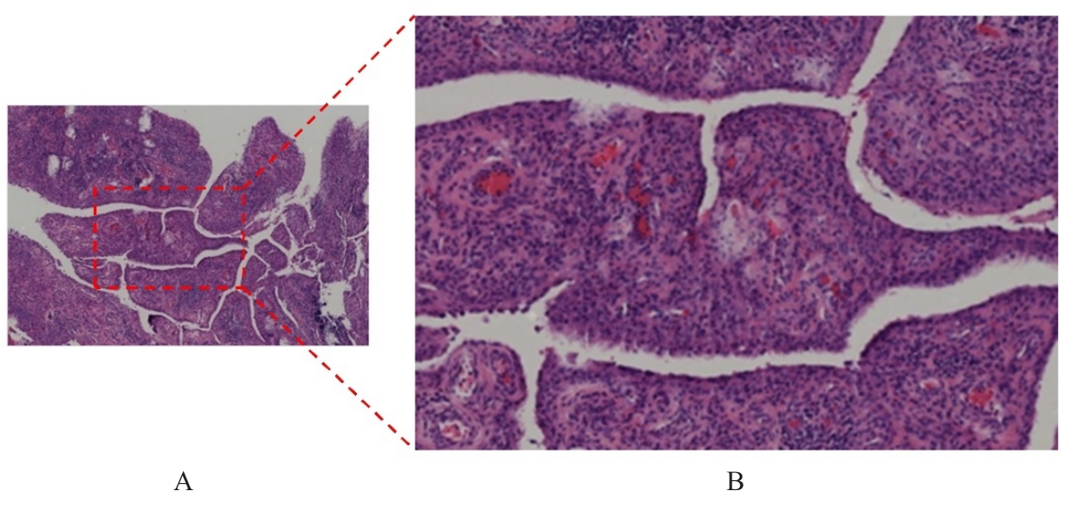
富于巨细胞的骨肉瘤(GCRO)是骨肉瘤中一种特殊的亚型,极为罕见。本文作者报道1例具有特殊影像学表现的GCRO患者的临床表现及影像学资料,为GCRO的临床诊治提供参考。患者,女性,69岁,因发现右小腿肿物1个月入院。X线影像表现为胫骨及腓骨相对缘皮质骨呈轻度虫蚀样改变。CT平扫影像表现为肌肉内肿物,胫骨和腓骨相对缘骨皮质破坏,肿瘤仅累及表面皮质骨,未侵及深部皮质骨及髓腔。磁共振成像(MRI)影像表现为肿瘤邻近的胫骨和腓骨骨质局部破坏,髓腔未受侵及。行肿物扩大切除术,术后病理诊断为GCRO。患者术后化疗,术后15个月右小腿肿物复发,术后21个月,患者去世。GCRO常因临床表现不特异导致误诊或漏诊,本文作者探讨该例GCRO患者的临床表现及影像学资料,以提高临床医生对GCRO的临床认识水平和诊疗水平。

关键词: 骨肉瘤, 巨细胞, X线计算机, 计算机断层扫描, 磁共振成像

Abstract:

Giant cell-rich osteosarcoma(GCRO) is a special subtype of osteosarcoma, and is rare. This paper reported the clinical and imaging data of the patient with GCRO who had special imaging manifestations, and to provide the reference for clinical diagnosis and treatment of GCRO. The patient, a 69-year-old female,was admitted to the hospital due to the discovery of a mass in the right lower leg for 1 month. The X-ray imaging manifestations showed that the cortical bones on the opposite edges of the tibia and fibula presented mild moth-eaten changes. The plain CT scan imaging manifestations showed that there was a mass within the muscle, and the cortical bones on the opposite edges of the tibia and fibula were destroyed. The tumor only involved the superficial cortical bones and did not invade the deep cortical bones and the medullary cavity. The MRI imaging manifestations showed that the local bones of the tibia and fibula adjacent to the tumor were destroyed, and the medullary cavity was not invaded. An extended resection of the mass was performed, and the postoperative pathological diagnosis was GCRO. The patient received chemotherapy after the operation. However, a tumor in the right lower leg recurred 15 months after the operation. The patient passed away 21 months after the operation. Giant cell-rich osteosarcoma often leads to misdiagnosis or missed diagnosis due to its non-specific clinical manifestations. This paper explored the clinical manifestations and imaging data of the patient with GCRO in order to improve the clinical understanding level and the level of diagnosis and treatment of the clinicians about this disease.

Key words: Osteosarcoma, Giant cell, X-ray computed, Computed tomography, Magnetic resonance imaging

中图分类号: 

  • R738.1