吉林大学学报(医学版) ›› 2024, Vol. 50 ›› Issue (6): 1654-1663.doi: 10.13481/j.1671-587X.20240619

• 基础研究 • 上一篇    

扶正软坚抗癌方调控Akt/MDM2/P53信号通路对肝癌HepG2细胞恶性生物学行为的影响

娄静1,赵雷1,朱岩洁1,袁帅强1,王菲1,张杭洲1,徐娇娇1,余晓珂2,侯留法1()   

  1. 1.河南省中医药研究院附属医院肝胆脾胃科,河南 郑州 450003
    2.河南中医药大学第三附属医院老年病内科,河南 郑州 450003
  • 收稿日期:2023-04-27 出版日期:2024-11-28 发布日期:2024-12-10
  • 通讯作者: 侯留法 E-mail:houliufa@126.com
  • 作者简介:娄 静(1981-),女,河南省新乡市人,副主任医师,医学硕士,主要从事中医内科消化病诊断和治疗方面的研究。
  • 基金资助:
    河南省中医药科学研究专项课题(2023ZY2139)

Effect of Fuzheng Ruanjian Anticancer Formula on malignant biological behaviors of hepatocellulars carcinoma HepG2 cells by regulating Akt/MDM2/P53 signaling pathway

Jing LOU1,Lei ZHAO1,Yanjie ZHU1,Shuaiqiang YUAN1,Fei WANG1,Hangzhou ZHANG1,Jiaojiao XU1,Xiaoke YU2,Liufa HOU1()   

  1. 1.Department of Hepatobiliary Splenogastric,Affiliated Hospital,Henan Provincial Academy of Traditional Chinese Medicine,Zhengzhou 450003,China
    2.Department of Geriatrics,Third Affiliated Hospital,Henan University of Traditional Chinese Medicine,Zhengzhou 450003,China
  • Received:2023-04-27 Online:2024-11-28 Published:2024-12-10
  • Contact: Liufa HOU E-mail:houliufa@126.com

摘要:

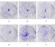
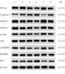
目的 探讨扶正软坚抗癌方调控蛋白激酶B(Akt)/鼠双微体2(MDM2)/P53信号通路对肝癌HepG2细胞恶性生物学行为的影响。 方法 采用0、0.05、0.10、0.20、0.40、0.80、1.60、3.20和6.40 g·mL-1扶正软坚抗癌方分别处理HepG2细胞48 h,CCK-8法检测HepG2细胞存活率,筛选扶正软坚抗癌方浓度用于后续实验。将HepG2细胞分为对照组、低剂量扶正软坚抗癌方组(0.2 g·mL-1)、中剂量扶正软坚抗癌方组(0.4 g·mL-1)、高剂量扶正软坚抗癌方组(0.8 g·mL-1)、SC79组(8 mg·L-1 SC79)和高剂量扶正软坚抗癌方+SC79组(0.8 g·mL-1 扶正软坚抗癌方+ 8 mg·L-1 SC79)。CCK-8法检测各组HepG2细胞增殖活性,克隆形成实验检测各组HepG2细胞克隆形成率,流式细胞术检测各组HepG2细胞凋亡率,Transwell小室实验检测各组HepG2细胞迁移和侵袭细胞数,Western blotting法检测各组HepG2细胞中增殖细胞核抗原(PCNA)、含半胱氨酸的天冬氨酸蛋白酶3(Caspase-3)、基质金属蛋白酶(MMP)-2、MMP-9、磷酸化Akt(p-Akt)、磷酸化MDM2(p-MDM2)和P53蛋白表达水平。 结果 随着扶正软坚抗癌方浓度(0、0.05、0.10、0.20、0.40、0.80、1.60、3.20和6.40 g·mL-1)的升高,HepG2细胞存活率逐渐降低(P<0.05),选取0.2、0.4和0.8 g·mL-1 扶正软坚抗癌方用于后续实验。CCK-8法检测,与对照组比较,低、中和高剂量扶正软坚抗癌方组HepG2细胞增殖活性均明显降低(P<0.05),并呈剂量依赖性,SC79组HepG2细胞增殖活性明显升高(P<0.05);与高剂量扶正软坚抗癌方组比较,高剂量扶正软坚抗癌方+SC79组HepG2细胞增殖活性明显升高(P<0.05)。克隆形成实验检测,与对照组比较,低、中和高剂量扶正软坚抗癌方组HepG2细胞克隆形成率均明显降低(P<0.05),并呈剂量依赖性,SC79组HepG2细胞克隆形成率明显升高(P<0.05);与高剂量扶正软坚抗癌方组比较,高剂量扶正软坚抗癌方+SC79组HepG2细胞克隆形成率明显升高(P<0.05)。流式细胞术检测,与对照组比较,低、中和高剂量扶正软坚抗癌方组HepG2细胞凋亡率均明显降低(P<0.05),并呈剂量依赖性,SC79组HepG2细胞凋亡率明显升高(P<0.05);与高剂量扶正软坚抗癌方组比较,高剂量扶正软坚抗癌方+SC79组HepG2细胞凋亡率明显升高(P<0.05)。Transwell小室实验检测,与对照组比较,低、中和高剂量扶正软坚抗癌方组HepG2细胞迁移和侵袭细胞数均明显降低(P<0.05),并呈剂量依赖性,SC79组HepG2细胞迁移和侵袭细胞数均明显升高(P<0.05);与高剂量扶正软坚抗癌方组比较,高剂量扶正软坚抗癌方+SC79组HepG2细胞迁移和侵袭细胞数均明显升高(P<0.05)。Western blotting法检测,与对照组比较,低、中和高剂量扶正软坚抗癌方组HepG2细胞中PCNA、MMP-2、MMP-9、p-Akt和p-MDM2蛋白表达水平均明显降低(P<0.05),并呈剂量依赖性;Caspase-3和P53蛋白表达水平均明显升高(P<0.05),并呈剂量依赖性;SC79组HepG2细胞中PCNA、MMP-2、MMP-9、p-Akt和p-MDM2蛋白表达水平均明显升高(P<0.05),Caspase-3和P53蛋白表达水平均明显降低(P<0.05);与高剂量扶正软坚抗癌方组比较,高剂量扶正软坚抗癌方+SC79组HepG2细胞中PCNA、MMP-2、MMP-9、p-Akt和p-MDM2蛋白表达水平均明显升高(P<0.05),Caspase-3和P53蛋白表达水平均明显降低(P<0.05)。 结论 扶正软坚抗癌方抑制HepG2细胞增殖、迁移和侵袭,促进细胞凋亡,其作用机制与抑制Akt/MDM2信号通路、上调P53蛋白表达有关。

关键词: 扶正软坚抗癌方, 蛋白激酶B, 鼠双微体2, P53, 肝肿瘤, 细胞增殖, 细胞凋亡

Abstract:

Objective To discuss the effect of Fuzheng Ruanjian Anticancer Formula on the malignant biological behaviors of the hepatocellular carcinoma HepG2 cells by requlating protein kinase B(Akt)/murine double minute 2(MDM2)/P53 signaling pathway. Methods The HepG2 cells were treated with 0, 0.05, 0.10, 0.20, 0.40, 0.80, 1.60, 3.20, and 6.40 g·mL-1 Fuzheng Ruanjian Anticancer Formula for 48 h. CCK-8 method was used to detect the survival rates of the HepG2 cells in various groups, and the concentrations of Fuzheng Ruanjian Anticancer Formula for the subsequent experiments were screened. The HepG2 cells were divided into control group, low dose of Fuzheng Ruanjian Anticancer Formula group (0.2 g·mL-1), medium dose of Fuzheng Ruanjian Anticancer Formula group (0.4 g·mL-1), high dose of Fuzheng Ruanjian Anticancer Formula group (0.8 g·mL-1), SC79 group (8 mg·L-1 SC79), and high dose of Fuzheng Ruanjian Anticancer Formula+SC79 group (0.8 g·mL-1 Fuzheng Ruijian Anticancer Formula+8 mg·L-1 SC79). CCK-8 method was used to detect the proliferation activities of the HepG2 cells in various groups; clone formation assay was used to detect the clone formation rates of the HepG2 cells in various groups; flow cytometry was used to detect the apoptotic rates of the HepG2 cells in various groups; Transwell chamber assay was used to detect the numbers of migration and invasion HepG2 cells in various groups; Western blotting method was used to detect the expression levels of proliferating cell nuclear antigen (PCNA), cysteine aspartate specific proteinase (Caspase-3), matrix metalloproteinase (MMP)-2, MMP-9, phosphorylated Akt (p-Akt), phosphorylated MDM2 (p-MDM2), and P53 proteins in the HepG2 cells in various groups. Results As the increasing of concentrations of Fuzheng Ruanjian Anticancer Formula (0, 0.05, 0.10, 0.20, 0.40, 0.80, 1.60, 3.20, and 6.40 g·mL-1), the surival rates of the HepG2 cells were gradually decreased (P<0.05), and 0.2, 0.4, and 0.8 g·mL-1 Fuzheng Ruanjian Anticancer Formula were selected for the subsequent experiments. The CCK-8 assay results showed that compared with control group, the proliferation activities of the HepG2 cells in low, medium, and high doses of Fuzheng Ruanjian Anticancer Formula groups were significantly decreased (P<0.05), in a dose-dependent manner, while the proliferation activity of the cells in SC79 group was significantly increased (P<0.05). Compared with high dose of Fuzheng Ruanjian Anticancer Formula group, the proliferation activity of the HepG2 cells in high dose of Fuzheng Ruanjian Anticancer Formula+SC79 group was significantly increased (P<0.05). The clone formation assay results showed that compared with control group, the clone formation rates of the HepG2 cells in low, medium, and high doses of Fuzheng Ruanjian Anticancer Formula groups were significantly decreased (P<0.05) in a dose-dependent manner, while the clone formation rate of the cells in SC79 group was significantly increased (P<0.05); compared with high dose of Fuzheng Ruanjian Anticancer Formula group, the clone formation rate of the cells in high dose of Fuzheng Ruanjian Anticancer Formula+SC79 group was significantly increased (P<0.05). The flow cytometry results showed that compared with control group, the apoptotic rates of the HepG2 cells in low, medium, and high doses of Fuzheng Ruijian Anticancer Formula groups were significantly increased (P<0.05) in a dose-dependent manner, while the apoptotic rate of the cells in SC79 group was significantly decreased (P<0.05); compared with high dose of Fuzheng Ruanjian Anticancer Formula group, the apoptotic rate of the cells in high dose of Fuzheng Ruanjian Anticancer Formula+SC79 group was significantly decreased(P<0.05). The Transwell chamber assay results showed that compared with control group, the numbers of migration and invasion HepG2 cells in low, medium, and high doses of Fuzheng Ruanjian Anticancer Formula groups were significantly decreased (P<0.05) in a dose-dependent manner, while the numbers of migration and invasion cells in SC79 group were significantly increased (P<0.05); compared with high dose of Fuzheng Ruanjian Anticancer Formula group, the numbers of migration and invasion HepG2 cells in high dose of Fuzheng Ruanjian Anticancer Formula+SC79 group were significantly increased (P<0.05). The Western blotting results showed that compared with control group, the expression levels of PCNA, MMP-2, MMP-9, p-Akt, and p-MDM2 proteins in the cells in low, medium, and high doses of Fuzheng Ruanjian Anticancer Formula groups were significantly decreased (P<0.05) in a dose-dependent manner, while the expression levels of Caspase-3 and P53 proteins were significantly increased (P<0.05) in a dose-dependent manner, while the expression levels of PCNA, MMP-2, MMP-9, p-Akt, and p-MDM2 proteins in the cells in SC79 group were significantly increased (P<0.05), and the expression levels of Caspase-3 and P53 proteins were significantly decreased (P<0.05); compared with high dose of Fuzheng Ruanjian Anticancer Formula group, the expression levels of PCNA, MMP-2, MMP-9, p-Akt, and p-MDM2 proteins in the cells in high dose of Fuzheng Ruanjian Anticancer Formula+SC79 group were significantly increased (P<0.05), while the expression levels of Caspase-3 and P53 proteins were significantly decreased (P<0.05). Conclusion Fuzheng Ruanjian Anticancer Formula may inhibit the proliferation, migration, and invasion of the HepG2 cells and promote the apoptosis, and its mechanism may be related to suppressing the Akt/MDM2 signaling pathway and upregulating the P53 proteim expression.

Key words: Fuzheng Ruanjian Anticancer Formula, Protein kinase B, Mouse double minute 2, P53, Liver neoplasm, Cell proliferation, Apoptosis

中图分类号: 

  • R735.7