吉林大学学报(医学版) ›› 2025, Vol. 51 ›› Issue (1): 115-123.doi: 10.13481/j.1671-587X.20250114

• 基础研究 • 上一篇    

沉默DDX39A基因对食管癌TE-1细胞增殖、迁移和侵袭的作用及其机制

武鹏立1,李凤玉2,刘博3,吕洋4()   

  1. 1.河北北方学院研究生学院,河北 张家口 075000
    2.陆军第八十一集团军医院肿瘤内科,河北 张家口 075000
    3.河北北方学院附属第一医院病理科,河北 张家口 075000
    4.河北北方学院 基础医学院病理教研室,河北 张家口 075000
  • 收稿日期:2024-03-12 接受日期:2024-03-22 出版日期:2025-01-28 发布日期:2025-03-06
  • 通讯作者: 吕洋 E-mail:yyang2bb@163.com
  • 作者简介:武鹏立(1998-),女,北京市人,在读硕士研究生,主要从事消化道肿瘤临床病理方面的研究。
  • 基金资助:
    河北省科技厅重点研发计划项目卫生健康创新专项(22377765D);河北省卫健委2024年度医学科学研究指导性课题(20240286);河北北方学院自然科学项目(XJ2023040)

Effect of silencing DDX39A gene on proliferation, migration and invasion of esophageal cancer TE-1 cells and its mechanism

Pengli WU1,Fengyu LI2,Bo LIU3,Yang LYU4()   

  1. 1.Graduate School,Hebei North University,Zhangjiakou 075000,China
    2.Department of Medical Oncology,Army 81st Group Army Hospital,Zhangjiakou 075000,China
    3.Department of Pathology,First Affiliated Hospital,Hebei North University,Zhangjiakou 075000,China
    4.Department of Pathology,Hebei North University,Zhangjiakou 075000,China
  • Received:2024-03-12 Accepted:2024-03-22 Online:2025-01-28 Published:2025-03-06
  • Contact: Yang LYU E-mail:yyang2bb@163.com

摘要:

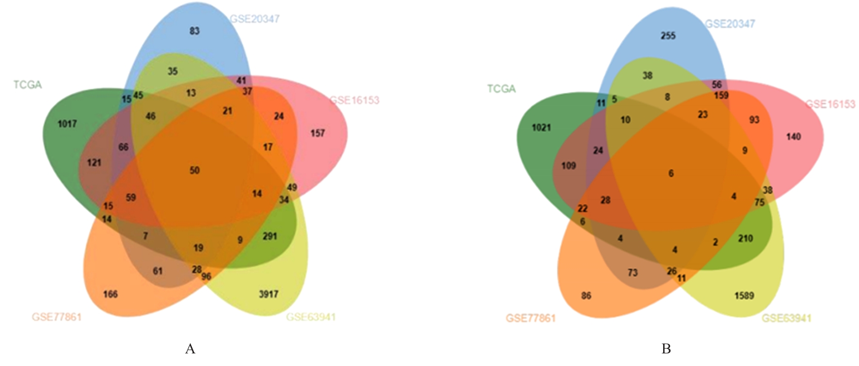
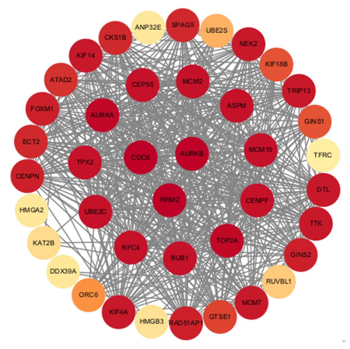
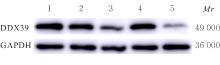
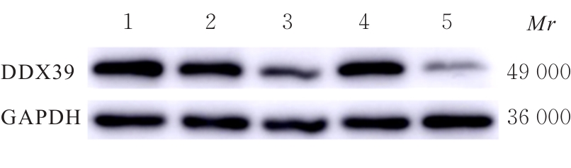
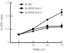
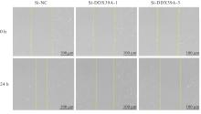
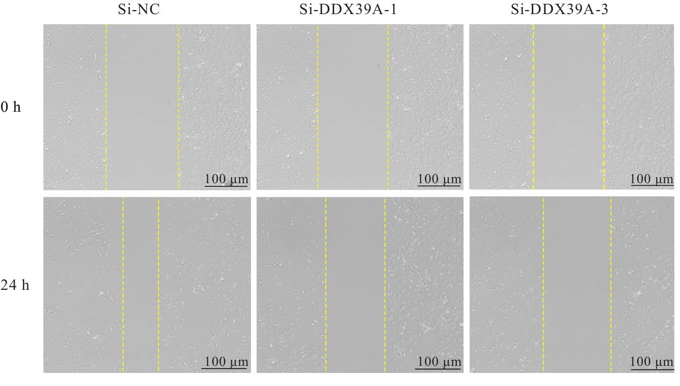
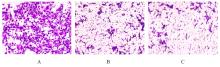
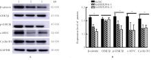
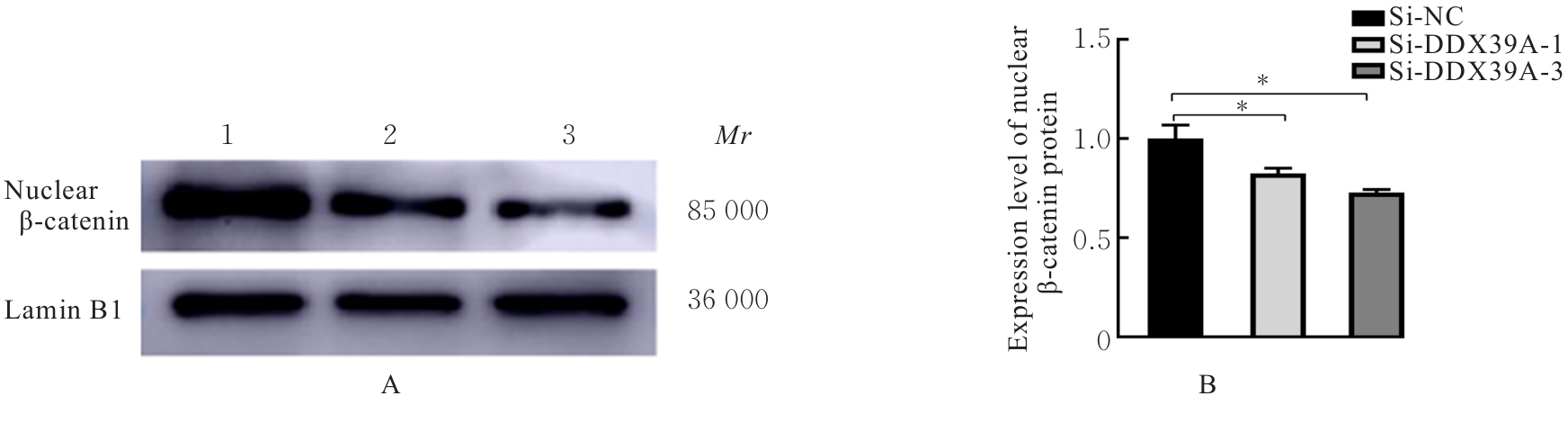
目的 探讨沉默DEAD-box RNA解旋酶39A(DDX39A)对食管癌TE-1细胞增殖、迁移和侵袭的调控作用,并阐明其可能的作用机制。 方法 通过基因表达汇编(GEO)数据库下载GSE63941、GSE77861、GSE20347和GSE16153芯片数据,利用癌症基因组图谱(TCGA)数据库筛选并下载食管癌相关数据;采用R软件分析差异表达基因;通过基因与蛋白质相互作用关系检索工具(STRING)构建蛋白-蛋白相互作用(PPI)网络数据,并采用Cytoscape软件中MCODE插件整合数据,鉴别出相关性强的关键基因,用基因表达谱数据动态分析网页工具(GEPIA 2)分析正常食管黏膜和食管癌组织中关键基因的表达情况,用卡普兰-迈尔曲线图(Kaplan-Meier Plotter)将筛查出来的关键基因进行生存分析并绘图。细胞学实验,选择食管癌TE-1细胞作为研究对象,采用小干扰RNA技术(siRNA)沉默DDX39A基因表达;取对数生长期TE-1细胞,将细胞分为空白组(MOCK组)、阴性对照组(si-NC组)和沉默组(si-DDX39A组);采用实时荧光定量PCR(RT-qPCR)和Western blotting法检测各组细胞中DDX39A mRNA及蛋白表达水平,CCK-8法检测各组细胞增殖活性,细胞划痕愈合实验检测各组细胞迁移率,Transwell小室实验检测各组TE-1细胞的侵袭细胞数,Western blotting法检测各组细胞中β-黏连蛋白(β-catenin)、糖原合成酶激酶-3β(GSK3β)、磷酸化糖原合成酶激酶-3β(p-GSK3β)、原癌基因(c-MYC)和细胞周期蛋白D1(Cyclin D1)及核内β-catenin蛋白表达水平。 结果 TCGA数据库与GEO数据库相结合共得到56个差异表达基因。Cytoscape软件MCODE插件共鉴别出41个相关性强的关键基因,通过GEPIA 2和Kaplan-Meier plotter数据库分析41个基因得到DDX39A。RT-qPCR和Western blotting法,与si-NC组比较,si-DDX39A组细胞中DDX39A mRNA和蛋白表达水平均降低(P<0.05)。CCK-8法,si-DDX39A组细胞增殖活性低于si-NC组(P<0.05);细胞划痕实验,24 h后,si-DDX39A组细胞迁移率低于si-NC组(P<0.05);Transwell小室实验,si-DDX39A组侵袭细胞数低于si-NC组(P<0.05)。与si-NC组比较,si-DDX39A-1组和si-DDX39A-3组TE-1细胞中β-catenin、p-GSK3β、c-MYC及Cyclin D1以及核内β-catenin蛋白表达水平均降低(P<0.01),而GSK3β蛋白表达水平差异无统计学意义(P>0.05)。 结论 DDX39A基因沉默可以抑制食管癌TE-1细胞的增殖、迁移和侵袭能力,其作用机制可能与调控Wnt/β-catenin信号通路有关。

关键词: 食管肿瘤, DEAD-box RNA解旋酶39A, 生物信息学, 信号通路, 细胞侵袭

Abstract:

Objective To discuss the effect of DEAD-box RNA helicase 39A(DDX39A) gene silencing on the proliferation,migration and invasion of the esophageal cancer TE-1 cells,and to clarify its possible mechanism. Methods For bioinformatics analysis, GSE63941, GSE77861, GSE20347, and GSE16153 chip data were downloaded from the GEO database. The esophagel cancer-related data were selected from the TCGA Database.R software was used to analyze the differentially expressed genes.STRING Database was used to construct the protein-protein interaction (PPI) network.Identification of key genes of high relevance was achieved using the MCODE plugin in Cytoscape.The expression of key genes in normal esophageal tissue and esophageal cancer tissue were analyzed with the GEPIA 2 database. Kaplan-Meier Plotter was used to perform survived analysis and plotting for the screened key genes.Cytological experiments were carried out on esophageal cancer TE-1 cells, and small interfering RNA (siRNA)technology was used to silence the expression of DDX39A gene.The TE-1 cells in the logarithmic growth phase were selected and divided into blank (MOCK) group, negative control (si-NC) group, and silencing (si-DDX39A) group. Real-time fluorescence quantitative PCR (RT-qPCR) and Western blotting methods were used to detect the expression levels of DDX39A mRNA and protein in the TE-1 cells in various groups;CCK-8 assay was conducted to detect the proliferation activity of cells in various groups, and the cell scratch assay was used to measure the migration rate of cells in various groups; Transwell chamber assay was used to detect the number of invasion cells in various groups;Western blotting method was used to detect the expression levels of β-catenin, glycogen synthase kinase-3β(GSK3β), phosphorylated glycogen synthase kinase-3β(p-GSK3β), c-MYC, Cyclin D1 and nuclear β-catenin proteins in the cells in various groups. Results Analyses using TCGA database combined with the GEO Database yielded a total of 56 differentially expressed genes. MCODE plugin in Cytoscape software identified 41 key genes of high relevance; DDX39A was screened by analyzing 41 genes through the GEPIA 2 and Kaplan-Meier plotter Databases. The results of RT-qPCR and Western blotting methods showed that compared with si-NC group, the expression levels of DDX39A mRNA and protein in the cells in si-DDX39A group were decreased (P<0.05). The CCK-8 results showed that the proliferation activity of the cells in si-DDX39A group was lower than that in si-NC group (P<0.05). The cell scratch assay results showed that the cell migration rate in si-DDX39A group after 24 h was lower than that in si-NC group (P<0.05).The results of Transwell chamber assay showed that the number of invasion cells in si-DDX39A group was lower than that in si-NC group (P<0.05). Compared with si-NC group, the expression levels of β-catenin, p-GSK3β, c-MYC, Cyclin D1, and nuclear β-catenin in the TE-1 cells in si-DDX39A-1 group and si-DDX39A-3 group were decreased (P<0.01), but the expression levels of GSK3β protein had no significant differences (P>0.05). Conclusion Silencing of DDX39A gene could inhibit the proliferation, migration and invasion of TE-1 cells, and the mechanism may be related to the regulation of Wnt/β-catenin signaling pathway.

Key words: Esophageal neoplasms, DEAD-box RNA Helicase 39A, Bioinformatics, Signaling pathway, Cell invasion

中图分类号: 

  • R735.1