吉林大学学报(医学版) ›› 2023, Vol. 49 ›› Issue (4): 1008-1017.doi: 10.13481/j.1671-587X.20230423

• 基础研究 • 上一篇    

钙黏蛋白17对结直肠癌细胞增殖和凋亡的影响及其PI3K/AKT/mTOR信号通路调节机制

刘蒙,黄晓东,韩峥,朱庆曦,谭洁,田霞()   

  1. 湖北省武汉市第三医院消化内科,湖北 武汉 430060
  • 收稿日期:2022-07-11 出版日期:2023-07-28 发布日期:2023-07-26
  • 通讯作者: 田霞 E-mail:xtian16@sina.cn
  • 作者简介:刘 蒙(1981-),男,湖北省荆州市人,副主任医师,医学博士,主要从事消化道肿瘤信号通路和抗癌药物耐药方面的研究。
  • 基金资助:
    湖北省科技厅中央引导地方科技发展专项项目(2019ZYYD067);湖北省科技厅自然科学基金项目(2019CFB749);武汉市卫健委重大项目(WX20M01)

Effect of cadherin-17 on proliferation and apoptosis of colorectal cancer cells and its PI3K/AKT/mTOR signaling pathway regulatory mechanism

Meng LIU,Xiaodong HUANG,Zheng HAN,Qingxi ZHU,Jie TAN,Xia TIAN()   

  1. Department of Gastroenterology,Third Hospital,Wuhan City,Hubei Province,Wuhan 430060,China
  • Received:2022-07-11 Online:2023-07-28 Published:2023-07-26
  • Contact: Xia TIAN E-mail:xtian16@sina.cn

摘要:

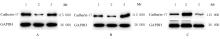
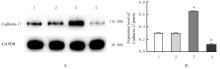
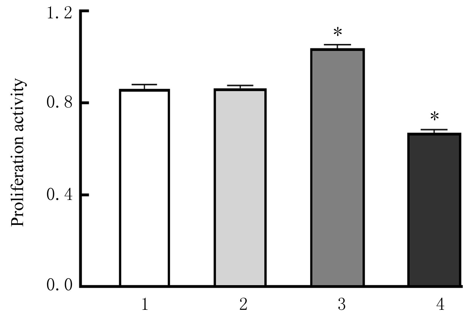
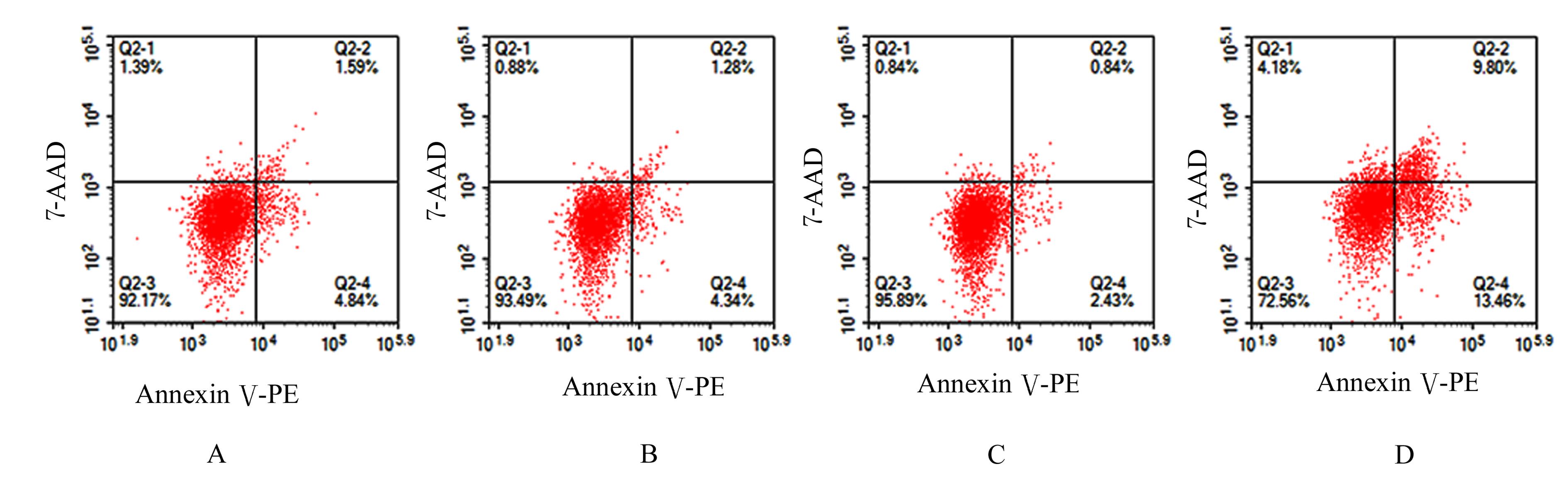
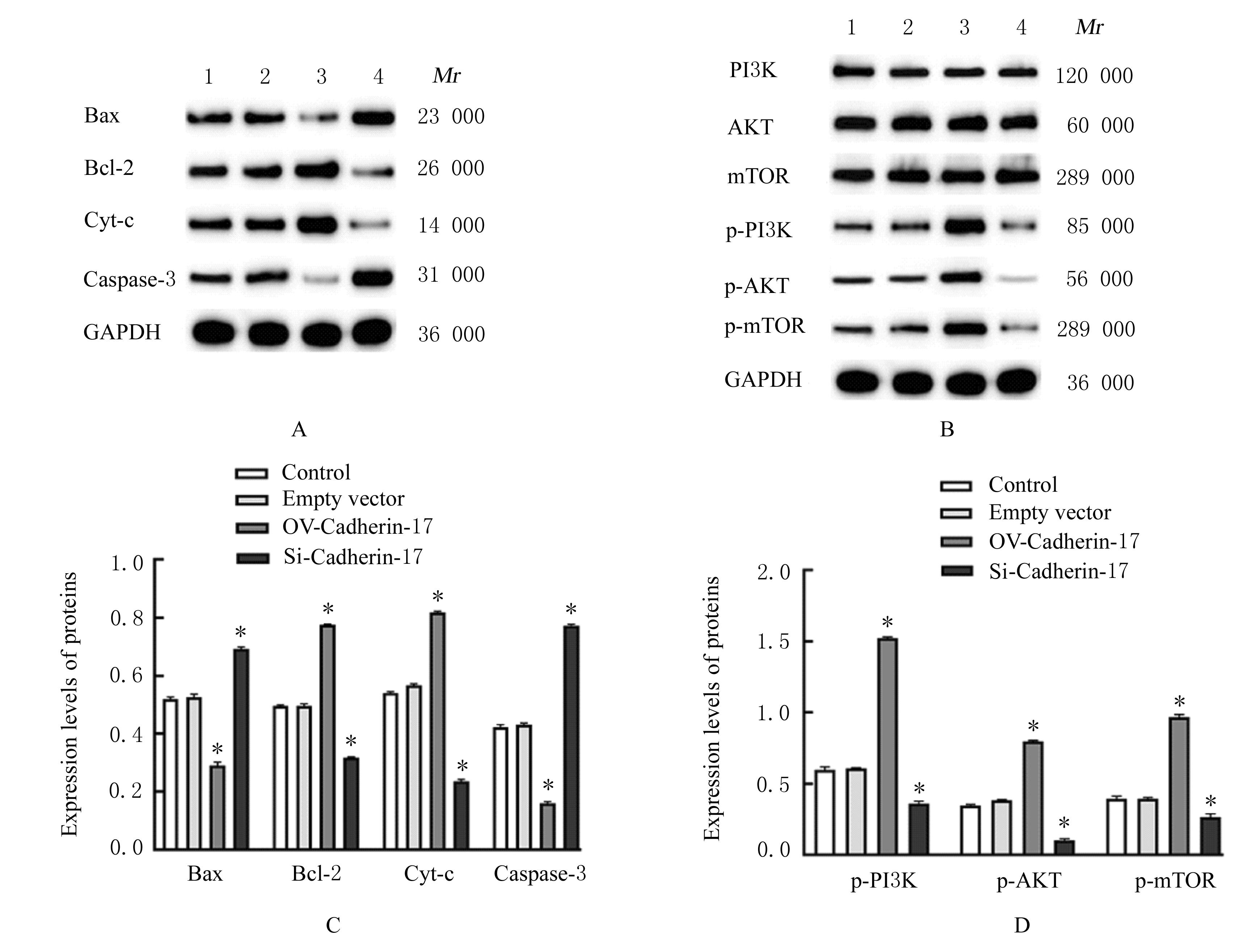
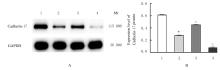
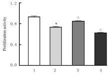
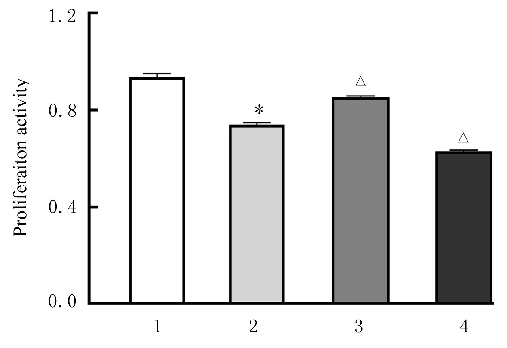
目的 探讨钙黏蛋白17(Cadherin-17)对结直肠癌(CRC)细胞增殖和凋亡的影响,阐明其可能的机制。 方法 构建Cadherin-17基因过表达及小干扰质粒,包装成慢病毒,转染至SW480细胞,构建过表达和干扰病毒稳转株。采用实时荧光定量PCR(RT-qPCR)法和Western blotting法检测细胞中Cadherin-17 mRNA和蛋白表达水平,验证转染效率并鉴定稳转株。SW480细胞分为对照组、空载体组、Cadherin-17过表达质粒(OV-Cadherin-17)组和Cadherin-17小干扰质粒(si-Cadherin-17)组,CCK-8法检测各组细胞增殖活性,流式细胞术检测各组细胞凋亡率,Western blotting法检测各组细胞中Cadherin-17、B细胞淋巴瘤2(Bcl-2)、Bcl-2相关X蛋白(Bax)、细胞色素c(Cyt-c)、含半胱氨酸的天冬氨酸蛋白水解酶3(Caspase-3)和磷脂酰肌醇3-激酶/蛋白激酶B/哺乳动物雷帕霉素靶蛋白(PI3K/AKT/mTOR)信号通路相关蛋白表达水平。采用PI3K抑制剂LY294002处理细胞,将细胞分为对照组、LY294002组、OV-Cadherin-17+LY294002组和si-Cadherin-17+LY294002组,检测各组细胞增殖活性、细胞凋亡率及细胞中Bcl-2、Bax、Cyt-c、Caspase-3和PI3K/AKT/mTOR信号通路相关蛋白表达水平。 结果 RT-qPCR和Western blotting法检测,OV-Cadherin-17和si-Cadherin-17转染和稳转株构建成功。与对照组比较,OV-Cadherin-17组细胞增殖活性明显升高(P<0.01),细胞凋亡率明显降低(P<0.01),细胞中Bax和Caspase-3蛋白表达水平明显降低(P<0.01),Bcl-2和Cyt-c蛋白表达水平明显升高(P<0.01),磷酸化PI3K(p-PI3K)、磷酸化AKT(p-AKT)和磷酸化mTOR(p-mTOR)蛋白表达水平明显升高(P<0.01),si-Cadherin-17组和LY294002组细胞增殖活性明显降低(P<0.01),细胞凋亡率明显升高(P<0.01),细胞中Bax和Caspase-3蛋白表达水平明显升高(P<0.01),Bcl-2和Cyt-c蛋白表达水平明显降低(P<0.01),p-PI3K、p-AKT和p-mTOR蛋白表达水平明显降低(P<0.01);与LY294002组比较,OV-Cadherin-17+LY294002组细胞增殖活性明显升高(P<0.01),细胞凋亡率明显降低(P<0.01),细胞中Bax和Caspase-3蛋白表达水平明显降低(P<0.01),Bcl-2和Cyt-c蛋白表达水平明显升高(P<0.01),p-PI3K、p-AKT和p-mTOR蛋白表达水平明显升高(P<0.01),si-Cadherin-17+LY294002组细胞增殖活性明显降低(P<0.01),细胞凋亡率明显升高(P<0.01),细胞中Bax和Caspase-3蛋白表达水平明显升高(P<0.01),Bcl-2和Cyt-c蛋白表达水平明显降低(P<0.01),p-PI3K、p-AKT和p-mTOR蛋白表达水平明显降低(P<0.01)。 结论 Cadherin-17可促进CRC细胞增殖,抑制CRC细胞凋亡,其机制可能与Cadherin-17调控PI3K/AKT/mTOR信号通路的激活有关。

关键词: 钙黏蛋白17, 结直肠肿瘤, 磷脂酰肌醇3-激酶/蛋白激酶B/哺乳动物雷帕霉素靶蛋白信号通路, 细胞增殖, 细胞凋亡

Abstract:

Objective To discuss the effect of Cadherin-17 on the proliferation and apoptosis of the colorectal cancer (CRC) cells,and to clarify its possible mechanism. Methods The Cadherin-17 gene over-expression and small interference plasmids were constructed and packaged as the lentivirus and transfected into the SW480 cells to construct the stable transfection strain of over-expression and interference virus. The expression levels of Cadherin-17 mRNA and protein in the cells were detected by real-time fluorescence quantitative PCR (RT-qPCR) and Western blotting methods, and the transfection efficiency was verified and the stable transfection strain was identified. The SW480 cells were divided into control group, empty vector group, Cadherin-17 over-expression plasmid (OV-Cadherin-17) group and Cadherin-17 small interference plasmid (si-Cadherin-17) group. The activities of cells in various groups were detected by CCK-8 assay;the apoptotic rates of cells in various groups were detected by flow cytometry; the expression levels of Cadherin-17,B-cell lymphoma-2 (Bcl-2), Bcl2-associated X (Bax), cytochrome c (Cyt-c) and cysteinyl aspartate specific proteinase-3 (Caspase-3),and the phosphatidylinosital-3-kinase/protein kinase B/mamalian target of repamycin (PI3K/AKT/mTOR) signaling pathway-related proteins in the cells in various groups were detected by Western blotting methods. The cells were treated with PI3K inhibitor LY294002 and divided into control group, LY294002 group, OV-Cadherin-17+LY294002 group,and si-Cadherin-17+LY294002 group; the proliferation activities and apoptotic rates of cells in various groups and the expression levels of Bcl-2,Bax,Cyt-c,Caspase-3 and the expression levels of PI3K/AKT/mTOR signaling pathway-related proteins in the cells in various groups were detected. Results The RT-qPCR and Western blotting results showed that the OV-Cadherin-17 and si-Cadherin-17 transfection and stable transfection stain were successfully constructed.Compared with control group, the proliferation ability of the cells in OV-Cadherin-17 group was increased (P<0.01), the apoptotic rate was decreased (P<0.01), the expression levels of Bax and Caspase-3 proteins in the cells were decreased (P<0.01), the expression levels of Bcl-2 and Cyt-c proteins in the cells were increased (P<0.01), and the expression levels of phosphorylated PI3K(p-PI3K),phosphorylated AKT(p-Akt), and phosphorylated mTOR(p-mTOR) proteins were increased (P<0.01); the proliferation abilities of the cells in si-Cadherin-17 and LY294002 groups were decreased (P<0.01), the apoptotic rates were increased (P<0.01), the expression levels of Bax and Caspase-3 proteins in the cells were increased (P<0.01), the expression levels of Bcl-2 and Cyt-c proteins in the cells were decreased (P<0.01),and the expression levels of p-PI3K, p-AKT, and p-mTOR proteins in the cells were decreased (P<0.01); compared with LY294002 group, the proliferation ability of the cells in OV-Cadherin-17+LY294002 group was increased (P<0.01), the apoptotic rate was decreased (P<0.01), the expression levels of Bax and Caspase-3 proteins in the cells were decreased (P<0.01), the expression levels of Bcl-2 and Cyt-c proteins in the cells were increased (P<0.01), the expression levels of p-PI3K, p-AKT, and p-mTOR proteins were increased (P<0.01), the proliferation activity of the cells in si-Cadherin-17+LY294002 group was decreased (P<0.01), the apoptotic rate was increased (P<0.01), the expression levels of Bax and Caspase-3 proteins in the cells were increased (P<0.01), the expression levels of Bcl-2 and Cyt-c proteins in the cells were decreased (P<0.01),and the expression levels of p-PI3K, p-AKT, and p-mTOR proteins were decreased (P<0.01). Conclusion Cadherin-17 can promote the proliferation and inhibit the apoptosis of the CRC cells, and its mechanism may be related to the activition of PI3K/AKT/mTOR signaling pathway regulated by Cadherin-17.

Key words: Cadherin-17, Colorectal neoplasm, Phosphatidylinosital-3-kinase/protein kinase B/mamalian target of repamycin signaling pathway, Cell proliferation, Apoptosis

中图分类号: 

  • R735.34