吉林大学学报(医学版) ›› 2024, Vol. 50 ›› Issue (6): 1572-1586.doi: 10.13481/j.1671-587X.20240611

• 基础研究 • 上一篇    

沉默CD147基因对姜黄素抑制前列腺癌细胞增殖、迁移、侵袭和诱导凋亡的影响

王馨1,赵杰瑞2,郭玉苗3,陈姝彤1,侯宗昊1,张若文1()   

  1. 1.北华大学基础医学院病原生物学教研室,吉林 吉林 132000
    2.澳门科技大学中医药学院附属医院骨外科,广东 珠海 519003
    3.延边大学医学院生物化学教研室,吉林 延吉 133000
  • 收稿日期:2023-12-02 出版日期:2024-11-28 发布日期:2024-12-10
  • 通讯作者: 张若文 E-mail:zrwa1828@163.com
  • 作者简介:王 馨(1998-),女,黑龙江省齐齐哈尔市人,在读硕士研究生,主要从事肿瘤分子生物学方面的研究。
  • 基金资助:
    吉林省科技厅科技发展计划项目(YDZJ202101ZYTS123);北华大学研究生创新项目(研创合字〔2022〕012)

Effect of silencing CD147 gene on proliferation, migration, invasion, and inducing apoptosis of prostate cancer cells inhibited by curcumin

Xin WANG1,Jierui ZHAO2,Yumiao GUO3,Shutong CHEN1,Zonghao HOU1,Ruowen ZHANG1()   

  1. 1.Department of Pathogenic Biology,School of Basic Medical Sciences,Beihua University,Jilin 132000,China
    2.Department of Orthopedics,Affiliated Hospital,Traditional Chinese Medicine,Macao University of Science and Technology,Zhuhai 519000,China
    3.Department of Biochemistry,School of Medical Sciences,Yanbian University,Yanji 133000,China
  • Received:2023-12-02 Online:2024-11-28 Published:2024-12-10
  • Contact: Ruowen ZHANG E-mail:zrwa1828@163.com

摘要:

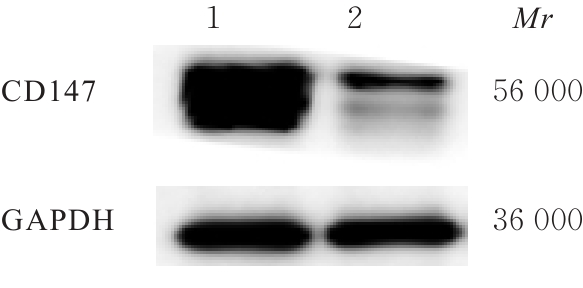
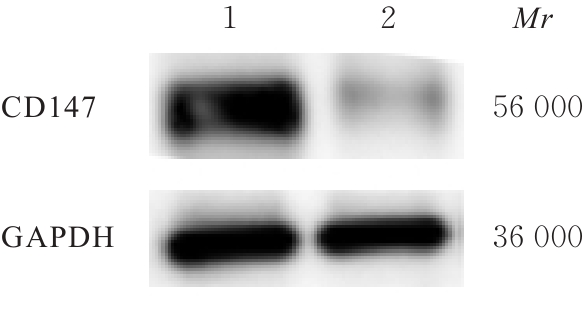
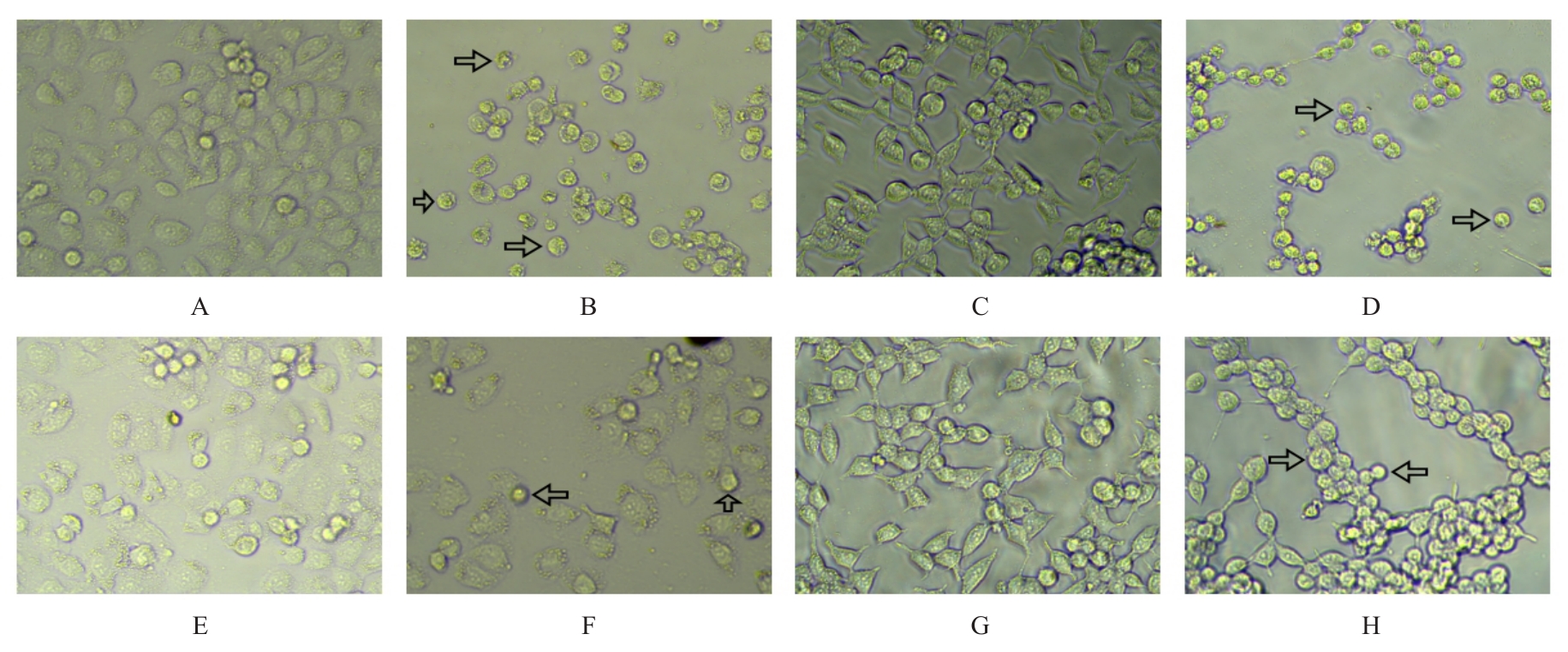
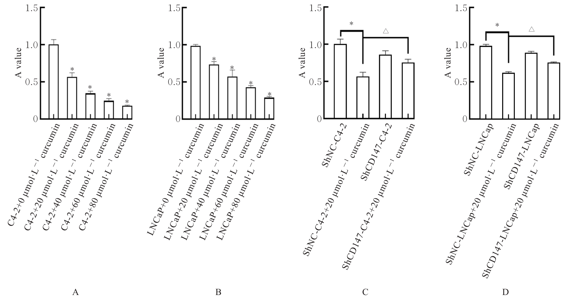
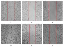
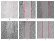
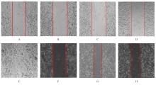
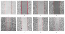
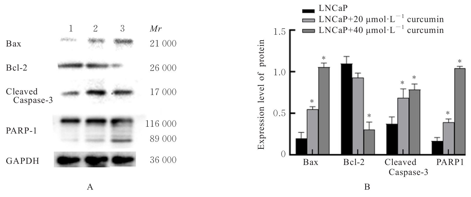
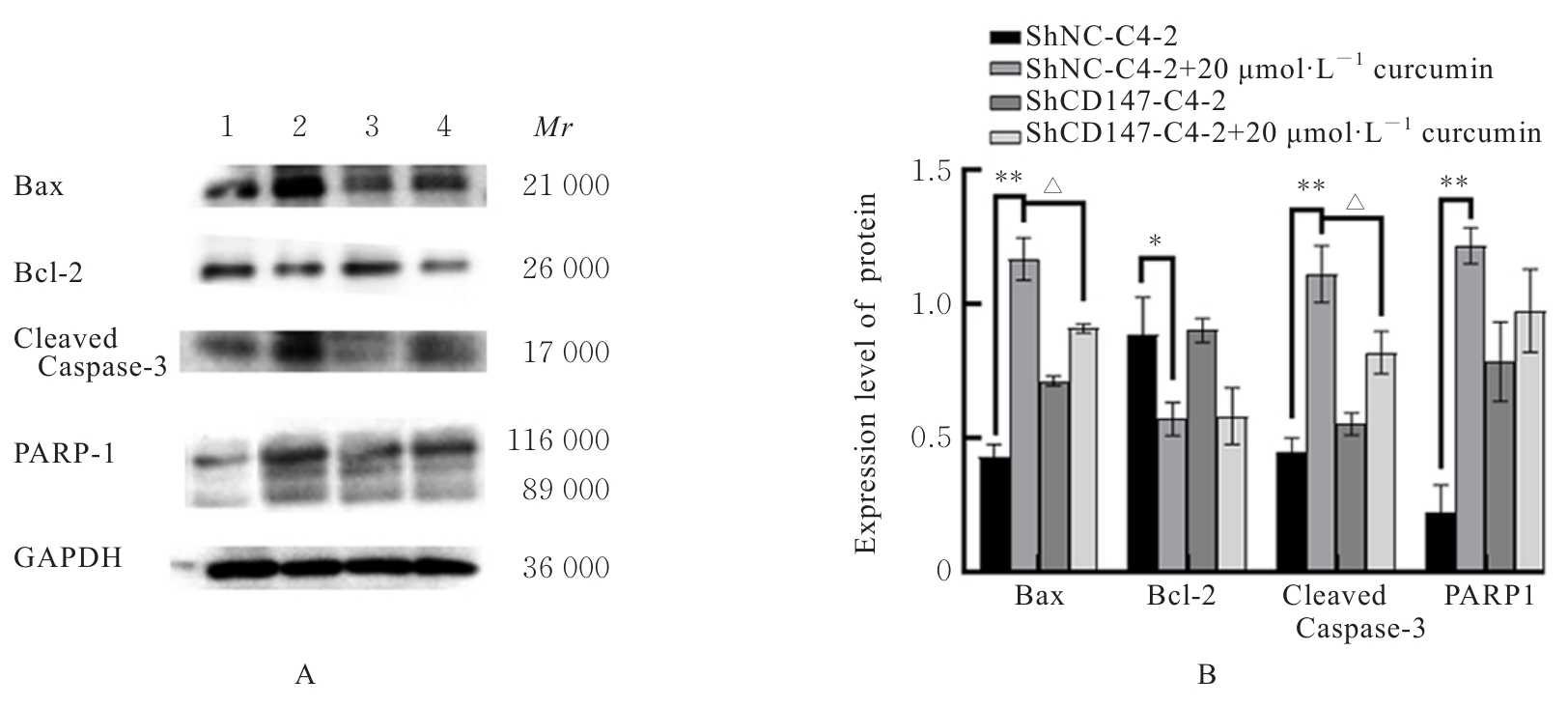
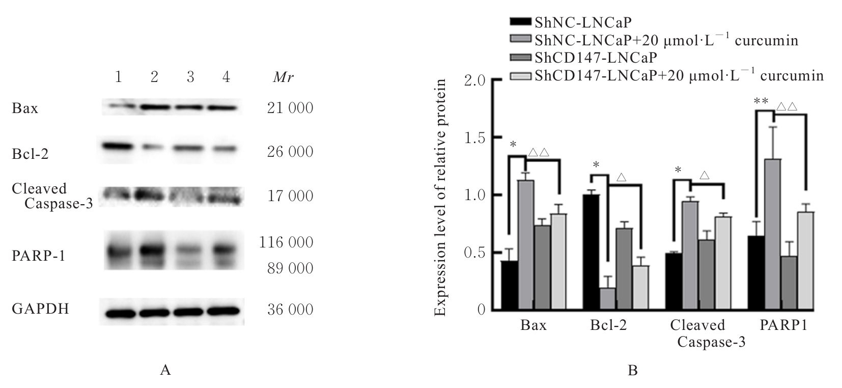
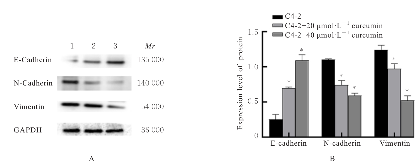
目的 探讨姜黄素对人前列腺癌C4-2细胞和LNCaP细胞增殖、迁移及侵袭的影响,并阐明其可能的作用机制。 方法 采用慢病毒转染系统分别转染C4-2细胞和LNCaP细胞,作为shCD147-C4-2组和shCD147-LNCaP组。采用RNA干扰技术制备沉默CD147基因细胞,以转入空载体的细胞作为阴性对照,分为shNC-C4-2组(shNC-C4-2细胞)和shNC-LNCaP组(shNC-LNCaP细胞)。取生长对数期C4-2、LNCap、shCD147-C4-2和shCD147-LNCaP细胞,加入20 μmol·L-1姜黄素,处理0和24 h时,显微镜观察各组细胞形态表现。噻唑蓝(MTT)法检测各组细胞增殖活性,细胞划痕实验检测各组细胞迁移率,Western blotting法检测各组细胞中凋亡、侵袭和迁移相关蛋白表达水平。 结果 与C4-2组比较,沉默CD147基因后shCD147-C4-2组细胞中CD147蛋白表达量明显减少;与LNCaP组比较,沉默CD147基因后shCD147-LNCaP组细胞中CD147蛋白表达量明显减少。与处理0 h比较,20 μmol·L-1姜黄素处理24 h后C4-2组和LNCaP组部分细胞出现凋亡征象,且有典型凋亡小体存在;shCD147-C4-2组和shCD147-LNCaP组细胞凋亡现象减弱。MTT法检测,与C4-2+ 0 μmol·L-1姜黄素组比较,C4-2+20 μmol·L-1姜黄素组、C4-2+40 μmol·L-1姜黄素组、C4-2+ 60 μmol·L-1姜黄素组和C4-2+80 μmol·L-1姜黄素组细胞增殖活性均明显降低(P<0.01);与LNCaP+ 0 μmol·L-1姜黄素组比较,LNCaP+20 μmol·L-1姜黄素组、LNCaP+40 μmol·L-1姜黄素组、LNCaP+60 μmol·L-1姜黄素组和LNCaP+80 μmol·L-1姜黄素组细胞增殖活性均明显降低(P<0.01);与shNC-C4-2组比较,shNC-C4-2+20 μmol·L-1姜黄素组细胞增殖活性明显降低(P<0.01);与shNC-C4-2+20 μmol·L-1姜黄素组比较,shCD147-C4-2+20 μmol·L-1姜黄素组细胞增殖活性明显升高(P<0.01);与shNC-LNCaP组比较,shNC-LNCaP+20 μmol·L-1姜黄素组细胞增殖活性明显降低(P<0.01);与shNC-LNCaP+20 μmol·L-1姜黄素组比较,shCD147-LNCaP+20 μmol·L-1姜黄素组细胞增殖活性明显升高(P<0.01)。细胞划痕愈合实验检测,姜黄素处理24 h,与C4-2组比较,C4-2+20 μmol·L-1姜黄素组和C4-2+40 μmol·L-1姜黄素组细胞迁移率均明显降低(P<0.01);与LNCaP组比较,LNCaP+20 μmol·L-1 姜黄素组和LNCaP+40 μmol·L-1 姜黄素组细胞迁移率均明显降低(P<0.01);与shNC-C4-2组比较,shNC-C4-2+20 μmol·L-1姜黄素组细胞迁移率明显降低(P<0.01);与shNC-C4-2+20 μmol·L-1姜黄素组比较,shCD147-C4-2+20 μmol·L-1 姜黄素组细胞迁移率明显升高(P<0.05);与shNC-LNCaP组比较,shNC-LNCaP+20 μmol·L-1姜黄素组细胞迁移率明显降低(P<0.01);与shNC-LNCaP+20 μmol·L-1姜黄素组比较,shCD147-LNCaP+20 μmol·L-1姜黄素组细胞迁移率明显升高(P<0.05)。Western blotting法检测,与C4-2组比较,C4-2+20 μmol·L-1姜黄素组和C4-2+40 μmol·L-1姜黄素组细胞中B细胞淋巴瘤2(Bcl-2)相关X蛋白(Bax)、裂解的含半胱氨酸的天冬氨酸蛋白酶3(cleaved Caspase-3)和聚二磷酸腺苷(ADP)-核糖聚合酶1(PARP1)蛋白表达水平均明显升高(P<0.01),Bcl-2蛋白表达水平均明显降低(P<0.05或P<0.01);与LNCaP组比较,LNCaP+20 μmol·L-1姜黄素组和LNCaP+40 μmol·L-1姜黄素组细胞中Bax、cleaved Caspase-3和PARP1蛋白表达水平均明显升高(P<0.01),LNCaP+40 μmol·L-1 姜黄素组Bcl-2蛋白表达水平明显降低(P<0.01);与shNC-C4-2组比较,shNC-C4-2+20 μmol·L-1 姜黄素组细胞中Bax、cleaved Caspase-3和PARP1蛋白表达水平均明显升高(P<0.01),Bcl-2蛋白表达水平明显降低(P<0.05);与shNC-C4-2+20 μmol·L-1姜黄素组比较,shCD147-C4-2+20 μmol·L-1姜黄素组细胞中Bax和cleaved Caspase-3蛋白表达水平均明显降低(P<0.01)。与shNC-LNCaP组比较,shNC-LNCaP+20 μmol·L-1姜黄素组细胞中Bax、cleaved Caspase-3和PARP1蛋白表达水平均明显升高(P<0.05或P<0.01),Bcl-2蛋白表达水平明显降低(P<0.05);与shNC-LNCaP+20 μmol·L-1姜黄素组比较,shCD147-LNCaP+20 μmol·L-1姜黄素组细胞中Bax、cleaved Caspase-3和PARP1蛋白表达水平均明显降低(P<0.05或P<0.01),Bcl-2蛋白表达水平明显升高(P<0.05)。与C4-2组比较,C4-2+20 μmol·L-1姜黄素组和C4-2+40 μmol·L-1姜黄素组细胞中E-钙黏蛋白(E-cadherin)蛋白表达水平均明显升高(P<0.01),神经钙黏蛋白(N-cadherin)和波形蛋白(Vimentin)蛋白表达水平均明显降低(P<0.01);与LNCaP组比较,LNCaP+20 μmol·L-1姜黄素组和LNCaP+40 μmol·L-1姜黄素组细胞中E-cadherin蛋白表达水平均明显升高(P<0.01),LNCaP+40 μmol·L-1姜黄素组细胞中N-cadherin和Vimentin蛋白表达水平均明显降低(P<0.01);与shNC-C4-2组比较,shNC-C4-2+ 20 μmol·L-1姜黄素组细胞中N-cadherin和Vimentin蛋白表达水平均明显降低(P<0.01);与shNC-C4-2+20 μmol·L-1姜黄素组比较,shCD147-C4-2+20 μmol·L-1姜黄素组细胞中E-cadherin蛋白表达水平明显降低(P<0.01),N-cadherin和Vimentin蛋白表达水平均明显升高(P<0.01);与shNC-LNCaP组比较,shNC-LNCaP+20 μmol·L-1姜黄素组细胞中E-cadherin蛋白表达水平明显升高(P<0.01),N-cadherin和Vimentin蛋白表达水平均明显降低(P<0.01);与shNC-LNCaP+20 μmol·L-1姜黄素组比较,shCD147-LNCaP+20 μmol·L-1姜黄素组细胞中E-cadherin蛋白表达水平明显降低(P<0.01),N-cadherin表达水平明显升高(P<0.05)。 结论 姜黄素对体外前列腺癌细胞增殖、迁移和侵袭有抑制作用,并诱导细胞凋亡,沉默CD147基因可在一定程度上降低其抑制作用和诱导细胞凋亡能力。

关键词: 姜黄素, CD147, 前列腺肿瘤, 细胞侵袭, 细胞迁移

Abstract:

Objective To discuss the effect of curcumin on the proliferation, migration, and invasion of the human prostate cancer C4-2 and LNCaP cells, and to clarify its possible mechanism. Methods The lentiviral transfection system was used to transfect the C4-2 and LNCaP cells, regarded as shCD147-C4-2 group and shCD147-LNCaP group. RNA interference technology was used to prepare the CD147-silenced cells; the cells transfected with an empty vector were regarded as negative control and divided into shNC-C4-2 group (shNC-C4-2 cells) and shNC-LNCaP group (shNC-LNCaP cells). The C4-2 and LNCaP cells at logarithmic growth phase, as well as shCD147-C4-2 and shCD147-LNCaP cells, were treated with 20 μmol·L-1 curcumin.The morphology of the cells in various groups was observed under microscope at 0 and 24 h of treatment; MTT method was used to detect the proliferation activities of the cells in various groups; cell scratch assay was used to detect the migration rates of the cells in various groups;Western blotting method was used to detect the expression levels of apoptosis, invasion, and migration-related proteins in the cells in various groups. Results Compared with C4-2 group, the expression of CD147 protein in the cells in shCD147-C4-2 group was significantly decreased after CD147 gene silenting.Compared with LNCaP group, the expression level of CD147 protein in the cells in shCD147-LNCaP group was significantly decreased after CD147 gene silenting. Compared with 0 h of treatment, some cells in C4-2 and LNCaP groups after 24 h of treatment with 20 μmol·L-1 curcumin, showed apoptosis signs with the presence of typical apoptotic bodies. The apoptotic phenomena in shCD147-C4-2 and shCD147-LNCaP groups was reduced.The MTT assay results showed that compared with C4-2+0 μmol·L-1 curcumin group, the proliferation activities of the cells in C4-2+20 μmol·L-1 curcumin group, C4-2+40 μmol·L-1 curcumin group, C4-2+60 μmol·L-1 curcumin group, and C4-2+80 μmol·L-1 curcumin group were decreased (P<0.01). Compared with LNCaP+0 μmol·L-1 curcumin group, the proliferation activity of the cells in LNCaP+20 μmol·L-1 curcumin group, LNCaP+ 40 μmol·L-1 curcumin group, LNCaP+60 μmol·L-1 curcumin group, and LNCaP+80 μmol·L-1 curcumin group were decreased (P<0.01). Compared with shNC-C4-2 group, the proliferation activity of the cells in shNC-C4-2+20 μmol·L-1 curcumin group was decreased (P<0.01). Compared with shNC-C4-2+20 μmol·L-1 curcumin group, the proliferation activity of the cells in shCD147-C4-2+20 μmol·L-1 curcumin group was increased (P<0.01). Compared with shNC-LNCaP group, the proliferation activity of the cells in shNC-LNCaP+20 μmol·L-1 curcumin group was decreased (P<0.01); compared with shNC-LNCaP+20 μmol·L-1 curcumin group, the proliferation activity of the cells in shCD147-LNCaP+20 μmol·L-1 curcumin group was significantly increased (P<0.01). The cell scratch healing assay results showed that compared with C4-2 group, the migration rates of the cells in C4-2+20 μmol·L-1 curcumin group and C4-2+40 μmol·L-1 curcumin group after 24 h of treatment were decreased (P<0.01); compared with LNCaP group, the migration rates of the cells in LNCaP+20 μmol·L-1 curcumin group and LNCaP+40 μmol·L-1 curcumin group were increased(P<0.01); compared with shNC-C4-2 group, the migration rate of the cells in shNC-C4-2+20 μmol·L-1 curcumin group was decreased (P<0.01); compared with shNC-C4-2+20 μmol·L-1 curcumin group, the migration rate of the cells in shCD147-C4-2+20 μmol·L-1 curcumin group was significantly increased (P<0.05); compared with shNC-LNCaP group, the migration rate of the cells in shNC-LNCaP+20 μmol·L-1 curcumin group was decreased (P<0.01); compared with shNC-LNCaP+20 μmol·L-1 curcumin group, the garation rate of the cells in shCD147-LNCaP+20 μmol·L-1 curcumin group was significantly increased (P<0.05).The Western blotting results showed that compared with C4-2 group, the expression levels of Bcl-2-associated X protein (Bax), cleaved Caspase-3, and poly ADP-ribose polymerase 1 (PARP1) proteins in the cells in C4-2+20 μmol·L-1 curcumin group and C4-2+40 μmol·L-1 curcumin group were significantly increased (P<0.01), and the expression levels of Bcl-2 protein was significantly decreased (P<0.05 or P<0.01); compared with LNCaP group, the expression levels of Bax, cleaved Caspase-3, and PARP1 proteins in the cells in LNCaP+20 μmol·L-1 curcumin group and LNCaP+40 μmol·L-1 curcumin group were significantly increased (P<0.01), and the expression level of Bcl-2 protein in the cells in LNCaP+40 μmol·L-1 curcumin group was decreased (P<0.01); compared with shNC-C4-2 group, the expression levels of Bax, cleaved Caspase-3, and PARP1 proteins in the cells in shNC-C4-2+20 μmol·L-1 curcumin group were significantly increased (P<0.05 or P<0.01), and the expression level of Bcl-2 protein was significantly decreased (P<0.05); compared with shNC-C4-2+20 μmol·L-1 curcumin group, the expression levels of Bax and cleaved Caspase-3 proteins in the cells in shCD147-C4-2+20 μmol·L-1 curcumin group were significantly decreased (P<0.01); compared with shNC-LNCaP group, the expression levels of Bax, cleaved Caspase-3, and PARP1 proteins in the cells in shNC-LNCaP+20 μmol·L-1 curcumin group were significantly increased (P<0.05 or P<0.01), and the expression level of Bcl-2 protein was significantly decreased (P<0.05); compared with shNC-LNCaP+20 μmol·L-1 curcumin group, the expression levels of Bax, cleaved Caspase-3, and PARP1 proteins in the cells in shCD147-LNCaP+20 μmol·L-1 curcumin group were significantly decreased (P<0.05 or P<0.01), and the expression level of Bcl-2 protein was significantly increased (P<0.05). Compared with C4-2 group, the expression levels of E-cadherin protein in the cells in C4-2+20 μmol·L-1 curcumin group and C4-2+40 μmol·L-1 curcumin group were significantly increased (P<0.01), and the expression levels of N-cadherin and Vimentin proteins were significantly decreased (P<0.01); compared with LNCaP group, the expression levels of E-cadherin protein in the cells in LNCaP+20 μmol·L-1 curcumin group and LNCaP+40 μmol·L-1 curcumin group were significantly increased (P<0.01), and the expression levels of N-cadherin and Vimentin proteins in the cells in LNCaP+40 μmol·L-1 curcumin group were significantly decreased (P<0.01); compared with shNC-C4-2 group, the expression levels of N-cadherin and Vimentin proteins in the cells in shNC-C4-2+20 μmol·L-1 curcumin group were significantly decreased (P<0.01); compared with shNC-C4-2+20 μmol·L-1 curcumin group, the expression level of E-cadherin protein in the cells in shCD147-C4-2+20 μmol·L-1 curcumin group was significantly decreased(P<0.01), and the expression levels of N-cadherin and Vimentin proteins were significantly increased (P<0.01); compared with shNC-LNCaP group, the expression level of E-cadherin protein in the cells in shNC-LNCaP+20 μmol·L-1 curcumin group was significantly increased (P<0.01), and the expression levels of N-cadherin and Vimentin proteins were significantly decreased (P<0.01); compared with shNC-LNCaP+20 μmol·L-1 curcumin group, the expression level of E-cadherin protein in the cells in shCD147-LNCaP+20 μmol·L-1 curcumin group was significantly decreased (P<0.01), and the expression level of N-cadherin was significantly increased (P<0.05). Conclusion Curcumin inhibits the proliferation, migration, and invasion of the prostate cancer cells in vitro and induces the apoptosis; silencing the CD147 gene partially reduces its inhibitory effect and its ability to induce the apoptosis.

Key words: Curcumin, CD147, Prostate neoplasm, Cell invasion, Cell migration

中图分类号: 

  • R735.25